A new genus and species of treefrog from Medog, southeastern Tibet, China (Anura, Rhacophoridae)
2016-03-22 03:35KeJIANGFangYANKaiWANGDaHuZOUChengLIJingCHE
Zoological Research 2016年1期
Ke JIANG, Fang YAN, Kai WANG, Da-Hu ZOU, Cheng LI, Jing CHE
1State Key Laboratory of Genetic Resources and Evolution, Kunming Institute of Zoology, Chinese Academy of Sciences, Kunming Yunnan 650223, China2Sam Noble Oklahoma Museum of Natural History and Department of Biology, University of Oklahoma, Norman OK 73072-7029, U.S.A.3Tibet University, Lhasa Tibet 850000, China4Imaging Biodiversity Expedition, Beijing 100107, China
A new genus and species of treefrog from Medog, southeastern Tibet, China (Anura, Rhacophoridae)
Ke JIANG1,#, Fang YAN1,#, Kai WANG2,1, Da-Hu ZOU3,1, Cheng LI4, Jing CHE1, *
1State Key Laboratory of Genetic Resources and Evolution, Kunming Institute of Zoology, Chinese Academy of Sciences, Kunming Yunnan 650223, China
2Sam Noble Oklahoma Museum of Natural History and Department of Biology, University of Oklahoma, Norman OK 73072-7029, U.S.A.3Tibet University, Lhasa Tibet 850000, China
4Imaging Biodiversity Expedition, Beijing 100107, China
Received: 27 October 2015; Accepted: 15 December 2015
Foundation items: This study was supported by the Ministry of Science and Technology of China (2014FY210200, 2011FY120200) and the Animal Branch of the Germplasm Bank of Wild Species of Chinese Academy of Sciences (the Large Research Infrastructure Funding).
#Authors contributed equally to this work
ABSTRACT
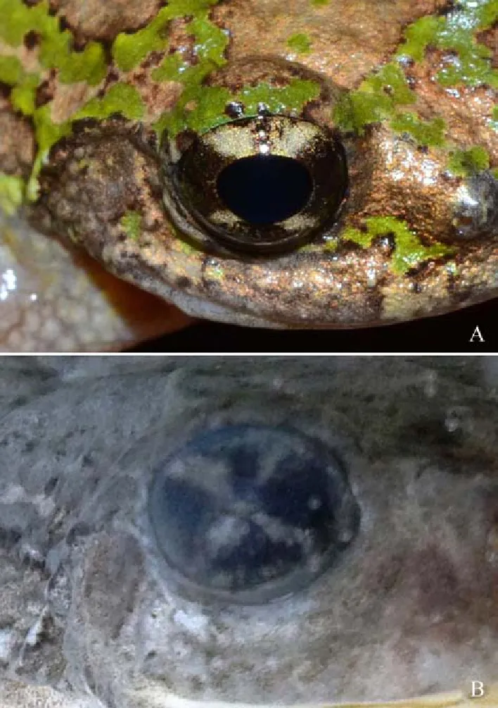
A new genus and species of threefrog is described from Medog, southeastern Tibet, China based on morphological and phylogenetic data. The new genus can be distinguished from other treefrog genera by the following combination of characters: (1) body size moderate, 45.0 mm in male; (2) snout rounded; (3) canthus rostralis obtuse and raised prominently, forming a ridge from nostril to anterior corner of eyes; (4) web rudimentary on fingers; (5) web moderately developed on toes; (6) phalange “Y” shaped, visible from dorsal side of fingers and toes; (7) skin of dorsal surfaces relatively smooth, scatted with small tubercles; (8) iris with a pale yellow, “X” shaped pattern of pigmentation.
Keywords:Taxonomy; New genus; New species; Theloderma moloch; Nasutixalus medogensis sp. nov.
INTRODUCTION
The Old World Treefrogs in the family Rhacophoridae are arboreal, occupying different ecological niches from low shrub to tree clown habitats (Wells, 2010). Currently the family consists of 393 recognized species in 17 genera (Frost, 2015), of which 105 species in 11 genera are found in southern and southwestern China (AmphibiaChina, 2015; Frost, 2015). Within China, the Medog (=Motuo) County at the southern slope of the Himalaya in southeastern Tibet harbors 16 known species of Rhacophoridae treefrogs from eight genera, about 15% of the total diversity of the family in China (AmphibiaChina, 2015).
However, despite the rich treefrog diversity of Medog, few detailed surveys have been done in the region, and much is unknown about the treefrog diversity, and the taxonomy of many species of the region remained unclear. For example, the endemic Mossy Treefrog Theloderma moloch (Annandale, 1912) was described based on two specimens from southern Medog. For nearly a century, there are no further reports or re-description of the species, and its species boundary is solely delimited based on the original description.1
During a herpetological survey of southeastern Tibet in 2015, a male treefrog was collected from the tree crown in the tropical rain forest at Medog. Phylogentic analysis revealed that this specimen shared the same haplotype with a specimen (6255 RAO) also from Medog that was identified as T. moloch in Li et al. (2009). However, morphological comparisons reveal that the treefrog we collected from Medog is distinguished readily from the true T. moloch by a suit of morphological characters, and both our specimen and the specimen (6255 RAO) in Li et al. (2009) formerly identified T. moloch formed a distinct lineage and diverged from the genus Theloderma and all other known genera in the family Rhacophoridae. Therefore, according to the morphological and molecular phylogenetic data of mitochondrial DNA, we describe a new species and a new genus based on our treefrog specimen from Medog. Phylogenetic position of the previously identified Theloderma specimen (6255 RAO) in Li et al. (2009) is also discussed.
MATERIALS AND METHODS
A single male specimen was collected from Gelin, Medog, southeastern Tibet, China. Following euthanasia, liver tissues was taken and preserved in 95% ethanol, and the specimen was fixed in 10% formalin solution and was transferred to 75% ethanol after fieldwork. The male specimen (KIZ 016395) wasdesignated as the holotype, and was deposited in Kunming Institute of Zoology, Chinese Academy of Sciences.
Morphological comparisons: All measurements were carried out with slide calipers to the nearest 0.1 mm. Morphological characters used and their measurement methods followed Fei et al. (2009), webbing formula followed Savage & Heyer (1997). The morphological characters and their abbreviations as: SVL, snout-vent length; HL, head length; HW, head width; SL, snout length; INS, internarial distance; IOS, interorbital distance; EHD, eye horizontal diameter; UEW, maximum width of upper eyelid; TD, tympanum diameter; FAHL, forearm and hand length; FAW, maximum width of forearm; HAL, hand length; FML, femur (thigh) length; TBL, tibia (shank) length; TFL, length of tarsus and foot; FOL, foot length.
Morphological data of congeners were obtained from vouchered specimens (Appendix) as well as from literatures (Annandale, 1912; Fei et al., 2009). The following museum abbreviations were used: CIB-Chengdu Institute of Biology, Chinese Academy of Sciences, Chengdu, China. KIZ-Kunming Institute of Zoology, Chinese Academy of Sciences, Kunming, China.
Molecular analysis: Total DNA of the single treefrog specimen (KIZ016395) from Gelin and two other known species (Theloderma beibengensis and T. moloch) were extracted with a standard three-step phenol-chloroform extraction method (Sambrook et al., 1989). A 1 999 base pair DNA sequence of mitochondrial gene 12S rRNA, tRNAVAL, and 16S rRNA (12S-16S) was sequenced using primers L2519 and 16Sbr (Table 1). Amplifications were conducted in a 25 uL volume reaction, involved initial denaturing step at 94 °C for 5 min; then 35 cycles of denaturing at 94 °C for 45 sec, annealing at 55 °C for 45 sec, and extending at 72 °C for 45 sec; and a final extending step of 72 °C for 7 min. The novel sequences were deposited in GenBank (Table 1). The 12S-16S sequences of other 38 specimens were downloaded from GenBank (Table 1).
All dataset were aligned and edited using MEGA 5 (Tamura et al., 2011). The best model of nucleotide substitution was calculated in Modeltest v1.0.1 (Posada, 1998). The phylogenetic relationship was conducted using Bayesian inference (BI) method with software MrBayes 3.1.2 (Ronquist & Huelsenbeck, 2003).
RESULTS
Morphological comparison
The male specimen is moderate body size, snout rounded, canthus rostralis obtuse and raised prominently, discs on fingers and toes moderate, rudimentary web on fingers and moderately developed web on toes, skin of dorsal surfaces relatively smooth, scatted some small tubercles, iris with a pale yellow, “X” shaped pattern of pigmentation. The specimen distinguished mainly from the genus Theloderma by the absence of large tubercles and jagged skin ridge on dorsum, the presence of prominently raised canthus rostralis, and iris with a pale yellow, “X” shaped pattern of pigmentation.
Phylogenetic analysis
Currently recognized genera of the family Rhacophoridae were recovered as monophyletic groups in our phylogenetic analysis (Figure 1). However, similar to previous studies (Li et al., 2009), our data cannot resolve phylogenetic relationships among different genera. The Medog treefrog was clustered and share the haplotype with a formerly identified T. moloch (specimen voucher number 6255 RAO) in Li et al. (2009), and both of them form a distinct clade from all other species of genus Theloderma, including the true T. moloch. Such result is similar to the phylogenetic topography recovered in Li et al. (2013) using more datasets.
Therefore, according to a combination of morphological characters and phylogenetic data of mitochondrial genes, we conclude that the male treefrog specimen (KIZ016395) consisted an independent evolutionary lineage and concordant evidence confirm species status (Hou et al., 2014; Wu & Murphy, 2015), which is described as a new species and a new genus in family Rhacophoridae.
Nasutixalus gen. nov. Jiang, Yan, Wang and Che
Type species: Nasutixalus medogensis sp. nov.
Diagnosis: (1) Body size moderate (45.0 mm in male); (2) snout rounded; (3) canthus rostralis obtuse and raised prominently, forming a ridge from nostril to anterior corner of eyes; (4) web rudimentary on fingers; (5) web moderately developed on toes; (6) phalange “Y” shaped, visible from dorsal side of fingers and toes; (7) skin of dorsal surfaces relatively smooth, scatted with small tubercles; (8) iris with a pale yellow, “X” shaped pattern of pigmentation, especially distinct in preservative.
Distribution: Currently known only from the type locality, Medog, Tibet, China.
Etymology: The generic nomen Nasutixalus is derived from the Latin adjective nasutus (“large-nosed” in English), means the prominent ridge from nostril to the anterior corner of eye, and ixalus, a common generic root for treefrogs. We suggest the common name of the new genus be “ridged-nose treefrog”in English, and “Leng Bi Shu Wa” (棱鼻树蛙) in Chinese.
Content: The new genus currently contains a single species, Nasutixalus medogensis sp. nov. which is described below.
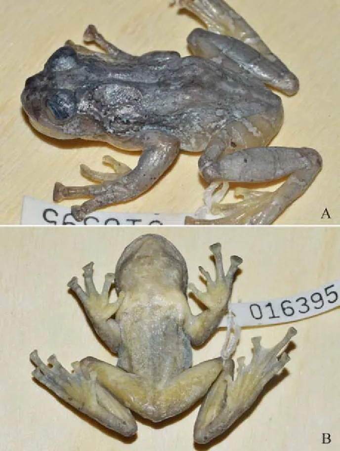
Nasutixalus medogensis sp. nov. Jiang, Wang, Yan and Che (Figures 2-4)
Synonyms
Thermoderma moloch: Li et al., 2009
Holotype: KIZ 016395, adult male, collected from Gelin (N29.21665°, E95.17571°, elevation 1 619 m), Beibeng, Medog, southeastern Tibet, China, on 28 April 2015, by Ke JIANG.
Diagnosis: As for the generic diagnosis.
Description of holotype: Body size moderate, SVL 45.0 mm; body stout, moderately slender at waist. Head width nearly as equal to length (HW/HL=1.04); snout rounded, slightly projecting beyond jaw; canthus rostralis distinct, obtuse, and raised prominently, forming a ridge from nostril to anterior
corner of eye; loreal region oblique, concave; internarial distance 1.14 times larger than interorbital distance. Eyes large, prominent, eye diameter 0.39 times of head length; pupil rounded. Interorbital region flat, interorbital distance 1.1 times larger than upper eyelid width. Tympanum distinct, 0.4 times of orbit diameter. Tongue pyriform, deeply notch behind, papillae absent; choanae large, visible viewed from below; a pair of vomerine teeth on inner sides of choanae; single, external, subgular vocal sac present, with a pair of small openings near inner corners of mouth; supratympanic fold from posterior corner of orbit to previous shoulder, distinctly developed but slender.

Table 1 Information of samples used in molecular analysis

Figure 1 Bayesian inference tree based on molecular data of Rhacophoridae and two outgroups

Table 2 Morphological measurements (mm) of the holotype of Nasutixalus medogensis sp. nov.
Fore-limbs long and strong; forearm and hand slightly longer than half of body length; lower arm thick; fingers compressed with discs; circummarginal grooves present; relative length of fingers: I Hind limbs relatively long, tibiotarsal articulation reaching the eye when adpressed; heels much overlapped when flexed and held perpendicular to body; shank nearly as equal to thigh (TBL/FML=1.02); foot length nearly as equal to shank (FOL/TBL=1.04); relative toe lengths: I Figure 2 Different views of the holotype (KIZ 016395) of Nasutixalus medogensis sp. nov. in life (Photos by Ke JIANG) Figure 3 Different views of the holotype (KIZ 016395) of Nasutixalus medogensis sp. nov. in preservative (Photos by Ke JIANG) Skin of dorsal surfaces of head, body and limbs relatively smooth, with small tubercles scattered; loreal and temporal region, and lateral body rough, with distinct tubercles; ventral surface with serried flat tubercles, relatively small on throat, chest and ventral forelimbs, relatively large on belly and ventral thigh; tubercles on basal ventral thigh prominent. Figure 4 Right eye of the holotype, showing the iris with a pale yellow, “X” shaped pattern of pigmentation, in life (A) and preservative (B) (Photos by Ke JIANG) Coloration of holotype in life Coloration of the dorsal and lateral surfaces of head and body are camouflage of pistachio and creamy brown. A distinct, creamy brown, reversed triangular pattern of pigmentation is observed on the dorsal surface of the head, with its base positioned between the orbits. The tip of the triangular pattern of pigmentation extends posteriorly to the pectoral region and connects with the large, creamy brown, “X”shaped pattern of pigmentation that extends further posteriorly and laterally to the pelvis. The coloration of the iris is dark blackish, with a distinct pale yellow, “X” shaped pattern of pigmentation; pupil is jet black. Dorsal surfaces of the limbs are creamy brown, which is more saturated on the disc of fingers and toes. Distinct, pistachio transverse bands were observed on the dorsal surfaces of limbs from the proximal end to the fingers/toes. Lateral surfaces of hind limbs and are yellowish. Coloration of the ventral surfaces of the head, body and limbs are pale flesh color. A single patch of light creamy yellow pigmentations is observed on the chest ventral to axilla on each side. The abdominal region and the ventral surfaces of feet are slightly creamy yellowish. Coloration in preservative The patterns of pigmentation in preservative closely resemble the patterns in life. However, the following coloration changes after preservation process: (1) The pistachio and creamy brown coloration of the dorsal and lateral surfaces of head, body, and limbs become light and dark gray respectively; (2) the patches on the chest, ventral surfaces hind limbs, and ventral surfaces of hands and feet are light yellow, and remaining parts of the ventral body and limbs become light gray. Etymology The species name “medogensis” is named after the type locality, Medog, Tibet, China. According to the Latin name, we suggest the English common name as “Medog Ridged-nose Treefrog”, and the Chinese formal name as “Muo Tuo Leng Bi Shu Wa”(墨脱棱鼻树蛙). In the recent phylogenetic study, Li et al. (2009) showed that a single specimen (6225 RAO) of treefrog from Medog that was formerly identified as T. moloch, is remotely related to the genus Theloderma and represents a distinct clade. Therefore, Li et al. (2009) concluded T. moloch was not a member of the genus Theloderma. However, our phylogenetic results show that the true T. moloch from Medog (Hou et al., unpublished data) is nested within the genus Theloderma and is distantly related to the specimen (6225 RAO) from Li et al. (2009). Furthermore, our results show that the specimen in Li et al. (2009) is clustered and share the haplotype with our new species of the new genus with well-supported, hence it is a conspecific of our new species. Therefore, we recommend further surveys focus on the high tree crown and collect additional specimens of species from the new genus to provide further data to resolve the remaining taxonomic and phylogenetic issues. We thank to Dr. Pi-Peng LI (Shenyang Normal University), Dr. Guang-Pu GUO (Tongji University), and the volunteers, Mr. Tao LIANG, Mr. Duan YOU, and Mr. Ya-Di HUANG, who helped with fieldwork in Tibet. AmphibiaChina. 2015. The database of Chinese amphibians. Kunming, Yunnan. Available at http: //www.amphibiachina.org/. Annandale N. 1912. Zoological results of the Abor Expedition, 1911–1912. I. Amphibia. Records of the Indian Museum, 8: 7-36. Fei L, Hu SQ, Ye CY, Huang YZ. 2009. Fauna Sinica, Amphibia. Vol. 2. Beijing, Science Press, 1-957. (in Chinese) Frost DR. 2015. Amphibian Species of the World: an Online Reference. Version 6.0 (Nov 26, 2015). Electronic Database accessible at http: // research.amnh.org/herpetology/amphibia/index.html. American Museum of Natural History, New York, USA. Hou M, Wu YK, Yang KL, Zheng S, Yuan ZY, Li PP. 2014. A missing geographic link in the distribution of the genus Echinotriton (Caudata: Salamandridae) with description of a new species from southern China. Zootaxa, 3895: 89-102 Li JT, Che J, Murphy WR, Zhao H, Zhao EM, Rao DQ, Zhang YP. 2009. New insights to the molecular phylogenetics and generic assessment in the Rhacophoridae (Amphibia: Anura) based on five nuclear and three mitochondrial genes, with comments on the evolution of reproduction. Molecular Phylogenetics and Evolution, 53 (2): 509-522. Li JT, Li Y, Klaus S, Rao DQ, Hillis MD, Zhang YP. 2013. Diversification of rhacophorid frogs provides evidence for accelerated faunal exchange between India and Eurasia during the Oligocene. Proceedings of the National Academy of Sciences of the United States of America, 110 (9): 3441-3446. Posada D, Crandall KA. 1998. MODELTEST: testing the model of DNA substitution. Bioinformatics, 14 (9): 817-818. Ronquist F, Huelsenbeck JP. 2003. MrBayes 3: Bayesian phylogenetic inference under mixed models. Bioinformatics, 19 (12): 1572-1574. Sambrook J, Fritsch E, Maniatis T. 1989. Molecular Cloning: A Laboratory Manual (2nd edn). New York: Cold Spring Harbor Laboratory Press. Savage JM, Heyer WR. 1997. Digital webbing formulae for anurans: a refinement. Herpetological Review, 28 (3): 131. Tamura K, Peterson D, Peterson N, Stecher G, Nei M, Kumar S. 2011. MEGA5: molecular evolutionary genetics analysis using maximum likelihood, evolutionary distance, and maximum parsimony methods. Molecular Biology and Evolution, 28 (10): 2731-2739. Wells KD. 2010. The Ecology and Behavior of Amphibians. Chicago and London: The University of Chicago Press. Wu YK, Murphy RW. 2015. Concordant species delimitation from multiple independent evidence: A case study with the Pachytriton brevipes complex (Caudata: Salamandridae). Molecular Phylogenetic and Evolution, 92: 108-117. The following specimens were examined: Polypedates cf. braueri: KIZ 06994, KIZ 0697-98, KIZ 07001, KIZ 010987-89, KIZ 010991, KIZ 010993, KIZ 011034 (10♂♂); KIZ 06993, KIZ 07000, KIZ 07002, KIZ 010990, KIZ 010992, KIZ 011016, KIZ 011029-30 (8♀♀). Medog, Tibet. Gracixalus medogensis: KIZ 010956 (1♂). Medog, Tibet. Kurixalus naso: KIZ 010962-64, KIZ 010967, KIZ 010976, KIZ 011003, KIZ 011005, KIZ 011009-10, KIZ 011023 (1010♂♂); KIZ 010966, KIZ 010977, KIZ 011006, KIZ 011028 (4♀♀). Medog, Tibet. Rhacophorus bipunctatus: KIZ 06999, KIZ 07003-007, KIZ 010979, KIZ 010983, KIZ 011044, KIZ 011047 (10♂♂); KIZ 010960, KIZ 010980 (2♀♀). Medog, Tibet. Rhacophorus burmanus: KIZ 016379-80, KIZ 016408-09, KIZ 016441-44, KIZ 016446-47 (10♂♂); KIZ 010957, KIZ 016376-78, KIZ 016410, KIZ 016445 (6♀♀). Medog, Tibet. Rhacophorus maximus: KIZ 016384 (1♂); KIZ 016385, KIZ 013859 (2♀♀). Medog, Tibet. Rhacophorus translineatus: KIZ 06648-52, KIZ 011161-62, KIZ 011167-70 (11♂♂); KIZ 012683 (1♀). Medog, Tibet. Feihyla vittata: KIZ 07352, KIZ 011171-72, KIZ 012708-10 (6♂♂); KIZ 07353-35 (3♀♀). Medog, Tibet. Theloderma beibengensis: KIZ 020453 (1♂). Medog, Tibet. DOI:10.13918/j.issn.2095-8137.2016.1.15 *Corresponding author, E-mail: chej@mail.kiz.ac.cn
DISCUSSION
ACKNOWLEDGEMENTS
REFERENCES
APPENDIX
猜你喜欢
Asian Herpetological Research(2020年4期)2020-12-30作文评点报·高中版(2020年38期)2020-11-06小哥白尼(野生动物)(2020年5期)2020-09-24课外生活(小学1-3年级)(2020年8期)2020-08-14中国记者(2020年2期)2020-03-28小学生必读(低年级版)(2019年3期)2019-07-08小学科学(学生版)(2018年7期)2018-08-13Asian Herpetological Research(2018年2期)2018-06-28台港文学选刊(2016年5期)2017-01-07华人时刊(2016年19期)2016-04-05