
End-to-side neurorrhaphy repairs peripheral nerve injury: sensory nerve induces motor nerve regeneration
2017-11-08 11:49QingYuShehongZhangTaowangFengPengDongJanYudongGu
中国神经再生研究(英文版) 2017年10期
Qing Yu, She-hong Zhang, Tao wang, Feng Peng, Dong Jan, Yu-dong Gu,
1 Department of Hand Surgery, Huashan Hospital of Fudan University, Key Laboratory of Hand Reconstruction, the Ministry of Health, Key
Laboratory of Peripheral Nerve and Microsurgery, Shanghai, China
2 Department of Rehabilitation Medicine, Huashan Hospital of Fudan University, Shanghai, China
How to cite this article: Yu Q, Zhang SH, Wang T, Peng F, Han D, Gu YD (2017) End-to-side neurorrhaphy repairs peripheral nerve injury:sensory nerve induces motor nerve regeneration. Neural Regen Res 12(10):1703-1707.
End-to-side neurorrhaphy repairs peripheral nerve injury: sensory nerve induces motor nerve regeneration
Qing Yu1, She-hong Zhang2, Tao wang1, Feng Peng1, Dong Jan1, Yu-dong Gu1,*
1 Department of Hand Surgery, Huashan Hospital of Fudan University, Key Laboratory of Hand Reconstruction, the Ministry of Health, Key
Laboratory of Peripheral Nerve and Microsurgery, Shanghai, China
2 Department of Rehabilitation Medicine, Huashan Hospital of Fudan University, Shanghai, China
How to cite this article: Yu Q, Zhang SH, Wang T, Peng F, Han D, Gu YD (2017) End-to-side neurorrhaphy repairs peripheral nerve injury:sensory nerve induces motor nerve regeneration. Neural Regen Res 12(10):1703-1707.
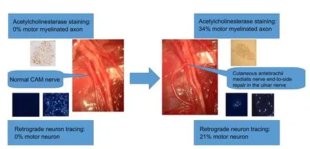
End-to-side neurorrhaphy is an option in the treatment of the long segment defects of a nerve. It involves suturing the distal stump of the disconnected nerve (recipient nerve) to the side of the intimate adjacent nerve (donor nerve). However, the motor-sensory specificity aer end-to-side neurorrhaphy remains unclear.is study sought to evaluate whether cutaneous sensory nerve regeneration induces motor nerves aer end-to-side neurorrhaphy.irty rats were randomized into three groups: (1) end-to-side neurorrhaphy using the ulnar nerve(mixed sensory and motor) as the donor nerve and the cutaneous antebrachii medialis nerve as the recipient nerve; (2) the sham group:ulnar nerve and cutaneous antebrachii medialis nerve were just exposed; and (3) the transected nerve group: cutaneous antebrachii medialis nerve was transected and the stumps were turned over and tied. At 5 months, acetylcholinesterase staining results showed that 34% ±16% of the myelinated axons were stained in the end-to-side group, and none of the myelinated axons were stained in either the sham or transected nerve groups. Retrograde fluorescent tracing of spinal motor neurons and dorsal root ganglion showed the proportion of motor neurons from the cutaneous antebrachii medialis nerve of the end-to-side group was 21% ± 5%. In contrast, no motor neurons from the cutaneous antebrachii medialis nerve of the sham group and transected nerve group were found in the spinal cord segment.ese results confirmed that motor neuron regeneration occurred aer cutaneous nerve end-to-side neurorrhaphy.
nerve regeneration; peripheral nerve injury; end-to-side neurorrhaphy; motor-sensory specificity; rat; dorsal root ganglion; motor neuron; axon; cutaneous antebrachii medialis nerve; ulnar nerve; acetylcholinesterase staining; retrograde neuron tracing; neural regeneration
Introduction
Peripheral nerve injury is a common result of trauma (Reyes et al., 2005; Chen et al., 2007; Sullivan et al., 2016).e incidence of peripheral nerve injury in patients with multiple injuries was 2–2.8% (Selecki et al., 1982; Noble et al., 1998),and this injury may cause severe disability. End-to-side neurorrhaphy is an alternative procedure for some nerve injury repairs (Lutz et al., 2000; Hanna and Dempsey, 2013; Isaacs,2013; Gao et al., 2015; Li et al., 2015). It consists of suturing the distal stump of the disconnected nerve (recipient nerve)to the side of the intimate adjacent nerve (donor nerve)(Viterbo et al., 1998; Al-Qattan, 2001; Yan et al., 2002; Ka-kibuchi et al., 2004; Baltzer et al., 2016).is technique was used a long time ago, but was recently revived by Viterbo et al. (1992) in an experimental study of rats. Although the value of end-to-side nerve repair is still controversial, experimental evidence suggests that it promotes axonal growth and nerve regeneration in the receptors of motor or sensory nerves(Yuksel et al., 1999; De Sa et al., 2004; Hayashi et al., 2004;Haastert et al., 2010; Gao et al., 2012; Zheng et al., 2012; Hanna and Dempsey, 2013; Kettle et al., 2013; Civi et al., 2017).
We have concentrated our investigations on the motor-sensory specificity after end-to-side neurorrhaphy. In previous studies, we found that the motor or sensory specificity was not clear when a mixed nerve was the recipient nerve (Yu et al., 2011, 2013). In this study, we used a rat ulnar nerve (mixed sensory and motor) as the donor nerve and the cutaneous antebrachii medialis nerve as the recipient sensory nerve.e aim of this study was to assess whether a cutaneous nerve can induce motor nerve regeneration after end-to-side neurorrhaphy, and explore the sensory/motor specificity aer end-to-side suturing.
Materials and Methods
Animals and surgery procedures
Study protocols were approved by the Animal Ethics Committee of Wenzhou Medical University of China (approval No. 201601-005). The experimental procedure followed the United States National Institutes of Health Guide for the Care and Use of Laboratory Animals (NIH Publication No. 85–23, revised 1986).
Rats were randomly divided into end-to-side group (n =12), sham group (n = 12) and the transected nerve group (n =6). Surgery was performed as described previously (Yu et al.,2011). Briefly, rats were anesthetized by intraperitoneal injection of ketamine (40 mg/kg, Shanghai Ziyuan Pharmaceutical Co., Ltd., Shanghai, China) and atropine (0.04 mg/kg, Shanghai Ziyuan Pharmaceutical Co., Ltd.). The right cutaneous antebrachii medialis nerve and ulnar nerves were exposed.For rats in the end-to-side group, we cut a window (diameter,1 mm) in the ulnar nerve epineurium.e cutaneous antebrachii medialis nerve was transected at the root level, and the distal stump was then sutured to the ulnar nerve medial window with 10/0 non-invasive monofilament polyamide(Ethicon, San Angelo, TX, USA) to form an end-to-side junction. The proximal stump of the cutaneous antebrachii medialis nerve was ligated tightly and turned back to prevent spontaneous innervation (Figure 1). In the sham group, we exposed the cutaneous antebrachii medialis nerve and ulnar nerve, and then closed the skin. In the transected nerve group, we cut the cutaneous antebrachii medialis nerve, ligated the two ends, and turned them back to prevent spontaneous innervation. Aer regaining consciousness, the rats were housed separately in boxes in an approved animal house facility.
Acetylcholinesterase staining
Application of neuron tracer
Five months after surgery, the fluorescence retrograde axon tracer Fluoro-Gold (Sigma, St. Louis, MO, USA) was used to demonstrate axonal growth after end-to-side neurorrhaphy(McBride et al., 1990; Byers and Lin, 2003; Chiu et al., 2008;Chen et al., 2012). As described above, the animals were re-anesthetized, and 1 μL 3% Fluoro-Gold dyes were injected into the cutaneous antebrachii medialis nerve (end-to-side and sham groups, n = 6) using a Hamilton syringe. Retrograde neuron tracing was not used in the transected nerve group because the nerve had been cut off and could not transport the tracer retrogradely. To avoid contamination of adjacent tissues or leakage of dyes, the surrounding tissue was covered with filter paper infused by Vaseline (Sigma) and the filter paper was removed aer injection.e wound was closed with 5/0 sutures.e retrograde transport of the dyes took 6 days.
Retrogradely labeled neurons
Animals survived for 6 days and were sacrificed by pentobarbital overdose.e rats were then perfused through the heart with 500 mL of warm Ringer solution + heparin 1,000 IE/kg/rat, followed by 500 mL of ice-cold 4% paraformaldehyde in 0.1 M phosphate buffer. The spinal cord segments(C5-T1) and the corresponding dorsal root ganglion (DRG)were removed and placed in 30% sucrose/4% paraformaldehyde solution for 24 hours. Continuous frozen sections (50 μm for motor neurons in the spinal cord segment and 40 μm for DRG neurons) were cut with a cryostat and mounted on slides. Specimens were observed under a fluorescence microscope (DMLB2) with a filter (Leica G/R; Leica, Wetzlar,Germany) to observe Fluoro-Gold fluorescence staining.Neurons marked with yellow-white fluorescence with clearly visible nuclei were counted in the ventral horn of the spinal cord segment (C5-T1) and the corresponding DRG.
Statistical analysis
All data are consistent with the normal distribution and presented as the mean ± SD. One-way analysis of variance followed by the least significant difference post hoc test were used to analyze the data statistically with STATA 7.0 software (StataCorp, TX, USA). A value of P less than 0.05 was considered statistically significant.
Results
Acetylcholinesterase stain
Retrograde neuron tracing
Discussion
In the present study, motor and sensory nerve fibers were differentiated by Karnovsky and Roots’ histochemical method (Engel et al., 1980; He and Zhong, 1988). In the sham group, the myelinated nerve fibers were unstained, but a great many unmyelinated fibers were stained for acetylcholine esterase in amongst the unstained myelinated fibers.With end-to-side neurorrhaphy, we found significantly increased numbers of myelinated axons showing acetylcholinesterase activity. A total of 34% of the myelinated fibers of the recipient cutaneous antebrachii medialis nerve were stained, and percentages were similar to the donor ulnar nerve demonstrated by our previous report (Yu et al., 2011).e results confirmed that the regenerated axons showed the same acetylcholinesterase activity as the donor ulnar nerve aer end-to-side neurorrhaphy.
Retrograde neuron tracing demonstrated that no motor neurons for the cutaneous antebrachii medialis nerve were found in the spinal cord segment in the sham group. In contrast, the proportion of motor neurons for the cutaneous antebrachii medialis nerve of the end-to-side group was 21%.The result is consistent with the acetylcholinesterase stain findings.e Fluoro-Gold tracer was injected into the nerve and would be incorporated into the axon when the axon was damaged during injection (Prodanov and Feirabend, 2008).As a result, the cell population labeled and quantified at DRGs and ventral horns would be underestimated.
End-to-side nerve repair is an alternative to the treatment of certain nerve damage, especially for surgeons confronted with a defect of a long segment of nerve (Koh et al., 2002;Kovacic et al., 2003, 2012; Barbour et al., 2012; Celis-Aguilar et al., 2013; Civi et al., 2017). Experimental studies published by Viterbo et al. (1992, 1994) and Lundborg et al. (1994) are generally considered to represent the rediscovery and development of the concept of end-to-side neurorrhaphy. They used the rat model to demonstrate that the denervated nerve stem connected to the donor nerve in an end-to-side manner stimulates the collateral sprouting of the axons from the intact donor nerve.
Consistent with our findings, Kalliainen et al. (1999) reported a good motor recovery after end-to-side suture. In their model, the distal stump of the peroneal nerve in the rat was sutured to the side of the tibial nerve. Despite the lower muscle mass and the higher percentage of denervated fibers,these authors did not observe any significant differences between the end-to-end and end-to-side groups associated with force contraction.
治疗前西医治疗组、中西医联合治疗组症状积分水平、生存质量水平相近,P>0.05;治疗后中西医联合治疗组症状积分水平、生存质量水平变化幅度更大,P<0.05。如表2。
A good motor recovery after peripheral nerve injury requires the precise regeneration of the motor axons to its original target tissue and structure (Tessier-Lavigne and Goodman, 1996; Hoke et al., 2006; Wright et al., 2014).Brushart et al. (1988, 1998, 2005) found that motor axons regenerating aer transection of mixed nerve preferentially reinnervate distal muscle branches and termed it preferential motor reinnervation. Madison et al. (1996) suggest that preferential motor reinnervation is potentially regulated by distal nerve pathways, terminal organs and motor neurons.e motor-sensory specificity aer end-to-side neurorrhaphy is currently unclear. Our experiments show that cutaneous nerve can induce motor nerve regeneration aer end-to-side neurorrhaphy and confirmed that motor-sensory specificity following end-to-side neurorrhaphy is not significant.
In conclusion, end-to-side nerve repair of a sensory nerve gives rise to successful motor nerve reinnervation. We also show no marked motor-sensory specificity aer end-to-side neurorrhaphy of a sensory nerve recipient.

Figure 2 Cutaneous antebrachii medialis nerve motor axons stained for acetylcholinesterase utilizing Karnovsky and Roots’ histochemical method, viewed through a Leica DM3000 microscope.
Figure 3 Regeneration of motor neuron cells in nerve tissue proximal to the site of end-to-side neurorrhaphy.
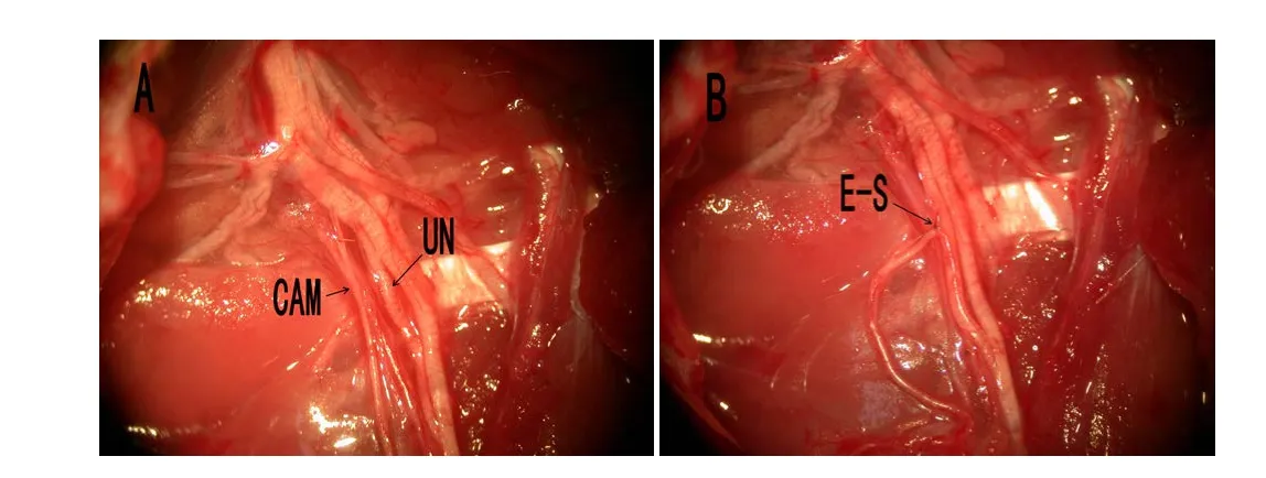
Figure 1 Surgical operation of the end-to-side neurorrhaphy.
Author contributions: YDG conceived and designed the study. QY and DH performed the experiments. QY and TW wrote the paper. FP and SHZ reviewed and edited the paper. All authors read and approved the final version of the paper.
Conflicts of interest:None declared.
Research ethics:The study protocol was approved by Animal Ethics Committee of Wenzhou Medical University of China (approval No.201601-005). The experimental procedure followed the United States National Institutes of Health Guide for the Care and Use of Laboratory Animals (NIH Publication No. 85-23, revised 1986).
Data sharing statement: Datasets analyzed during the current study are available from the corresponding author on reasonable request.
Plagiarism check:Checked twice by ienticate.
Peer review:Externally peer reviewed.
Open access statement:is is an open access article distributed under the terms of the Creative Commons Attribution-NonCommercial-ShareAlike 3.0 License, which allows others to remix, tweak, and build upon the work non-commercially, as long as the author is credited and the new creations are licensed under identical terms.
Al-Qattan MM (2001) Terminolateral neurorrhaphy: review of experimental and clinical studies. J Reconstr Microsurg 17:99-108.
Baltzer H, Woo A, Oh C, Moran SL (2016) Comparison of Ulnar Intrinsic function following supercharge end-to-side anterior interosseous-to-ulnar motor nerve transfer: a matched cohort study of proximal ulnar nerve injury patients. Plastic Reconstrucsurg 138:1264-1272.
Barbour J, Yee A, Kahn LC, Mackinnon SE (2012) Supercharged endto-side anterior interosseous to ulnar motor nerve transfer for intrinsic musculature reinnervation. J Hand Surgery 37:2150-2159.
Brushart TM (1988) Preferential reinnervation of motor nerves by regenerating motor axons. J Neurosci 8:1026-1031.
Brushart TM, Gerber J, Kessens P, Chen YG, Royall RM (1998) Contributions of pathway and neuron to preferential motor reinnervation.J Neurosci 18:8674-8681.
Brushart TM, Jari R, Verge V, Rohde C, Gordon T (2005) Electrical stimulation restores the specificity of sensory axon regeneration. Exp Neurol 194:221-229.
Byers MR, Lin KJ (2003) Patterns of fluoro-gold entry into rat molar enamel, dentin, and pulp. J Dent Res 82:312-317.
Celis-Aguilar E, Lassaletta L, Roda JM, Gavilan J (2013) End-to-Side interposed donor graing as a facial nerve reinforcement technique after vestibular schwannoma surgery. Ann Otol Rhinol Laryngol 122:520-523.
Chen L, Hu M, Zhang L, Liu S, Luo J, Deng T, Tao Y (2012) Motor fiber organization in the extratemporal trunk of the facial nerve in rats: a retrograde Fluoro-Gold study. Exper Med 4:844-848.
Chen ZL, Yu WM, Strickland S (2007) Peripheral regeneration. Annu Rev Neurosci 30:209-233.
Chiu K, Lau WM, Yeung SC, Chang RC, So KF (2008) Retrograde labeling of retinal ganglion cells by application of fluoro-gold on the surface of superior colliculus. J Vis Exp doi: 10.3791/819.
Civi S, Durdag E, Aytar MH, Kardes O, Kaymaz F, Aykol S (2017) Usefulness of end-to-side bridging anastomosis of sural nerve to tibial nerve: an experimental research. J Korean Neurosurg Soc 60:417-423.
De Sa JM, Mazzer N, Barbieri CH, Barreira AA (2004)e end-to-side peripheral nerve repair. Functional and morphometric study using the peroneal nerve of rats. J Neurosci Methods 136:45-53.
Engel J, Ganel A, Melamed R, Rimon S, Farine I (1980) Choline acetyltransferase for differentiation between human motor and sensory nerve fibers. Ann Plastic Surg 4:376-380.
Gao W, Liu Q, Li S, Zhang J, Li Y (2015) End-to-side neurorrhaphy for nerve repair and function rehabilitation. J Surg Res 197:427-435.
Gao WS, Dong CJ, Li SQ, Kunwar KJ, Li B (2012) Re-innervation of the bladder through end-to-side neurorrhaphy of autonomic nerve and somatic nerve in rats. J Neurotrauma 29:1704-1713.
Haastert K, Joswig H, Jaschke KA, Samii M, Grothe C (2010) Nerve repair by end-to-side nerve coaptation: histologic and morphometric evaluation of axonal origin in a rat sciatic nerve model. Neurosurgery 66:567-576.
Hanna A, Dempsey R (2013) Nerve conduits used for end-to-side graing aer nerve injury. Neurosurgery 72:N15-16.
Hayashi A, Yanai A, Komuro Y, Nishida M, Inoue M, Seki T (2004)Collateral sprouting occurs following end-to-side neurorrhaphy.Plast Reconstr Surg 114:129-137.
He YS, Zhong SZ (1988) Acetylcholinesterase: a histochemical identification of motor and sensory fascicles in human peripheral nerve and its use during operation. Plast Reconstr Surg 82:125-132.
Hoke A, Redett R, Hameed H, Jari R, Zhou C, Li ZB, Griffin JW,Brushart TM (2006) Schwann cells express motor and sensory phenotypes that regulate axon regeneration. J Neurosci 26:9646-9655.
Isaacs J (2013) Supercharged end-to-side nerve transfer: too soon for“prime time”? J Hand Surg 38:617-618.
Kakibuchi M, Tuji K, Fukuda K, Terada T, Yamada N, Matsuda K,Kawai K, Sakagami M (2004) End-to-side nerve grafor facial nerve reconstruction. Ann Plastic Surg 53:496-500.
Kalliainen LK, Cederna PS, Kuzon WM, Jr. (1999) Mechanical function of muscle reinnervated by end-to-side neurorrhaphy. Plast Reconstr Surg 103:1919-1927.
Kettle SJ, Starritt NE, Glasby MA, Hems TE (2013) End-to-side nerve repair in a large animal model: how does it compare with conventional methods of nerve repair? J Hand Surg Eur Vol 38:192-202.
Koh KS, Kim J, Kim CJ, Kwun BD, Kim SY (2002) Hypoglossal-facial crossover in facial-nerve palsy: pure end-to-side anastomosis technique. Br J Plastic Surg 55:25-31.
Kovacic U, Sketelj J, Bajrovic FF (2003) Sex-related difference in collateral sprouting of nociceptive axons aer peripheral nerve injury in the rat. Exp Neurol 184:479-488.
Kovacic U, Zele T, Tomsic M, Sketelj J, Bajrovic FF (2012) Influence of breaching the connective sheaths of the donor nerve on its myelinated sensory axons and on their sprouting into the end-to-side coapted nerve in the rat. J Neurotrauma 29:2805-2815.
Li Q, Zhang P, Yin X, Jiang B (2015) Early nerve protection with anterior interosseous nerve in modified end-to-side neurorrhaphy repairs high ulnar nerve injury: a hypothesis of a novel surgical technique. Artif Cells Nanomed Biotechnol 43:103-105.
Lundborg G, Zhao Q, Kanje M, Danielsen N, Kerns JM (1994) Can sensory and motor collateral sprouting be induced from intact peripheral nerve by end-to-side anastomosis? J Hand Surg 19:277-282.
Lutz BS, Ma SF, Chuang DC, Wei FC (2000) Role of the target in endto-side neurorrhaphy: reinnervation of a single muscle vs. multiple muscles. J Reconstr Microsurg 16:443-448.
Madison DL, Kruger WH, Kim T, Pfeiffer SE (1996) Differential expression of rab3 isoforms in oligodendrocytes and astrocytes. J Neurosci Res 45:258-268.
McBride RL, Feringa ER, Garver MK, Williams JK, Jr. (1990) Retrograde transport of fluoro-gold in corticospinal and rubrospinal neurons 10 and 20 weeks aer T-9 spinal cord transection. Exp Neurol 108:83-85.
Naumann T, Hartig W, Frotscher M (2000) Retrograde tracing with Fluoro-Gold: different methods of tracer detection at the ultrastructural level and neurodegenerative changes of back-filled neurons in long-term studies. J Neurosci Methods 103:11-21.
Noble J, Munro CA, Prasad VS, Midha R (1998) Analysis of upper and lower extremity peripheral nerve injuries in a population of patients with multiple injuries. J Trauma 45:116-122.
Prodanov D, Feirabend HK (2008) Automated characterization of nerve fibers labeled fluorescently: determination of size, class and spatial distribution. Brain Res 1233:35-50.
Puigdellivol-Sanchez A, Valero-Cabre A, Prats-Galino A, Navarro X, Molander C (2002) On the use of fast blue, fluoro-gold and diamidino yellow for retrograde tracing aer peripheral nerve injury:uptake, fading, dye interactions, and toxicity. J Neurosci Methods 115:115-127.
Reyes O, Sosa I, Kuffler DP (2005) Promoting neurological recovery following a traumatic peripheral nerve injury. P R Health Sci J 24:215-223.
Selecki BR, Ring IT, Simpson DA, Vanderfield GK, Sewell MF (1982)Trauma to the central and peripheral nervous systems. Part II: a statistical profile of surgical treatment New South Wales 1977. Aust N Z J Surg 52:111-116.
Sullivan R, Dailey T, Duncan K, Abel N, Borlongan CV (2016) Peripheral nerve injury: stem cell therapy and peripheral nerve transfer. Int J Mol Sci 17:2101.
Tessier-Lavigne M, Goodman CS (1996)e molecular biology of axon guidance. Science 274:1123-1133.
Viterbo F, Trindade JC, Hoshino K, Mazzoni Neto A (1992) Latero-terminal neurorrhaphy without removal of the epineural sheath.Experimental study in rats. Rev Paul Med 110:267-275.
Viterbo F, Trindade JC, Hoshino K, Mazzoni Neto A (1994) End-toside neurorrhaphy with removal of the epineurial sheath: an experimental study in rats. Plast Reconstr Surg 94:1038-1047.
Viterbo F, Teixeira E, Hoshino K, Padovani CR (1998) End-to-side neurorrhaphy with and without perineurium. Sao Paulo Med J 116:1808-1814.
Wright MC, Mi R, Connor E, Reed N, Vyas A, Alspalter M, Coppola G,Geschwind DH, Brushart TM, Hoke A (2014) Novel roles for osteopontin and clusterin in peripheral motor and sensory axon regeneration. J Neurosci 34:1689-1700.
Yan JG, Matloub HS, Sanger JR, Zhang LL, Riley DA, Jaradeh SS (2002)A modified end-to-side method for peripheral nerve repair: large epineurial window helicoid technique versus small epineurial window standard end-to-side technique. J Hand Surg 27:484-492.
Yu Q, Lin ZK, Ding J, Wang T, Chi YL, Gao WY (2011) Functional motor nerve regeneration without motor-sensory specificity following end-to-side neurorrhaphy: an experimental study. J Hand Surg 36:2010-2016.
Yu Q, Chen C, Zhang X, Lv L, Lin K, Chi Y, Gao W (2013) Quantitative assessment of the motor-sensory specificity of the motor and primary sensory neurons aer the end-to-side neurorrhaphy. J Reconstr Microsurg 29:579-586.
Yuksel F, Karacaoglu E, Guler MM (1999) Nerve regeneration through side-to-side neurorrhaphy sites in a rat model: a new concept in peripheral nerve surgery. Plast Reconstr Surg 104:2092-2099.
Zheng MX, Xu WD, Shen YD, Xu JG, Gu YD (2012) Reconstruction of elbow flexion by end-to-side neurorrhaphy in phrenic nerve transfer. Plast Reconstr Surg 129:573e-575e.
Graphical Abstract
Cutaneous nerve induces motor nerve regeneration aer end-to-side neurorrhaphy
*Correspondence to:
Yu-dong Gu,yudonggu@hotmail.com.
orcid:
0000-0003-3143-8307
(Yu-dong Gu)
10.4103/1673-5374.217350
Accepted: 2017-09-17
Copyedited by Wang J, Li CH, Qiu Y, Song LP, Zhao M
猜你喜欢
股市动态分析(2021年25期)2021-12-30
学生天地(2020年14期)2020-08-25
科学导报·学术(2020年19期)2020-07-09
海峡姐妹(2020年3期)2020-04-21
宇航计测技术(2018年3期)2018-09-08
特别文摘(2018年3期)2018-08-08
诗选刊(2015年6期)2015-10-26
中国民族民间医药·下半月(2014年4期)2014-09-26
股市动态分析(2014年27期)2014-07-29
中国火炬(2012年2期)2012-07-24
- 中国神经再生研究(英文版)的其它文章
- Matrix bound vesicles and miRNA cargoes are bioactive factors within extracellular matrix bioscaffolds
- Diffusion tensor tractography studies on mechanisms of recovery of injured fornix
- Using 3D bioprinting to produce mini-brain
- Beta secretase activity in peripheral nerve regeneration
- Embracing oligodendrocyte diversity in the context of perinatal injury
- On the road towards the global analysis of human synapses