大菱鲆脾肾结节病病原菌的分离和鉴定*
2019-09-27 01:33刘耀宽陈四清阎永伟莫照兰
渔业科学进展 2019年5期
李 杰 刘耀宽,3 白 露,2 陈四清,2 阎永伟 莫照兰,2
大菱鲆脾肾结节病病原菌的分离和鉴定*
李 杰1刘耀宽1,3白 露1,2陈四清1,2阎永伟1莫照兰1,2①
(1. 中国水产科学研究院黄海水产研究所 农业农村部海水养殖病害防治重点实验室 青岛海洋科学与技术试点国家实验室海洋渔业科学与食物产出过程功能实验室 青岛 266071;2. 上海海洋大学水产与生命学院 上海 201306;3. 青岛农业大学海洋科学与工程学院 青岛 266109)
2017年夏季,江苏省连云港市某养殖场大菱鲆出现大规模死亡,发病鱼体表无明显症状,解剖可见脾脏、肾脏出现白色散在结节,从发病鱼的内脏中分离得到1株优势菌SM-Myco001。人工感染实验结果显示,SM-Myco001可以引起大菱鲆内脏结节症状并造成鱼类死亡,且在高养殖水温(22℃)条件下发病更为剧烈。生理生化鉴定和16S rRNA基因分析结果显示,SM-Myco001属于海分枝杆菌()。本研究首次报道了我国养殖大菱鲆感染海分枝杆菌的病例,可为大菱鲆养殖过程中的疾病防控提供参考。
大菱鲆;海分枝杆菌;分枝杆菌病;结节
大菱鲆()是我国北方海水工厂化养殖的主要品种之一,根据《2017年中国渔业统计年鉴》,我国鲆鲽类养殖产量合计131389 t,大菱鲆产量居首位。我国的大菱鲆养殖业经过20多年的发展,养殖成本不断降低,从高档酒店逐步进入大众市场,为人民生活水平的提高做出了贡献,但大菱鲆产业的发展也遇到了瓶颈。病害是影响大菱鲆养殖业的重要因素,不但引起鱼类死亡,造成直接经济损失,还可能导致药物滥用、药残超标,造成消费者恐慌,进而影响市场价格。对大菱鲆的病害已有较多的研究,目前已报道多种大菱鲆病原,包括鳗弧菌() (邹玉霞等, 2004)、哈维氏弧菌() (范文辉等, 2005)、溶藻弧菌() (张伟妮等, 2006)、迟缓爱德华氏菌() (李筠等, 2006)、杀鲑气单胞菌() (吕俊超等, 2009)、虹彩病毒(史成银等, 2005)和盾纤毛虫等(王印庚等, 2004),这些流行病学的研究为大菱鲆养殖疾病防控提供了理论依据。
2017年,本实验室从江苏省连云港市某养殖场患脾肾结节病的大菱鲆中分离到1株优势菌,鉴定为海分枝杆菌(),通过人工感染实验证明该菌株对大菱鲆的致病性,并对其发病温度进行初步分析,以期为我国养殖大菱鲆的病害防控提供参考。
1 材料与方法
1.1 病鱼来源
2017年7~9月,江苏省连云港市某养殖场工厂化养殖大菱鲆出现大量死亡事件。发病时,养殖水温为18℃~22℃,发病鱼体重为500~650 g,共养殖大菱鲆4万尾左右,发病鱼约为10000尾,其中约死亡5000尾。应用广谱抗生素进行治疗效果不明显,待水温下降至18℃以下,大菱鲆逐渐恢复正常。
1.2 细菌分离
取发病鱼的鳃、体表粘液和尾鳍,分别在显微镜下观察,检测寄生虫感染情况。取发病鱼的脾脏、肾脏组织分别在2216E和Middlebrook 7H10 (含10% OADC,青岛海博)固体培养基平板划线进行病原菌分离,28℃培养4周后,将优势菌落进行纯化培养,纯化培养后的细菌用甘油保存于-80℃冰箱备用。
1.3 人工感染实验
将细菌接种于酸性罗氏斜面培养基(青岛海博),28℃培养4周后,用PBS冲洗菌体,离心收集并重悬。健康大菱鲆购自烟台一大菱鲆养殖场,体重为100 g左右,实验前随机选取5%的大菱鲆进行解剖观察和内脏匀浆涂布,确定大菱鲆未携带病原。将大菱鲆随机分为2组,每组20尾,饲养水温分别为16℃和22℃,每尾大菱鲆腹腔注射0.1 ml 0.5×10⁷CFU/ml的菌液,设置对照组饲养于相同条件下,并注射等体积的PBS。注射感染后,正常饲养观察30 d,解剖和记录实验鱼死亡情况并进行病原分离。实验结束后,将实验组和对照组剩余的大菱鲆麻醉致死,解剖观察记录内脏结节情况。
1.4 细菌鉴定
采用引物27F(5¢-AGAGTTTGATCCTGGCTCAG- 3¢)和1492R (5¢-TACGGCTACCTTGTTACGACTT-3¢)对细菌的16S rRNA基因进行扩增并测序(Lane, 1991),获得的序列信息在GenBank和EzBioCloud进行同源性比对,用MEGA 5.0软件采用邻位相连法(Neighbor- Joining)构建系统发育进化树;根据Chiodini(1986)的方法,检测细菌的Tween-80水解、尿素酶、68℃下过氧化氢酶、硫酸酯酶、硝酸还原酶、色素产生、5% NaCl和麦康凯琼脂(MacConkey Agar)生长等生理生化指标。
2 结果
2.1 病鱼症状
发病鱼体表无明显症状,偶见白便。解剖可见脾脏、肾脏出现白色散在结节,直径为0.5~3 mm,肠道发红,有白色内容物(图1)。显微镜下未观察到寄生虫感染,利用Middlebrook 7H10培养基,从发病鱼的肝脏、脾脏和肾脏可以分离到大量形态一致的白色不透明圆形菌落,选择单菌株进行纯化后将其命名为SM-Myco001。将平板放置于可见光下12 h,菌落变为黄色。从2216E平板上未分离到明显优势菌。
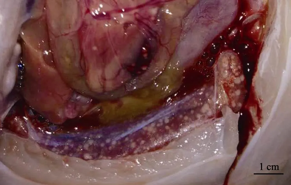
图1 发病大菱鲆症状
2.2 人工感染实验
人工感染实验结果如表1所示,在16℃水温养殖条件下,25%的感染鱼出现死亡,解剖可见明显脾肾结节症状,30 d后有结节症状的鱼合计占35%;当养殖水温为22℃时,65%的感染鱼出现死亡,解剖可见明显脾肾结节症状,30 d后有结节症状的鱼合计占90%。利用Middlebrook 7H10培养基,从结节组织中均可分离到与SM-Myco001菌落形态一致的优势菌。2组对照组实验鱼均未发生死亡或出现结节症状。
表1 SM-Myco001对大菱鲆人工感染实验
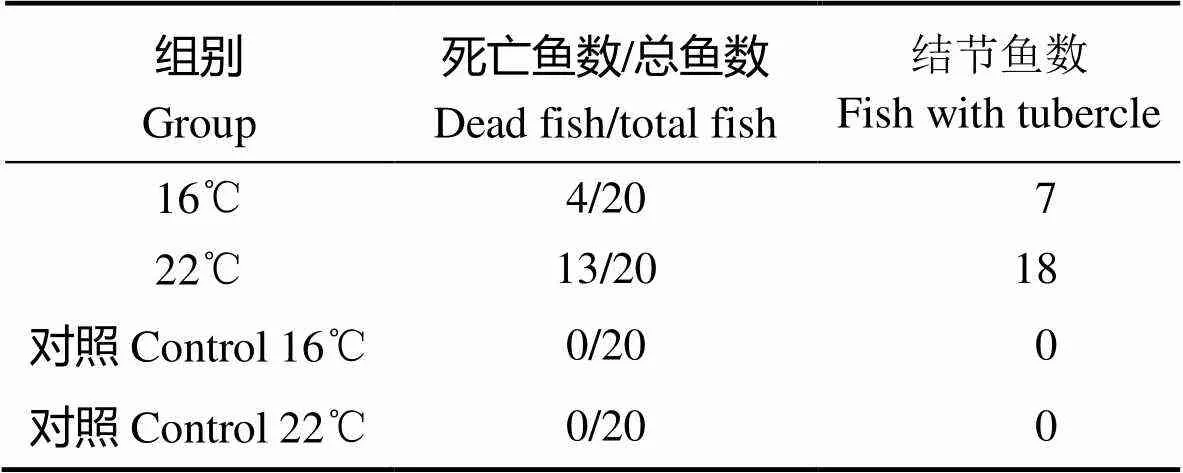
Tab.1 Experimental infection of S. maximus infected with SM-Myco001
2.3 病原菌鉴定
PCR扩增菌株SM-Myco001的16S rRNA基因序列(MH636826),测序结果经比对分析显示,在GenBank中同源性>99%的前15株菌均为海分枝杆菌(),EzBioCloud比对结果也显示其为海分枝杆菌。从NCBI数据库中选择不同分枝杆菌属()的细菌,使用MEGA 5.0构建系统进化树。结果显示,SM-Myco001与海分枝杆菌和溃疡分枝杆菌()聚为一支(图2)。
生理生化检测结果显示,SM-Myco001在Tween-80水解、尿素酶、68℃下过氧化氢酶、硫酸酯酶和色素产生等反应为阳性,硝酸还原酶、5% NaCl和麦康凯琼脂生长为阴性,与海分枝杆菌的生理生化指标相符。由于溃疡分枝杆菌Tween-80水解和硫酸酯酶反应为阴性,综合基因序列进化分析和生理生化结果,将病原菌SM-Myco001鉴定为海分枝杆菌。
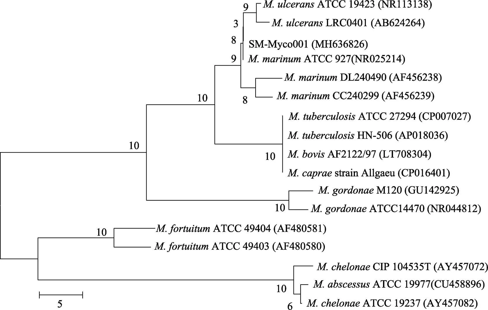
图2 基于16S rRNA基因序列构建的系统发育进化树
3 讨论
分枝杆菌是广泛存在于水体环境中的条件致病菌,龟分枝杆菌()、偶发分枝杆菌()和海分枝杆菌等多种分枝杆菌属的细菌引起野生或养殖鱼结核病,在世界各地均有报道(Frerichs, 1993; Belas,1995)。由于培养条件苛刻,生长周期较长,分枝杆菌感染的情况经常被忽略。本研究从患脾肾结节病的大菱鲆中分离得到优势菌SM-Myco001,经生理生化检测和16S rRNA基因分析,确定为海分枝杆菌。人工感染实验结果显示,SM-Myco001可以引起大菱鲆内脏结节症状,并导致感染鱼的死亡。当水温由16℃升高至22℃后,感染鱼的死亡率和出现结节的比例都有所提高,这与自然发病时的条件较为吻合,在夏季高温期间发病,水温降低后大菱鲆逐渐恢复正常。大菱鲆最适养殖温度在20℃以下,高温会影响大菱鲆的生长状态和免疫力,增加条件致病菌的感染几率,同时,分枝杆菌的生长速度也会加快,这可能是高温增加大菱鲆结核病发病率的原因。

分枝杆菌可以通过水体、养殖器械、饵料等多种渠道进行水平传播(Hedrick, 1987; Jacobs, 2009),在鱼体状态不佳或受到环境胁迫条件下发作(Zanoni, 2008)。除消毒和隔离外,目前尚无有效预防和治疗鱼类分枝杆菌感染的方法(Jacobs, 2009)。海分枝杆菌属于人鱼共患病原,也可以感染人类,引起游泳池肉芽肿(Fish tank granuloma)(Lewis, 2003),因此,有效的预防和应对海水养殖鱼类海分枝杆菌感染,对养殖鱼类、养殖从业者和消费者的安全都具有重要意义。
Belas R, Faloon P, Hannaford A. Potential applications of molecular biology to the study of fish mycobacteriosis. Annual Review of Fish Diseases, 1995, 5: 133–173
Brocklebank J, Raverty S, Robinson J. Mycobacteriosis in Atlantic salmon farmed in British Columbia. Canadian Veterinary Journal, 2003, 44(6): 486–489
Chiodini RJ. Biochemical characteristics of various strains of. American Journal of Veterinary Research, 1986, 47(7): 1442–1445
Fan WH, Huang J, Wang XH,. Identification and phylogenetic study of pathogenic bacteria causing ulcer disease of cultured turbot (). Acta Microbiologica Sinica, 2005, 45(5): 665–670 [范文辉, 黄倢, 王秀华, 等. 养殖大菱鲆溃疡症病原菌的分离鉴定及系统发育分析. 微生物学报, 2005, 45(5): 665–670]
Frerichs GN. Mycobacteriosis: Nocardiosis. In: Inglis V, Roberts RJ, Bromage NR (eds). Bacterial diseases of fish. New York: Halsted Press, 1993, 219–234
Hedrick RP, Mcdowell T, Groff J. Mycobacteriosis in cultured striped bass from California. Journal of Wildlife Diseases, 1987, 23(3): 391–395
Jacobs JM, Stine CB, Baya AM,. A review of mycobacteriosis in marine fish. Journal of Fish Diseases, 2009, 32(2): 119–130
Jeon SJ, Gonsalves LC, Jacobs JM,. Short-term infection of striped basswith. Diseases of Aquatic Organisms, 2011, 94(2): 117–124
Lane DJ. 16S/23S rRNA sequencing. Nucleic Acid Techniques in Bacterial Systematics, 1991: 125–175
Lewis FMT, Marsh BJ, von Reyn CF. Fish tank exposure and cutaneous infections due to: Tuberculin skin testing, treatment, and prevention. Clinical Infectious Diseases, 2003, 37(3): 390–397
Li Y, Yan XH, Chen JX,. Studies on the characteristics of pathogenicisolated from diseased. Periodical of Ocean University of China, 2006, 36(4): 649–654 [李筠, 颜显辉, 陈吉祥, 等. 养殖大菱鲆腹水病病原的研究. 中国海洋大学学报, 2006, 36(4): 649–654]
Luo Z, Li J, Zhang Z,.is the causative agent of splenic and renal granulomas in half-smooth tongue sole (, Günther) in China. Aquaculture, 2018, 490: 203–207
Lv JC, Zhang XH, Wang Y,. Isolation and identification of bacterial pathogen:subsp.in cultured turbot and histopathological study. Periodical of Ocean University of China, 2009, 39(1): 91–95 [吕俊超, 张晓华, 王燕, 等. 养殖大菱鲆病原菌——杀鲑气单胞菌无色亚种的分离鉴定和组织病理学研究. 中国海洋大学学报, 2009, 39(1): 91–95]
dos Santos NMS, Vale AD, Sousa MJ,. Mycobacterial infection in farmed turbot. Diseases of Aquatic Organisms, 2002, 52(1): 87–91
Shi CY, Wang YG, Qin L,. Investigation of pathogen and epidemiology of a novel disease, ‘Viral reddish body syndrome’, among farmedin China. Marine Fisheries Research, 2005, 26(1): 1–6 [史成银, 王印庚, 秦蕾, 等. 我国养殖大菱鲆病毒性红体病及其流行情况调查. 海洋水产研究, 2005, 26(1): 1–6]
Tortoli E, Bartoloni A, Bozzetta E,. Identification of the newly describedfrom tuberculous lesions of snakehead fish (). Comparative Immunology Microbiology and Infectious Diseases, 1996, 19(1): 25–29
Wang YG, Zhang Z, Qin L,. The main diseases of cultured turbot () and their prevention and treatment. Marine Fisheries Research, 2004, 25(6): 61–68 [王印庚, 张正, 秦蕾, 等. 养殖大菱鲆主要疾病及防治技术. 海洋水产研究, 2004, 25(6): 61–68]
Zanoni RG, Florio D, Fioravanti ML,. Occurrence ofspp. in ornamental fish in Italy. Journal of Fish Diseases, 2008, 31(6): 433–441
Zhang DF, Li AH, Gong XN. Research on the mycobacteriosis and its pathogen in sturgeons. Acta Hydrobiologica Sinica, 2014, 38(3): 495–504 [张德锋, 李爱华, 龚小宁. 鲟分枝杆菌病及其病原研究. 水生生物学报, 2014, 38(3): 495–504]
Zhang WN, Zhou L, Xing J,. Isolation and identification of pathogen SR1 associated with swollen abdomen of cultured turbot (). Journal of Fishery Sciences of China, 2006, 13(4): 603–609 [张伟妮, 周丽, 邢婧, 等. 养殖大菱鲆腹水症病原菌SR1的分离及鉴定. 中国水产科学, 2006, 13(4): 603–609]
Zou YX, Zhang PJ, Mo ZL,. Isolation and identification of the pathogenic bacteria from. Chinese High Technology Letters, 2004, 14(4): 89–93 [邹玉霞, 张培军, 莫照兰, 等. 大菱鲆出血症病原菌的分离和鉴定. 高技术通讯, 2004, 14(4): 89–93]
(编辑 马璀艳)
Isolation and Identification ofAssociated with Splenic and Renal Granuloma Disease of Cultured Turbot ()
LI Jie1, LIU Yaokuan1,3, BAI Lu1,2, CHEN Siqing1,2, YAN Yongwei1, MO Zhaolan1,2①
(1. Yellow Sea Fisheries Research Institute, Chinese Academy of Fishery Sciences, Key Laboratory of Maricultural Organism Disease Control, Ministry of Agriculture and Rural Affairs, Laboratory for Marine Fisheries Science and Food Production Processes, Pilot National Laboratory for Marine Science and Technology (Qingdao), Qingdao 266071; 2. College of Fisheries and Life Science, Shanghai Ocean University, Shanghai 201306; 3. Marine Science and Engineering College, Qingdao Agricultural University, Qingdao 266109)
With the highest annual production among flatfish, turbot () is reared as the major industrial aquaculture marine fish species in North China. In the culture process, members of this species are subject to infection with a variety of pathogens. In 2017, massive death of reared turbot occurred in a farm in Lianyungang City, Jiangsu Province. Externally, no clinical signs were observed for most of the diseased fish. However, after dissection, splenic and renal tubercles were found in all the diseased fish. Homogeneous colonies were isolated from diseased or moribund fish and were named as SM-Myco001. Healthy turbot were subjected to challenge tests by intraperitoneal injection using SM-Myco001. SM-Myco001 was found capable of causing death in turbot, especially at a higher water temperature (22℃). The diseased turbot displayed clinical signs, such as splenic and renal tubercles, similar to those observed in naturally infected fish. SM-Myco001 was identified asbased on bacterial morphology, analytical profile index identification, and 16S rRNA gene sequencing. This study suggests that a strain of.is the causal agent of splenic and renal tubercle disease in turbot. As a first report, to our knowledge, this study provides a new perspective on disease control in the flat fish aquaculture industry in China.
;; Mycobacteriosis; Granuloma
MO Zhaolan, E-mail: mozl@ysfri.ac.cn
* 国家重点研发计划(2018YFC0311300)、国家自然科学基金-山东省人民政府联合基金(U1706205)、中国水产科学研究院黄海水产研究所基本科研业务费(20603022017008)和鳌山科技创新计划(2015ASKJ02)共同资助[This work was supported by National Key R&D Program of China(2018YFC0311300), NFSC-Shandong Joint Fund (U1706205), the Central Public-Interest Scientific Institution Basal Research Fund, YSFRI, CAFS (20603022017008), and Aoshan Technology Innovation Program (2015ASKJ02) ]. 李 杰,E-mail: lijie@ysfri.ac.cn
莫照兰,研究员,E-mail: mozl@ysfri.ac.cn
2018-07-21,
2018-09-03
S943
A
2095-9869(2019)05-0195-05
10.19663/j.issn2095-9869.20180721001
http://www.yykxjz.cn/
李杰, 刘耀宽, 白露, 陈四清, 阎永伟, 莫照兰. 大菱鲆脾肾结节病病原菌的分离和鉴定. 渔业科学进展, 2019, 40(5): 195–199
Li J, Liu YK, Bai L, Chen SQ, Yan YW, Mo ZL. Isolation and identification ofassociated with splenic and renal granuloma disease of cultured turbot (). Progress in Fishery Sciences, 2019, 40(5): 195–199
猜你喜欢
湘潭大学自然科学学报(2022年2期)2022-07-28
昆明医科大学学报(2021年10期)2021-12-02
中老年保健(2021年6期)2021-08-24
中老年保健(2021年9期)2021-08-24
天津医科大学学报(2021年3期)2021-07-21
医学概论(2021年18期)2021-01-21
中国水产(2019年3期)2019-03-25
中国生殖健康(2019年10期)2019-01-07
农村百事通(2016年18期)2016-11-05
河北渔业(2016年5期)2016-09-08