
非小细胞肺癌中UHRF-1蛋白的表达及临床意义
2020-09-22 06:42张梦娇张明张旭
医学信息 2020年16期
张梦娇 张明 张旭
摘要:目的 探讨UHRF-1蛋白在非小细胞肺癌组织中的表达及临床意义。方法 选取2017年1月1日~12月31日在我院经手术病理检查确诊的非小细胞肺癌 110 例,另选正常肺组织64例设为对照。采用免疫组化 SP 法检测 UHRF-1 蛋白的表达,并分析其与临床病理参数的关系。结果 UHRF-1蛋白在肺癌组织中的表达(88.18%)高于正常组织(40.63%),差异有统计学意义(P<0.05);不同肺癌分化程度、淋巴结转移、临床分期UHRF-1蛋白表达比较,差异有统计学意义(P<0.05);不同性别、年龄及肿瘤大小、组织学类型UHRF-1蛋白比较,差异无统计学意义(P>0.05);其中低分化肺癌UHRF1表达高于中、高分化者,伴淋巴结转移肺癌UHRF1表达高于无淋巴结转移者,Ⅲ~Ⅳ期肺癌UHRF1表达高于Ⅰ~Ⅱ者,差异有统计学意义(P<0.05)。结论 UHRF-1蛋白在非小细胞肺癌组织中表达升高,其在非小细胞肺癌的发生和转移过程中可能发挥促进作用。
关键词:非小细胞肺癌;甲基化;UHRF-1;免疫组织化学
Abstract:Objective To investigate the expression and clinical significance of UHRF-1 protein in non-small cell lung cancer.Methods 110 cases of non-small cell lung cancer diagnosed by surgical pathology in our hospital from January 1st to December 31st, 2017 were selected, and 64 cases of normal lung tissue were selected as controls. The SP method of immunohistochemistry was used to detect the expression of UHRF-1 protein and analyze its relationship with clinicopathological parameters.Results The expression of UHRF-1 protein in lung cancer tissues (88.18%) was higher than that in normal tissues (40.63%),the difference was statistically significant (P<0.05); comparison of UHRF-1 protein expression in different lung cancer differentiation levels, lymph node metastasis, and clinical stages,the difference was statistically significant (P<0.05); the UHRF-1 protein of different gender, age, tumor size, and histological type were not statistically different (P>0.05); Among them, the expression of UHRF1 in poorly differentiated lung cancer was higher than that in the middle and well-differentiated patients, the expression of UHRF1 in lung cancer with lymph node metastasis was higher than that in those without lymph node metastasis, and the expression of UHRF1 in stage III-IV lung cancer was higher than that of I-II,the difference was statistically significant (P<0.05).Conclusion UHRF-1 protein expression is elevated in non-small cell lung cancer tissues, and it may play a role in promoting the occurrence and metastasis of non-small cell lung cancer.
Key words:Non-small cell lung cancer;Methylation;UHRF-1;Immunohistochemistry
肺癌(lung cancer)是一種常见的恶性肿瘤,所有恶性肿瘤中肺癌的发病率和病死率均是第1位[1],对人类生命健康造成了严重损害。肺癌的侵袭与转移是导致患者治疗效果不佳和死亡最重要的因素,因此研究肺癌侵袭和转移的机制,寻找抑制其侵袭和转移的靶点,对肺癌的治疗有着重要意义。近年来研究表明泛素样含PHD和环指域1(ubiquitin-like containing phd ring finger 1,UHRF-1)在膀胱癌、肝癌等多种恶性肿瘤呈高表达[2-6]。既往研究显示[7],抑制肺癌A549细胞中UHRF1的表达可诱导癌细胞的凋亡,本团队前期研究发现抑制肺癌A549细胞UHRF1表达,可降低癌细胞侵袭和迁移能力[8],但临床中UHRF1与肺癌的浸润和转移是否相关尚不明确。本研究通过检测肺腺癌、鳞癌、正常支气管和肺泡组织中UHRF-1蛋白表达,分析UHRF1与临床病理参数的关系,旨在进一步明确UHRF1与肺癌侵袭和转移的关系,现将结果报道如下。
1资料与方法
1.1一般资料 收集2017年1月1日~12月31日在西南医科大学附属医院胸外科经手术病理检查确诊的非小细胞肺癌患者110例为研究对象,诊断均采用2015 版 WHO 肺肿瘤分类诊断标准,所有患者手术前均未接受化疗或放疗。患者中男71 例、女39 例,年龄36~78岁,平均年龄(59.86±11.88)岁;腺癌74例、鳞癌36例;低分化46例,中高分化64例;有淋巴结转移65 例,无淋巴结转移45例。选取癌旁正常组织64例(肺泡35例,支气管29例)设为对照。
1.2检测方法
1.2.1试剂与染色方法 鼠抗人UHRF-1蛋白抗体、兔抗人GAPDH抗体购自英国ABcam公司,DAB试剂盒和免疫组化染色试剂盒购自北京中杉金桥生物技术有限公司,苏木素染液购自南京建成科技有限公司。全部组织标本采用10%甲醛溶液固定和石蜡包埋,连续切片,切片厚度4 μm,切片常规脱蜡、抗原修复、滴加抗体、DAB显色、苏木素复染、脱水、透明、封片,所有步骤均按照免疫组化试剂盒说明书操作。
1.2.2染色结果判定 显微镜下观察免疫组化染色结果,UHRF1表达量取决于染色强度和阳性细胞数。采用双盲法,两位病理科医生对UHRF1表达量进行评分。染色强度为0~3分,依次为无染色、浅黄色、黄色、棕黄色;阳性细胞构成比得分为0~4分,依次为<5%,6%~25%,26%~50%,51%~75%,>76%,两项相乘获得最后得分[9]。UHRF1表达程度分为阴性(-)、弱阳性(+)、中等阳性(++)、强阳性(+++),其对应分值为0分、1~4分、5~8分、9~12分。
1.3统计学方法 统计学分析使用SPSS 19.0软件进行。肺癌组织和癌旁正常组织中UHRF1蛋白的表达差异采用?字2检验分析;肺癌组织UHRF1蛋白的表达与临床病理参数间的关系采用Mann-Whitney U检验分析。检验水准α=0.05。
2结果
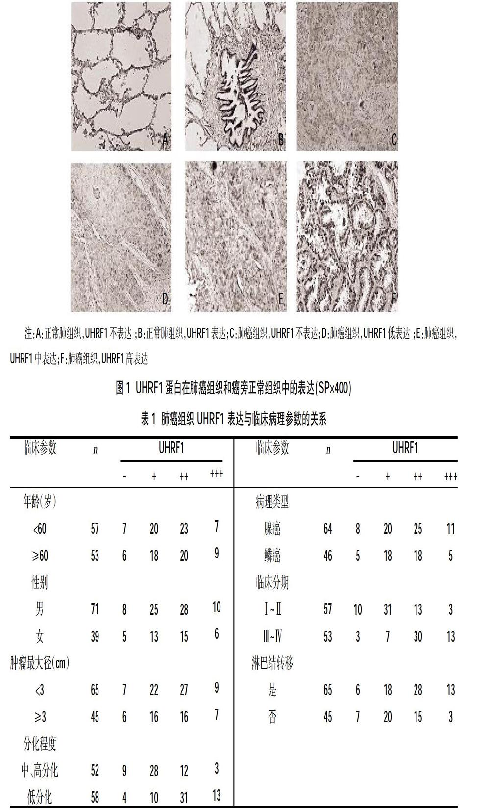
2.1 UHRF1蛋白在肺癌组织和癌旁正常组织中的表达 UHRF1主要定位于胞核中,在胞质中也有一定表达。在 110例肺癌组织中阳性表达 97 例(88.18%),其中弱阳性38例(34.55%),中等阳性43例(39.09%),强阳性16例(14.55%);而在64例癌旁组织中,阳性表达26例 (40.63%),其中弱阳性20例(31.25%),中等阳性4例(6.25%),强阳性2例(3.13%);肺癌组织中UFRF1表达高于正常组织(P<0.05),见图1。
2.2 UHRF-1与肺癌临床病理参数的关系 不同肺癌分化程度、淋巴结转移、临床分期UHRF-1蛋白表达比较,差异无统计学意义(P<0.05);不同性别、年龄及肿瘤大小、组织学类型UHRF-1蛋白比较,差异无统计学意义(P>0.05);低分化肺癌UHRF1表达高于中、高分化者,伴淋巴结转移肺癌UHRF1表达高于无淋巴结转移者,Ⅲ~Ⅳ期肺癌UHRF1表达高于Ⅰ~Ⅱ者,差异有统计学意义(P<0.05),见表1。
3讨论
UHRF1作为表观遗传学中重要的调节蛋白,可通过其SRA结构域识别半甲基化的DNA,招募DNA 甲基轉移酶1维持DNA 甲基化,进而对基因表达进行调控[10]。UHRF1与细胞增殖密切相关,在细胞分裂过程中可促进G1/S向G2/M期快速转换,有利于细胞增殖,在骨髓、脾脏及睾丸等增殖速度快的细胞中呈高表达。既往研究显示UHRF1在多种类型的癌症中均发现高表达,如肝癌、胰腺癌、膀胱癌及髓母细胞癌[2-5],且研究显示UHRF1高表达可沉默肿瘤抑制基因,促进结肠癌、胃癌等肿瘤细胞的增殖[11-13],而降低UHRF1的表达可抑制肺癌、乳腺癌细胞的增殖[7,14]。可见UHRF1的高表达和恶性肿瘤有着密切的关系。本研究结果显示肺癌组织UHRF1蛋白表达明显高于正常肺组织,这也提示UHRF1在肺癌发生发展中可能发挥着促进作用。
UHRF1表达与肿瘤患者分期和预后密切相关,Saidi S等[2]研究发现,高级别膀胱癌UHRF1表达明显高于低级别膀胱癌,UHRF1高表达的膀胱癌多为肌肉浸润型,病灶转移更为常见,治疗后易复发,预后相对更差。有关胃癌的研究显示[10],组织分化程度低、伴淋巴及远处转移、临床分期晚患者UHRF1表达相对更高,UHRF1表达越高,患者生存期越短。UHRF1高表达可提高肿瘤抑制基因(如:UBE2L6、CDH1、IGFBP3)甲基化程度,抑制肿瘤抑制基因转录表达,进而导致肿瘤的发生、侵袭及转移[3,15,16]。本团队前期研究通过慢病毒介导的shRNA 沉默肺癌A549细胞中UHRF1 基因表达,可降低A549 细胞侵袭和迁移能力[8]。本研究发现临床肺癌组织中UHRF1 表达与肿瘤分化、淋巴结转移和临床分期有关,高水平表达UHRF1者肿瘤常呈低分化,肿瘤分化越低,侵袭性越强,也更容易发生转移,UHRF1表达较高者,淋巴结转移明显,临床分期较晚。可见UHRF1对非小细胞肺癌的侵袭和转移可能产生促进作用。
综上所述,非小细胞肺癌组织中UHRF1呈高表达,与肿瘤分化程度、淋巴结转移和临床分期密切相关,对非小细胞肺癌的侵袭和转移起到促进作用。UHRF1可成为协助肺癌诊断及预后推测的较好标记物,同时可为肺癌个体化治疗提供新的思路。
参考文献:
[1]Freddie B,Jacques F,Isabelle S,et al.Global Cancer Statistics 2018: Globocan Estimates of Incidence and Mortality Worldwide for 36 Cancers in 185 Countries[J].CA,2018,68(6):394-424.
[2]Saidi S,Popov Z,Janevska V,et al.Overexpression of UHRF1 gene correlates with the major clinicopathological parameters in urinary bladder cancer[J].International Braz J Urol,2017,43(2):224-229.
[3]Liu X,Ou H,Xiang L,et al.Elevated UHRF1 expression contributes to poor prognosis by promoting cell proliferation and metastasis in hepatocellular carcinoma[J].Oncotarget,2017,8(6):10510-10522.
[4]Zhang ZY,Cai JJ,Hong J,et al.Clinicopathological analysis of UHRF1 expression in medulloblastoma tissues and its regulation on tumor cell proliferation[J].Medical Oncology,2016,33(9):99.
[5]Abu-Alainin W,Gana T,Liloglou T,et al.UHRF1 regulation of the Keap1-Nrf2 pathway in pancreatic cancer contributes to oncogenesis[J].The Journal of Pathology,2016,238(3):423-433.
[6]Chen J,Sheng X,Ma H,et al.WDR79 mediates the proliferation of non-small cell lung cancer cells by regulating the stability of UHRF1[J].Journal of Cellular and Molecular Medicine,2018,22(5):2857-2864.
[7]杨从容,赵学涛,王军,等.UHRF1基因沉默可促进人肺腺癌A549细胞的凋亡[J].肿瘤,2015,35(11):1222-1229.
[8]徐小会,张明,范贤明.UHRF1基因沉默对人肺腺癌A549细胞侵袭和迁移的影响及机制[J].山东医药,2018,58(34):17-21.
[9]张伟,周林,李乃义,等.胃癌组织UHRF1表达与预后相关性分析[J].中华肿瘤防治杂志,2015,22(2):37-40.
[10]Kent B,Magnani E,Walsh MJ,et al.UHRF1 regulation of Dnmt1 is required for pre-gastrula zebrafish development[J].Dev Biol,2016,412(1):99-113.
[11]Babbio F,Pistore C,Curti L,et al.The SRA protein UHRF1 promotes epigenetic crosstalks and is involved in prostate cancer progression[J].Oncogene,2012,31(46):4878-4887.
[12]Ying L,Lin J,Qiu F,et al.Epigenetic repression of regulator of G‐protein signaling 2 by ubiquitin‐like with PHD and ring‐finger domain1 promotes bladder cancer progression[J].The FEBS Journal,2015,282(1):174-182.
[13]Zhou L,Shang Y,Jin Z,et al.UHRF1 promotes proliferation of gastric cancer via mediating tumor suppressor gene hypermethylation[J].Cancer Biology&Therapy,2015,16(8):1241-1251.
[14]Fang L,Shanqu L,Ping G,et al.Gene therapy with RNAi targeting UHRF1 driven by tumor-specific promoter inhibits tumor growth and enhances the sensitivity of chemotherapeutic drug in breast cancer in vitro and in vivo[J].Cancer Chemotherapy And Pharmacology,2012,69(4):1079-1087.
[15]Magnani E,Macchi F,Mancini M,et al.UHRF1 regulates CDH1 via promoter associated non-coding RNAs in prostate cancer cells[J].Biochim Biophys Acta Gene Regul Mech,2018,1861(3):258-270.
[16]Beck A,Trippel F,Wagner A,et al.Overexpression of UHRF1 promotes silencing of tumor suppressor genes and predicts outcome in hepatoblastoma[J].Clinical Epigenetics,2018,10(27):1-11.
收稿日期:2020-05-12;修回日期:2020-06-12
編辑/肖婷婷
猜你喜欢
保健医苑(2023年2期)2023-03-15
中国临床医学影像杂志(2022年2期)2022-05-25
清华金融评论(2022年4期)2022-04-13
天津医科大学学报(2021年4期)2021-08-21
国际放射医学核医学杂志(2021年10期)2021-02-28
房地产导刊(2020年7期)2020-08-24
医学研究杂志(2015年12期)2015-06-10
郑州大学学报(医学版)(2015年1期)2015-02-27
郑州大学学报(医学版)(2015年1期)2015-02-27
中国药理学通报(2014年2期)2014-05-09
