
Effect of scraping therapy on Th1/Th2 balance in rats w ith lumbar disc herniation
2020-10-23 09:52Yangin杨敏XuGuihua徐桂华
Yang M in (杨敏), Xu Gui-hua (徐桂华)
School of Nursing, Nanjing University of Traditional ChineseMedicine, Nanjing 210000,China
Abstract
Keywords:Scraping Therapy; IntervertebralDisc Displacement; Low Back Pain; Helper T Cells; Interleukin; Rats
Lumbar disc herniation (LDH) is the most common cause of low back pain and radiculopathy[1], and it is also one of the main causes of disability and loss of production[2]. Therefore,the treatment of LDH has always been a hot topic in medical research. Scraping therapy (ST) is a unique external treatment technique of Chinese medicine, and has been w idely applied to treat common diseases in various clinical departments. One of the dom inant diseases in the application of ST is LDH[3-4], and its clinical efficacy has been confirmed by lots of studies[5-6]. Our Research team has found that[7-9]the levels of serum immunoglobulin(Ig)G and interleukin(IL)-1 in the ST group were significantly reduced,suggesting that ST can inhibit the nucleus pulposus-induced autoimmune response and autoimmune response-mediated inflammation.This research intends to study the effect of ST on the immune balance of serum helper T (Th) 1/Th2 cells in LDH model rats, aim ing to promote the further study of immune mechanism of ST in the treatment of LDH.
1 Experimental Materials and Methods
1.1 Experimental animals and groups
Seventy-two healthy adult male Sprague-Daw ley (SD)rats, SPF grade and weighing (300±50) g were provided by Zhejiang Provincial Laboratory Animal Center [Certificate No.: SCXK (Zhejiang) 2014-0001]. Before the beginning of formal experiment,7 d of adaptive ordinary feed was required. The ambient temperature was kept at (23±2)℃and the hum idity was maintained at (50±10)%. All the rats were given the even split of 12 h of light and 12 h of darkness and free water and food intake. The rats were fasted for 12 h before modeling. The 72 rats were random ly divided into a sham operation group, a model group and a ST group using the random number table method, w ith 24 rats in each group. This experiment strictly complied w ith the animal experiment use and protection regulations of Nanjing University of Traditional Chinese Medicine.
1.2 Experimental drugs and reagents
1.2.1 Experimental instruments
Microplate Reader (Victor X3,PerkinElmer,USA);desktop high-speed refrigerated centrifuge (Allegra 64R,Beckman, USA); constant temperature water incubator (GNP-9050,Shanghai Jinghong Instrument Company,China);intelligent hot plate YLS-6B(Jinan Yiyan Technology Development Co.,Ltd.,China);cryostat (Leica CM 1100, Germany); optical m icroscope (Eclipse Ti-SR, Nikon, Japan); inverted fluorescence m icroscope(Eclipse Ti-S, Nikon, Japan).
1.2.2 Experimental reagents
Interferon(IFN)-γ,interleukin(IL)-4,IL-10 enzymelinked immunosorbent assay (ELISA)detection kit (Nanjing Jinyibai Biotechnology Co.,Ltd.,China);penicillin sodium powder injection (Batch No.: B151215,Harbin Pharmaceutical General Factory, China).
1.3 Establishment of animal models
Referred the modeling method of Sham ji MF,et al[10],10%chloral hydrate[350 mg/(kg·bw)]was injected intraperitoneally for anesthesia. A longitudinal incision was made along the m idline of the back, 15 mm above and below the flat iliac crest. The muscles were bluntly separated, the L5-L6vertebral joints were exposed, and the resections of the right inferior articular process of L5lower articular process, superior articular process of L6,and the half lam ina of L5were performed, and finally the right L5dorsal root ganglion (DRG) was exposed. The tail vertebrae were cut at the intervertebral disc, and the nucleus pulposus tissue (about 0.5 mg) was taken.After that, the removed autologous nucleus pulposus was immediately placed on the exposed DRG,with attention not to cause mechanical compression. Finally,the wound was sutured layer by layer, 3-0 vicryl suture for muscle and fascia tissues and 3-0 nylon suture for skin.Each rat was intraperitoneally injected with penicillin 80 000 U/d for 3 consecutive days after operation to prevent surgical infection.
1.4 Intervention methods
1.4.1 Model group
Surgery was performed according to the abovementioned modeling method in the model group, and antibacterial and anti-inflammatory treatment was performed after modeling.All of the rats were fed normally w ithout any intervention.
1.4.2 Sham operation group
Surgery was also performed according to the abovementioned modeling method in the sham operation group,and antibacterial and anti-inflammatory treatment was performed after modeling.The difference was that the nucleus pulposus was not removed in the sham operation group.
1.4.3 ST group
Surgery was also performed according to the above-mentioned modeling method in the ST group,and antibacterial and anti-inflammatory treatment was performed after modeling. ST intervention started on the 5th day after the surgery[7].
The m inimal size of Hwato Brand scraping board(Suzhou Medical Products Factory Co., Ltd., China) was used. The sequence of scraping was as follows: first scraped the Governor Vessel on the back, then scraped the Bladder Meridian, and finally scraped the right hind limb of the rat. When scraping over the incision for the first time,the scraping force should be reduced appropriately. The frequency of scraping for each part was 10-20 times,and the standard of scraping was manifested as skin flushing, red m iliary or purplish red,even dark red ecchymosis. The treatment to bilateral Shenshu (BL 23), Weizhong (BL 40), Huantiao (GB 30)and Yanglingquan (GB 34) on the right hind limb should be intensified. Scraping was given once every other day for a total of 9 times (3 courses).
1.5 Sample collection and processing
1.5.1 Pain threshold determ ination
The pain threshold of rats was measured by YLS-6B intelligent hot plate instrument. The specific method was: the rat was fixed so that the right hind plantar touched the (52.0±0.2)℃hot plate, set the obvious back twitch caused by thermal stimulation as a pain response index,and the time required for the pain response was recorded. The paw w ithdrawal thermal latency (PWTL) was used as the pain threshold. The test was repeated 3 times, and the average value was taken as the PWTL, w ith an interval of 15 m in between two tests. PWTL determ ination timetable: before modeling(basic pain threshold),before treatment (4 d after modeling, that was, 1 d before scraping), and at the end of the first,second,and third treatment courses,respectively. In order to ensure the objectivity of the pain threshold measurement results, the pain threshold evaluators were blind to the grouping of experimental animals.
1.5.2 Sample collection
The peripheral blood samples of rats in each group were collected before treatment and at the end of the first, second and third treatment courses, respectively.Six rats were random ly selected from each group, and 2 m L blood was collected from the abdom inal aorta.The samples were kept at room temperature for 1 h,then centrifuged for 20 m in(3 000 r/m in),the supernatant was discarded and the samples were stored at-80℃.
After the third course of treatment, the sample of the right DRG of L5was collected.The rats were anesthetized by intraperitoneal injection of 10% chloral hydrate [350 mg/(kg·bw)]. Placed the rat in the supine position, opened the chest, fixed the left ventricle and the ascending part of the aorta,cut the right atrial appendage,used 400 m L normal saline and 400 m L 4% paraformaldehyde for cardiac perfusion, and then immediately took the DRG of L5. The samples were placed in 4% paraformaldehyde for fixation, and then stored at 4 ℃.
1.5.3 Hematoxylin-eosin (HE) staining
All samples of DRG were stained with HE, and then were sealed with neutral gum.The staining was observed under a m icroscope.
1.5.4 ELISA detection
The levels of serum IFN-γ,IL-4,and IL-10 in each group of rats were determ ined by ELISA method, and the experiment was strictly operated step by step in accordance w ith the kit instructions.
1.6 Statistical methods
All the experimental data were statistically processed by SPSS version 20.0 software. The measurement data were first tested for normality, and those conforming to the normal distribution were expressed as mean±standard deviation(±s).For multiple comparisons,the one-way analysis of variance(ANOVA)w ith a completely random design was used. For comparison between groups, the least significant difference (LSD)method was used,the strength of the association between the two variables was measured by Pearson's correlation coefficient.If the data were not normally distributed,non-parametric test was used to test for differences between groups,the strength of the association between the two variables was measured by Spearman's rank correlation coefficient.P<0.05 was considered statistically significant.
2 Results
2.1 PWTL test results
There were no significant differences among groups in the PWTL before modeling (P>0.05). The PWTL in the model group after modeling and the first, second, and third treatment courses was lower than that before modeling (P<0.05); the PWTL in the sham operation group had no significant change at each time point (P>0.05). Before treatment and after the first, second and third treatment courses, the PWTL in the model group was lower than that in the sham operation group(allP<0.05). Before treatment, the PWTL in the ST group was lower than that before modeling (P<0.05);and after the first course of treatment, the PWTL was still lower than that before modeling (P<0.05), but higher than that in the model group (P<0.05). After the second course of treatment, the PWTL in the ST group was still lower than that before modeling (P<0.05), but higher than that before treatment (P<0.05) and in the model group at the same time point (P<0.05); there was no statistically significant difference between the sham operation group and the ST group at this time point(P>0.05). At the end of the third course of treatment,there was no statistically significant difference in the PWTL in the ST group compared w ith that before modeling (P>0.05), (Table 1).
Table 1. Com parison of the PWTL (±s, s)
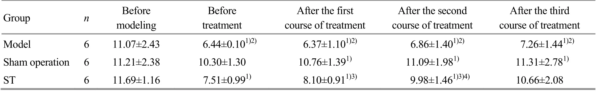
Table 1. Com parison of the PWTL (±s, s)
Note:Compared w ith the same group before modeling,1)P<0.05;compared w ith the sham operation group at the same time point,2) P<0.05;compared w ith the model group at the same time point,3) P<0.05;compared w ith the same group before treatment,4) P<0.05
Group n Before modeling Before treatment After the first course of treatment After the second course of treatment After the third course of treatment Model 6 11.07±2.43 6.44±0.101)2) 6.37±1.101)2) 6.86±1.401)2) 7.26±1.441)2) Sham operation 6 11.21±2.38 10.30±1.30 10.76±1.391) 11.09±1.981) 11.31±2.781) ST 6 11.69±1.16 7.51±0.991) 8.10±0.911)3) 9.98±1.461)3)4) 10.66±2.08
2.2 Morphological changes of L5 DRG
In the model group, the morphological changes in L5DRG included vacuolations,blebbing and highly irregular cell boundaries,disordered distribution of Nissl bodies and the staining of the Nissl substance weakened or even disappeared(retrograde chromatolysis).In the sham operation group,the membrane of DRG and the centered nucleolus was clearly observed,and the Nissl bodies were evenly distributed. In the ST group, the morphological changes in DRG were sim ilar to those in the model group, but the changes were less severe than those in the model group (Figure 1).
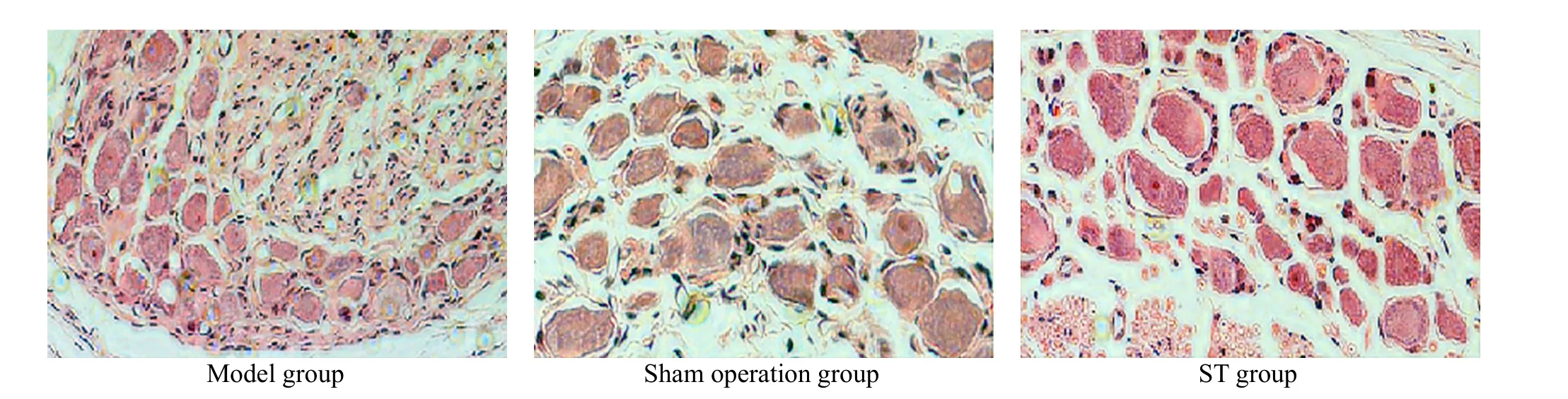
Figure 1.M icroscopic observation of L5 DRG in each group after the third course of treatment (HE, ×400)
2.3 Serum IFN-γ, IL-4 and IL-10 test results
In the sham operation group,there were no statistically significant changes in the serum IFN-γ level at each time point (P>0.05).At the beginning of treatment, the serum IFN-γ levels in the model group and ST group were higher than that in the sham operation group (bothP<0.05). After the second and third courses of treatment, the serum IFN-γ levels in the model group were significantly lower than that before treatment (bothP<0.05), but still higher than those in the sham operation group and ST group. In the ST group,the serum IFN-γ levels decreased significantly w ith the treatment time,and were lower than that before treatment and in the model group at the same time points,and the between-group differences were statistically significant (allP<0.05).There were no statistical difference between the ST group and the sham operation group (P>0.05), (Table 2).
There were no statistically significant differences in serum IL-4 levels at each time point in the sham operation group (P>0.05). Before treatment, the serum IL-4 levels in the model and ST groups were higher than those in the sham operation group(allP<0.05). The serum IL-4 level in the model group at each time point after treatment was lower than that before treatment(allP<0.05).After the second and third courses of treatment, the serum IL-4 levels in the ST group were significantly lower than that before treatment, and the differences were statistically significant (bothP<0.05),(Table 2).
There were no statistically significant differences in the serum IL-10 level at each time point in the sham operation group (P>0.05). Before treatment, the serum IL-10 levels in the model group and ST group were higher than that in the sham operation group(bothP<0.05). After the second course of treatment, the level of serum IL-10 in the model group was lower than that before treatment (P<0.05) and in the ST group (P<0.05),and there was no statistical difference compared w ith the sham operation group (P>0.05). At the end of the third course of treatment, the level of serum IL-10 in the ST group was significantly lower than that before treatment (P<0.05). There were no significant between- group differences comparing the model group and the sham operation group (bothP>0.05), (Table 2).
2.4 Correlation analysis between the PWTL and IFN-γ,IL-4 and IL-10
Pearson's correlation analysis showed that the PWTL in the three groups before treatment were negatively correlated w ith the levels of IFN-γ, IL-4, and IL-10 in the same period, and the correlation coefficient was -0.667(P=0.003),-0.537(P=0.22)and-0.579(P=0.052),respectively. At the end of the third course of treatment,the PWTL in the three groups were negatively correlated w ith the level of IFN-γ in the same period,w ith a correlation coefficient of -0.594 (P=0.009),w ithout significant correlation between the PWTL and cytokine levels at the other time points.
2.5 Effect of ST on the ratio of IFN-γ/IL-4 in rats'peripheral blood
The distributions of IFN-γ/IL-4 ratio in the peripheral blood in the three groups are shown in Figure 2. The data were tested by multiple independent samples of Kruskal-Wallis H. The results showed that the overall distributions in the three groups were partially the same(P<0.05).According to the average rank,the IFN-γ/IL-4 ratios in the three groups were the model group, the ST group and the sham operation group in the descending order.
Table 2. Com parison of the serum IFN-γ, IL-4 and IL-10(±s, pg/m L)
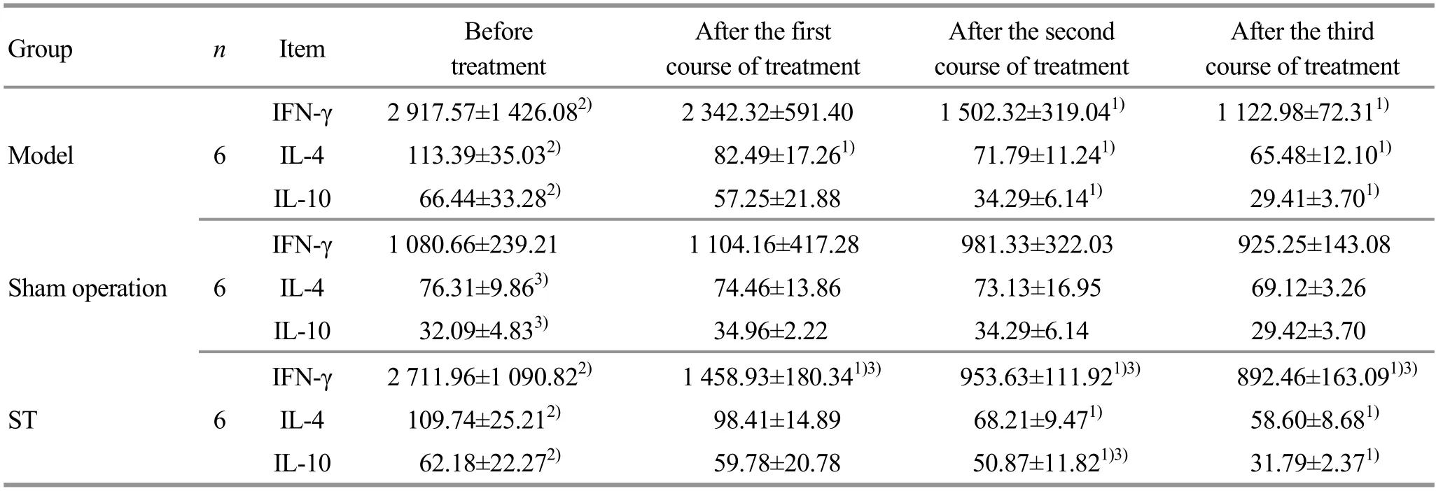
Table 2. Com parison of the serum IFN-γ, IL-4 and IL-10(±s, pg/m L)
Note:Compared w ith the same group before treatment, 1) P<0.05;compared w ith the sham operation group at the same time point,2) P<0.05;compared with the model group at the same time point,3) P<0.05
Group n Item Before treatment A fter the first course of treatment After the second course of treatment After the third course of treatment IFN-γ 2 917.57±1 426.082) 2 342.32±591.40 1 502.32±319.041) 1 122.98±72.311) Model 6 IL-4 113.39±35.032) 82.49±17.261) 71.79±11.241) 65.48±12.101) IL-10 66.44±33.282) 57.25±21.88 34.29±6.141) 29.41±3.701) IFN-γ 1 080.66±239.21 1 104.16±417.28 981.33±322.03 925.25±143.08 Sham operation 6 IL-4 76.31±9.863) 74.46±13.86 73.13±16.95 69.12±3.26 IL-10 32.09±4.833) 34.96±2.22 34.29±6.14 29.42±3.70 IFN-γ 2 711.96±1 090.822) 1 458.93±180.341)3) 953.63±111.921)3) 892.46±163.091)3) ST 6 IL-4 109.74±25.212) 98.41±14.89 68.21±9.471) 58.60±8.681) IL-10 62.18±22.272) 59.78±20.78 50.87±11.821)3) 31.79±2.371)
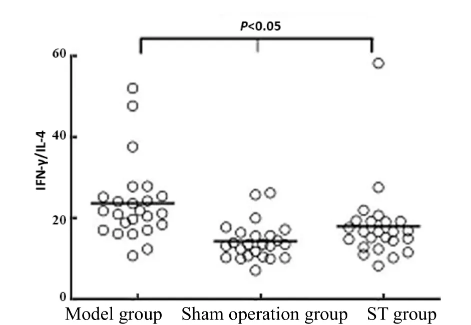
Figure 2. Distribution of IFN-γ/IL-4 ratio
2.6 Correlation analysis between the PWTL and IFN-γ/IL-4 ratio
Spearman's rank correlation analysis showed that the ratio of IFN-γ/IL-4 had no significant correlation w ith the PWTL before treatment and at the end of the first course of treatment. However, at the end of the second and third courses of treatment,the PWTLs were negatively correlated with IFN-γ/IL-4 ratio,and the correlation coefficient was -0.556 (P=0.057) and -0.476(P=0.046), respectively.
3 Discussion
LDH belongs to the category of Bi-impediment syndrome in traditional Chinese medicine (TCM). Kidney deficiency is the root cause, and blood stasis is the external manifestation. This condition is mainly related to bone, marrow, tendon and muscle. For a long time,the external treatment methods of TCM, including tuina,acupuncture and moxibustion, have shown significant efficacy,safety and convenience in the treatment of LDH. ST is based on the holistic concept, the theory of the twelve meridians and points, and the integration of the advantages of acupuncture,massage,cupping and other TCM external treatment methods[11].It has received more and more attention from hundreds of patientsand researchers.The current research on ST in the treatment of LDHmainly focuseson the observation of clinical efficacy,w ith few reports on basic research.Therefore,on the basis of previous research,this study explored the immunological mechanism of ST in reducing LDH pain from the perspective of Th1/Th2 immune function.
At present,themechanism of LDH pain hasnot been fully elucidated.Its generally recognized causes include compressive and non-compressive factors.Compressive factors mainly refer to the compression of nerve roots.Non-compressive factors are produced by the contact between nucleus pulposus and nerve fibers,including chem icaland immune factors (a variety of chem icaland immune inflammatory mediators).Studies have found that mechanical compression alone is not enough to stimulate pain and reduce the body's pain threshold[12];and when the chronic pain mechanism has been triggered,even if the nerve root compression factor is removed,the pain w ill still continue[13].Therefore,the chem icaland immune inflammatory mediators induced by contact between nucleus pulposus and nerve fibers may be the pathological basis of LDH back pain and sciatica.
The nucleus pulposus is a closed non-vascular tissue.Under normal circumstances,the nucleus pulposus is isolated from the host immune system.Once the nucleuspulposusprotrudes through the annulus fibrous,triggering an autoimmune response[14],generating autoantibodies,depositing antigen-antibody complexes and activating inflammation,nerve root damage develops[15].This study established a non-compressive LDH model of autologous nucleus pulposus transplantation,and found that the heat pain threshold of rats was significantly reduced after the modelwas established,confirm ing that the autoimmune inflammatory response of the nucleus pulposus should be one of the factors that cause LDH neurogenic pain. In addition, the results of HE staining showed that after the autologous nucleus pulposus was transplanted to the surface of the DRG, the cell morphology changed w ith obvious edema and degeneration. However, after receiving ST,the degree of ganglion damage was significantly reduced,indicating that ST can promote the repair of DRG damage.
In autoimmune diseases,Th cells proliferate and differentiate into either the Th1 or the Th2 subset to promote the occurrence and development of autoimmune diseases. Th1 cells participate in cellular immunity and are involved in the development of delayed hypersensitive inflammatory reactions.Th1 mainly secretes cytokines such as IFN-γ and TNF-β; Th2 can help B cells differentiate into antibody-secreting cells,participating in humoral immune response, and Th2 cells mainly secrete IL-4 and IL-10[16].
IFN-γ and IL-4 are representative cytokines of Th1 and Th2 cells, respectively. Therefore, most studies use the quantitative comparison of these two to represent the balance of Th1/Th2[17-18].The normal nucleus pulposus is isolated from the immune system. There is no expression of Th subtype cells in the nucleus pulposus, the levels of IFN-γ and IL-4 are m inimal, and Th1/Th2 is in a dynam ic equilibrium state to maintain the body's normal cellular and humoral immune functions[19],which is sim ilar to that in the sham operation group. After the nucleus pulposus protrudes,it activates the autoimmune response and promotes the differentiation of T cells, and the Th1/Th2 balance is finally broken. Therefore, adjusting Th1/Th2 drift and restoring its balance may be one of the immunological mechanisms in the treatment of LDH pain.
IFN-γ is a pro-inflammatory factor. It can induce pain by inducing neuronal hyperexcitability, and the level of IFN-γ increases in pathological conditions[20].IL-4 and IL-10 are effective anti-inflammatory factors.It can reduce neuropathic pain by promoting the high expression of these two cytokines[21-22]. The results of this experiment suggested that the Th1 type cytokine IFN-γ in the non-compression LDH model rats w ith autologous nucleus pulposus transplantation increased significantly compared w ith the sham operation group,and the ratio of IFN-γ/IL-4 was significantly higher than that in the sham operation group. In addition, the ratio of IFN-γ/IL-4 was negatively correlated w ith the thermal pain threshold at the last two detection time points,suggesting the advantage of Th1 cytokine.After modeling, the levels of IL-4 and IL-10 in the model group and the ST group were significantly higher than those in the sham operation group, suggesting that the body itself had an anti-inflammatory effect, which is manifested as an anti-inflammatory compensatory response.This may explain the negative correlation between the expression levels of the three cytokines,IL-4, IL-10, and IFN-γ, and the thermal pain threshold before treatment.
In addition,this study found that the Th1 type cytokine IFN-γof LDH rats in the ST group after treatment was significantly reduced compared w ith the model group, almost restored to the same level as in the sham operation group, but little effect was found on the expression of Th2 cytokines. Therefore, one of the effective mechanisms of ST may be to inhibit Th1 type cytokine IFN-γ,restore Th1 deviation to the normal balance of Th1/Th2, and achieve the effect of reducing pain.
In summary, this study initially explored the effect of ST on serum Th1/Th2 immune balance in non- compressive LDH rats, and found that the pain relief effect of ST on LDH may be closely related to its ability to promote the restoring of the dynam ic balance of Th1/Th2. The specific mechanism is worth to be further explored in the future.
Conflict of Interest The authors declare that there is no potential conflict of interest in thisarticle.Acknow ledgments This work was supported by National Natural Science Foundation of China (国家自然科学基金项目,No.81473791);Advantage Discipline Construction Project of Nursing in Jiangsu Universities (江苏高校护理学优势学科建设工程资助项目,No.2019YSHL028);Undergraduate Practice Innovation Training Program of Nanjing University of Traditional Chinese Medicine (南京中医药大学大学生实践创新训练计划项目,No.201810315121).Statem ent of Human and Animal Rights The treatment of animals conformed to the ethical criteria in thisexperiment.
Received:22 January 2020/Accepted:25 February 2020
猜你喜欢
华人时刊(2022年9期)2022-09-06
Chinese Physics B(2022年3期)2022-03-12
护理学报(2022年1期)2022-02-25
国际放射医学核医学杂志(2021年6期)2021-11-30
国际放射医学核医学杂志(2021年1期)2021-11-30
护理学杂志(2021年1期)2021-02-04
西安航空学院学报(2018年5期)2018-10-15
中国气象科学研究院年报(2017年0期)2017-07-19
伴侣(2015年10期)2015-09-10
中国中医药现代远程教育(2014年21期)2014-03-01
 Journal of Acupuncture and Tuina Science2020年5期
- Journal of Acupuncture and Tuina Science的其它文章
- Discussion of the prom ising effect of electroacupuncture on cognitive improvement in D-galactose-induced aging rats based on NLRP3-ASC-Caspase-1 signaling pathway
- Study on the body surface temperature variation patterns of the meridian acupoints related to the physiological status of the uterus
- Therapeutic efficacy observation on moxibustion w ith moxa of different storage years for moderate-to-severe primary knee osteoarthritis
- Acupuncture therapy w ith point selection based on syndrome differentiation along the meridians for functional dyspepsia: a random ized controlled trial
- Clinical observation of acupuncture plus Frenkel exercises for ataxia after cerebral stroke
- Efficacy evaluation of acupuncture plus rehabilitation training for post-stroke deglutition disorders of qi-deficiency blood stasis pattern