
Petroselinum crispum extract ameliorates scopolamine-induced cognitive dysfunction: role on apoptosis, inflammation and oxidative stress
2022-06-23 07:23kslnrKarakaogluDilkOzyliSrnfiyYanaragOzlmSaanAsliAyka
食品科学与人类健康(英文) 2022年5期
Göksl Şnr*, Göz Karakaıoglu, Dilk Ozyli, Srn E,R fiy Yanarag, Ozlm Saan, Asli Ayka
a Department of Pharmacology, Faculty of Pharmacy, Fenerbahce University, Istanbul 34758, Turkey
b Department of Pharmacology, Institude of Health Sciences, Marmara University, Istanbul 34854, Turkey
c Department of Medical Pathology Techniques, Vocational School of Health Services, Marmara University, Istanbul 34865, Turkey
d Department of Biochemistry, Faculty of Engineering, Istanbul Universityş-Cerrahpasa, Istanbul 34320, Turkey
e Department of Biophysics, Faculty of Medicine, Near East University, Nicosia 99138, Cyprus
Keywords:
Petroselinum crispum
Apoptosis
Spatial memory
Recognition memory
Oxidative stress
M1 receptor
A B S T R A C T
This study was designed to investigate whether Petroselinum crispum (PC) extract has protective effects on the brain in the scopolamine-induced Alzheimer’s disease (AD) rat model. The rats were divided into; control,scopolamine (1 mg/kg, i.p.), galantamine (1.5 mg/kg, i.p.) and PC extract (2 g/kg, p.o.)-treated scopolamine groups. On day 14, the novel object recognition test (NORT) and Morris water maze test (MWMT) were performed and then the rats were sacrificed. Scopolamine-induced cognitive impairments observed in the NORT and MWMT, significantly improved with PC extract and galantamine treatments. Scopolamine reduced M1 receptor expression, Bcl-2/Bax ratio, and glutathione levels in the hippocampus and frontal cortex, while malondialdehyde levels, caspase-3/9 expressions, and acetylcholinesterase (AChE) activity were found to be increased. On the other hand, PC and galantamine treatments reversed these changes. In conclusion, PC extract has shown an ameliorative effect on the spatial and recognition memory, M1 receptor expression,apoptosis, oxidative stress, and increased AChE activity. Thus, it was concluded that PC could prevent AD-like conditions and can be used as a functional food. However, since animal models do not completely mimic those of humans, based on the data obtained in this study, the importance of PC on human AD should be demonstrated in future studies.
1. Introduction
Alzheimer’s disease (AD), one of the most important causes of dementia is characterized by impairment in learning ability and memory formation caused by progressive neuronal loss [1].The pathogenesis of AD includes cholinergic synaptic disorder,mitochondrial dysfunction, oxidative damage, and apoptosis in the memory-sensitive brain regions [2-4]. Currently, acetylcholinesterase(AChE) inhibitors such as galantamine, rivastigmine, and donepezil are used which can increase the concentration and effect duration of acetylcholine (ACh) in memory sensitive brain regions, however,these drugs are not adequate to stop the progression of AD [5,6].Thus, research is in progress to develop potent drugs targeting the multiple pathological mechanisms and provide successful treatment of AD in humans.
It is known that the cholinergic system plays an important role in the regulation of memory function and ACh controls numerous cognitive processes in this system [7]. The subtype of muscarinic receptors (mAChRs) M1has a pivotal role in learning, memory,cognitive functions, and mood [7,8]. Indeed, the blockage of these receptors has been observed to cause memory errors [9].Scopolamine is a non-selective mAChR antagonist that negatively affects learning achievement and memory in animals and humans [10-12]. Scopolamine disrupts short-term novel object recognition memory [13], spatial memory [14], and passive avoidance memory [15]. Scopolamine-induced memory impairment has been reported to be associated with increased brain oxidative stress [12,15-17],apoptosis [4,12,18]and AChE activity [12]. Consequently,scopolamine-induced memory impairment is a useful animal model for investigating antioxidative, AChE inhibition, and anti-apoptotic therapy strategies in AD.
Parsley (Petroselinum crispum, family Apiaceae, PC) is a green plant with important medicinal properties such as antioxidant [19-24], antiapoptotic [20,21], anti-inflammatory [21], and anti-diabetic [22,23,25].In addition to its high nutritional properties, it is a natural vitamin and mineral source. It contains some antioxidant substances such as flavonoids (apigenin, luteolin), carotenoids, and ascorbic acid [25,26].PC components were found to be prominent suppressors to reactive oxygen species (ROS) in brain and other tissues by stimulating the generation of glutathione synthesis and by increasing cellular antioxidant defense [27]. PC has been reported to have AChE inhibitory activityin vitro[28,29]. Thus, the potential therapeutic benefit of this substance is very important especially for treatment in the early stage of AD [30]. However, the protective effects of PC against the Scopolamine-induced AD rat model are not known.
Therefore, this study aimed to investigate whether PC has protective effects on the brain in terms of oxidative damage, apoptosis, and cognitive function in the scopolamine-induced AD rat model.
2. Materials and methods
2.1 Preparation of PC aqueous extract
After the leaves of the PC plant harvested from the countryside of Istanbul were thoroughly washed, they were dried in the shade at(20 ± 2) °C for 3 days and then kept in cellophane bags. 100 g of dried PC leaves were extracted with 1 L distilled water and boiled for 30 min. The extract was then filtered, and the filtrate was evaporated, using a rotary evaporator under reduced pressure to dryness (at 45 °C) [22].The gained extract was kept in a dark container at 4 °C until it was used. The PC extract was dissolved in distilled water before the applied to the rats.
2.2 Animals and conditions
All protocols were approved by Marmara University Animal Care and Use Committee (no: 25.2019.mar). Wistar albino rats weighing 250-300 g of both sexes (n= 32) provided by Marmara University Experimental Animals Research and Application Center were used. The rats were housed at ambient temperature ((22 ± 2) °C)and of humidity (55 ± 5)% with 12 h : 12 h light-dark. During the experiment, the rats were freely allowed to drink water and standard pellet dietad libitum.
2.3 Drugs and chemicals
All chemicals were supplied by Merck KGaA (Darmstadt,Germany) and all antibodies were supplied from Santa Cruz Biotechnology, Inc (Santa Cruz, CA, USA).
2.4 Experimental design
After 10 days of habituation, the rats were randomly divided into 4 groups each containing 8 rats. Experimental groups were as follows:Control (C), scopolamine (Scop) and, galantamine (GAL), and PC treated-scopolamine groups.
To induce AD to the rat model, scopolamine hydrobromide(C17H21NO4·HBr) a mAChRs antagonist was administered at a dose of 1 mg/kg BW, i.p. The dose and duration of scopolamine treatment were based on previous studies [13,31]. Galantamine hydrobromide(C17H21NO3·HBr) an AChE inhibitor was administered at a dose of 1.5 mg/kg BW, i.p. [32]andPC extract was administered 2 g/kg BW orally [22]. An equal volume of physiological saline was given to the control group. All treatments were given at 9 am for 14 days (Fig. 1).

Fig. 1 An overview of the experimental design.
2.5 Cognition tests
To evaluate the cognitive status of rats, the novel object recognition test (NORT) and Morris water maze test (MWMT) were performed respectively at the end of the 14thday (Fig. 1).
2.5.1 NORT
The NORT is designed to evaluate short-term recognition memory and the results are affected by both hippocampal and cortical defects [33].The tests were performed in an apparatus and before the test, the rats were handled by the researchers. During all experiments, boxes and objects were wiped with 70% alcohol while passing to the next animal. Habituation, familiarization, and test phases were performed according to a previous study [34]. The evaluation of the results was performed with a comparison of the time spent with the familiar and novel objects. The results were calculated with the formula previously described and the result was expressed as a discrimination index [35,36].
2.5.2 MWMT
MWMT was performed after the NORT. MWM is a test for spatial learning and memory study in rodents. MWM test is based on the native reluctance of rats to float [37]. MWM task was performed with a black circular pool with a diameter of 1.2 m and a height of 0.47 m. The apparatus was filled with black coloured 24-25 °C water to 20 cm deep. Coloured cartons were placed on the walls around the pool for the rats to identify the environment. Two main axes of the pool were determined. Each splits the pool in two vertical directions to form an imaginary ‘+’. In this way, the pool was divided into 4 equal quadrants.
MWM task consists of the training period and probe trial. Each process was recorded with a video camera. The training period was performed in the first 4 days of the experiment. During the training period, an escape platform in the same colour of the water with a 10 cm diameter, was hidden 2 cm under the water and was placed in the middle of one of the quadrants (target quadrant) [38]. Animals were released into the pool from each quadrant with a random sequence. The rats were allowed to swim for 75 s to find the platform.Platform finding times of rats in 75 s were recorded. The animals that cannot find the platform within this period were directed to the platform and their platform finding times were saved as 75 s. They were allowed to observe around on the platform for 20 s. Then the animal was kept in its cage for 60 s before released to the pool again.On the fifth day of the MWM task, the probe trial was performed. By removing the hidden platform, rats were allowed to swim in the pool for 60 s. During this period, how much time was spent on the target quadrant was evaluated.
2.6 Dissection of the brain tissues
2.7 Biochemical analysis
The levels of malondialdehyde (MDA) and reduced glutathione(GSH) expressed as nmol/g and μmol/g in the hippocampal and frontal cortex tissues were measured with the method of Buege and Aust [40], and Beutler [41], respectively. The activity of the AChE responsible for the degradation of ACh was measured in tissue samples using a spectrophotometer with the Ellman method and was expressed as μmol/h/mg protein [42].
2.8 Immunoblotting analysis
For the immunoblotting analyses of both brain tissues of the rats, Tris-HCl buffer containing EDTA (1 mmol/L), glycerol (10%),protease inhibitors, NaCl, and DTT (50, 2 mmol/L, respectively)were added to the tissues and then centrifuged for 85 s. After the homogenization using a centrifuge (at 2 000 ×gfor 12 min), they were vortexed for 90 min for incubation with Tris-HCl buffer and 0.05% Triton X-100. The protein content of the samples was determined using the Lowry method [43]. Protein in gels obtained from the loading of samples containing 100 μg protein into electrophoresis was transferred to nitrocellulose membranes [13].After blocking with Tris containing 1% BSA, all membranes were incubated for 12 h with polyclonal primary antibodies [Bax(1:100) (sc-20067), Bcl-2 (1:200) (sc-7382), caspase-3 (1:200)(sc-56053), caspase-9 (1:200) (sc-56076), or M1(1:100)(sc-365966)]at 4 °C. After detection with the desired antibody against the protein of interest, the membranes were incubated with a secondary antibody (anti-goat IgG) (1:1 000) for 1 h at 20 °C.Finally, all membranes were washed with Tris buffer solution. The results were determined by densitometric analysis using the Bio-Rad Molecular Analyst software (free edition, www. totallab.com).
2.9 Statistical analysis
Statistical analysis was performed using Graphpad Prism 6.0(Graphpad Software, San Diego, CA, USA). All data are expressed as mean ± standard error (SEM). Biochemical data of groups were analyzed with variance analysis (ANOVA) followed by Tukey multiple comparison tests. The results of NORT and MWMTs were analyzed with the Mann-Whitney U nonparametric test. For all data,P< 0.05 was considered statistically significant.
3. Results
3.1 Assessment of scop-induced cognitive deficits in two different tests
The NORT was performed to evaluate the effect of galantamine and PC extract treatment to impairment in object recognition memory of rats due to scopolamine administration. It was determined that the administration of scopolamine significantly reduced the time spent with the novel object and elevated the time spent with the familiar object, thus reducing the discrimination index of the NORT (P< 0.01;Fig. 2). On the other hand, galantamine andPC treatment reversed the time spent exploring the novel object (P< 0.01).

Fig. 2 According to NORT, discrimination index of the 4 groups. **P <0.01,compared to control group; ++P < 0.01, compared to Scop group. (n = 8).
The effect of scopolamine on cognitive impairment was assessed by MWMT which is used to assess spatial memory ability. The comparison of each group according to the 1stand 4thdays is given in Fig. 3a and the comparison between the groups on the test day is given in Fig. 3b. While there was a platform in MWMT from day 1 to day 4, there was no platform in the tank on day 5 of the experiments.No difference in escape latency in all groups was observed on the first training days. When the escape latency of the control group was examined, a significant difference was determined in the 3rdand 4thdays compared to the 1stday of the MWMT (P< 0.05,P< 0.01,respectively; Fig. 3a) however there was no significant difference in escape latency day 1 to day 4 in Scop group. On the other hand,galantamine andPC-treated rats found the platform in a short time,and therefore escape latencies were decreased significantly.

Fig. 3 Cognitive function assessed by the MWM test of the 4 groups. (a) Mean escape latency time was monitored from day 1 to day 4 in each group. (b) Time spent in the target quadrant was monitored at day 5 in each group. (n = 8). *P < 0.05; **P < 0.01; ***P < 0.001 as compared to the control group; ++P < 0.01 vs Scop group.
In MWM probe test that was monitored on the 5thday, results showed that the saline-treated Scop group had significantly lower performance(P< 0.05) vs control group (Fig. 3b), but the PC-treated group displayed higher performance and the time spent in the target quadrant was found to be increased significantly compared to the Scop group (P< 0.01).
Although the galantamine-treated group tended to increase in time spent in the target quadrant, this increase was not statistically different when compared with the Scop group, and there was no significant difference between two treatment groups on the 5thday.
3.2 Biochemical analysis
The GSH levels of the hippocampal and frontal cortex tissues were significantly reduced in the Scop group compared to the control group (P< 0.001), but administration of galantamine andPC extract increased this antioxidant in both tissue samples significantly (P< 0.01 or 0.001, Figs. 4a and 4b).
枢纽及防洪配套工程年运行费按投资2.5%计,后续灌区配套工程按其投资4%计,防洪、养殖等配套工程按其投资5%计[2],则正常年份年运行费32.77万元。
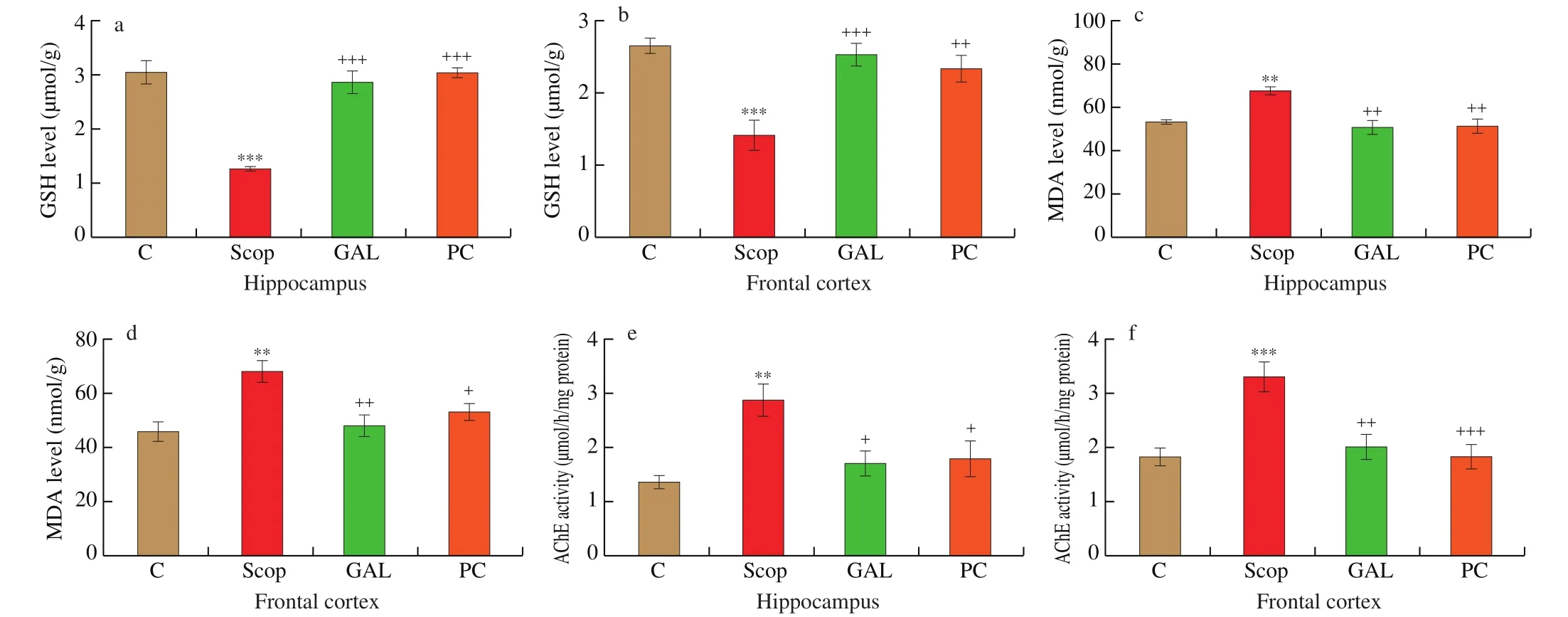
Fig. 4 The levels of (a-b) GSH, (c-d) MDA and (e-f) AChE activity obtained from the hippocampus and frontal cortex regions of the rats in the 4 groups (n = 8).
Malondialdehyde levels in the hippocampus and frontal cortex tissues were found to be significantly increased due to scopolamine(P< 0.01), and treatment of galantamine andPCextract significantly(P< 0.05 or 0.01) decreased MDA which were closer to the control levels (Figs. 4c and 4d).
Scopolamine administration for 14 days caused significant increase in AChE activity (P< 0.01 or 0.001) in the investigated regions, while in galantamine andPC treated groups, the enzyme activities were decreased significantly (P< 0.05 or 0.001; Figs. 4e and 4f).
3.3 The results of immunoblotting
We performed immunoblotting experiments in 8 rats per group for the determination of different protein expression levels. The images of the membranes obtained from western blotting assays for each protein are shown in Fig. 5.
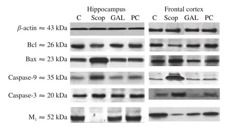
Fig. 5 The representative image of the membranes obtained from western blotting experiments in the Scop-induced AD rat model.
When evaluating the change of the Bcl-2/Bax ratio in the hippocampus, we found that the Bcl-2/Bax ratio was decreased in the Scop groups compared to the control groups (P< 0.001). In the hippocampal region, the Bcl-2/Bax ratio significantly increased with the administration of galantamine andPC treatment compared to the Scop group (P< 0.001, Fig. 6a).

Fig. 6 The density of immunoblotting of (a-b) Bcl-2/Bax ratio, (c-d) caspase-9, (e-f) caspase-3 and (g-h) M1 levels of the hippocampus and frontal cortex regions of the rats in the Scop-induced AD rat model (n = 8).
In the frontal cortex region, it was detected that the levels of the Bcl-2/Bax ratio were found to be decreased in rats treated with scopolamine compared to the control group (P< 0.001). In frontal cortex regions, expression levels of the Bcl-2/Bax ratio were markedly increased in GAL and PC groups compared with the Scop group(P< 0.001, Fig. 6b).
When we analyzed the expression changes of caspase-9 proteins effective in the apoptosis caspase pathway in the hippocampus, it was found that caspase-9 expression levels were increased in the Scop group compared with the control groups (P< 0.001). When we compared the effect of galantamine andPC treatments on the level of caspase-9 expression compared with the Scop group, we determined that both treatments significantly reduced the caspase-9 expression level compared with the Scop group (P< 0.001, Fig. 6c).
When the caspase-9 expression was analyzed in the frontal cortex region, it was determined that the caspase-9 expression levels of Scop group rats increased compared to the control groups (P< 0.01). The treatments of galantamine andPC were found to cause the caspase-9 expression levels to increase compared to Scop group (P< 0.001, Fig. 6d).
In the hippocampus, caspase-3 expressions were markedly elevated in the Scop group compared to the control group (P< 0.001).Galantamine treatment significantly decreased caspase-3 expression levels compared to the Scop group in the same region (P< 0.001).Similar to GAL treatment, it was determined that PC treatment has decreased the level of caspase-3 expression in the hippocampus(P< 0.001, Fig. 6e).
It was determined that the expressions of caspase-3 were raised in the frontal cortex of rats treated with scopolamine compared to the control group (P< 0.001). Additionally, galantamine treatment led to a significant decrease in caspase-3 expression levels in the frontal cortex region of rats compared with the Scop group (P< 0.001).Moreover, it was determined that PC-treatment was effective in decreasing caspase-3 expression levels in the frontal cortex (P< 0.01,Fig. 6f).
In the hippocampus, the expression levels of mACh M1subtype was found to be diminished in the rats treated with scopolamine compared to the expression in control rats treated with saline(P< 0.000 1). The expression level of M1was significantly increased in the galantamine treated groups compared to Scop groups(P< 0.001). The M1level was significantly increased in the PC-treated compared to the Scop group (P< 0.000 1, Fig. 6g).
When M1expression levels were examined in the frontal cortex region, it was determined that the expression level of Scop group diminished markedly compared with the control group (P< 0.001). In the same region, expression levels of M1were markedly raised in the galantamine treated group compared with Scop groups (P< 0.001).The M1level was significantly increased with thePCtreated group compared with the Scop group (P< 0.001, Fig. 6h).
4. Discussion
This study reveals a significant increase in the degree of apoptosis, oxidative stress, and AChE activity in the frontal cortex and hippocampus regions of scopolamine-induced cognitive deficient animals. There was also a significant decline in the level of M1receptor expression in the selected brain regions of the rats.The results of the cognitive experiments of this study showed that scopolamine injection resulted in learning and memory impairment in the formation of recognition memory of rats determined with NORT and MWMTs. On the other hand,PCextract has been shown to have an ameliorative effect on apoptosis, oxidative stress, M1receptor expression, and increased AChE activity.
The NORT was conducted to evaluate the effect of PC on hippocampus-dependent cognitive function. Similar to galantamine, PC extract caused improvement in short term working memory, and these results show that the reduction in scopolamine-induced recognition memory is reversed [44,45]. Although, there is no data in the literature for PC extract treatment on recognition memory function, luteolin an important flavonoid found in the PC, has been shown to improve new object recognition memory in the Ts65Dn mouse model [46]. Thus, the present results indicate for the first time, that PC has an enhancing effect on short term object recognition memory.
In the MWM experiments, the time spent on the target quadrant is considered a sign of memory rejuvenation. Scopolamine impairs place navigation ability and spatial memory in rats and humans [47,48]and recognition memory in rats [44,45]. Galantamine significantly mitigated the scopolamine-induced reduction of spatial memory in maze tests [49]. The enhancing effects of PC extract on cognitive functions have not been known in the literature. In the present study,scopolamine raised the escape latency in the acquisition phase and declined time spent in the target quadrant in the probe phase meliorated by the treatment with PC extract. Our MWMT results,in which we examined the spatial learning and working memory,indicated that in rats treated with scopolamine, PC extract has significant improvement in spatial memory deficits, similar to galantamine.
Scopolamine is associated with increased oxidative stress in the hippocampus and frontal cortex [12,50]. GSH a powerful cellular antioxidant clears ROS or free radicals directly, while MDA is one of the end products of fatty acid peroxidation in the membrane. Previous studies have reported that scopolamine caused an increase in MDA levels [13,48,51]and a reduction in GSH levels [13,48,52]in the rat brain. In agreement with previous studies, in the present study, MDA levels were found to be significantly increased while GSH levels were decreased, in the hippocampus and frontal cortex regions with 14 days of scopolamine treatment [53-55]. On the other hand, PC and galantamine treatment in the scopolamine treated rats reversed these changes. In our study, we used galantamine, one of the existing drugs used in AD, as a positive control, because it has been shown to have protective effects on various neurotoxic models in the brain via reducing oxidant damage. Although there is no study about the effects of PC extract in scopolamine-induced AD model, PC has been shown to suppress lipid peroxidation by diminishing MDA and enhancing GSH levels in rats in cisplatin-induced hepatic-cardiac injuries [21]and testicular oxidative stress [20]. Besides, Allam et al. [56],reported that low-dose parsley (P. crispum) juice leads to an increase in GSH levels and decreased oxidative stress when exposed to cadmium in newborn rat brain. Vora et al. [19]indicated that parsley has a protective effect from oxidative damage in the mouse brain on the d-galactose induced oxidative stress model. Taken together, our results indicate that the PC could have a protective effect on scopolamineinduced oxidative injury in the hippocampus and frontal cortex.
Acetylcholine plays a pivotal role in cognitive function and it is well known that in AD it is reduced. While scopolamine increases AChE’s protein expression level [51,57,58], it reduces ACh level [57]. In agreement with the previous reports, in the current study, scopolamine increasing AChE activity caused cognitive impairment. Accordingly, AChE inhibition is an effective strategy in the early phase of AD [30]. The efficacy of galantamine in AD is accompanied by AChE expression or activity [13,58]. Present results showed that similar to galantamine, PC extract has anti-AChE activity in the hippocampus and frontal cortex, and improve failure caused by a deficiency in the cholinergic system.
M1is the most expressed muscarinic receptor type in the hippocampus and prefrontal cortex [59]. Jahanshahi et al. [59]showed that M1-neuron density decreased in the entire hippocampal region depending on the dose of scopolamine. Sayyahi et al. [60]reported that M1muscarinic receptor-immunoreactive neurons decreased numerically with scopolamine. Similar to a previous report [13], in our study, the rats who received scopolamine exhibited a significant loss in the level of M1expression in the hippocampus and frontal cortex regions. This result may be related to neuronal oxidative damage or apoptosis caused by scopolamine [4,12,15,17,18]. On the other hand, PC extract increased M1receptor expression and this effect may be due to antioxidant [19-24]and anti-apoptotic [20,21]effects of PC. In a recent study, PC has been shown to protect the number of neurons and dendrite structure in morphine-induced prefrontal cortex damage by reducing oxidative and nitro-oxidative stress [23].
In our study, Bcl-2/Bax expression ratio was decreased in sensitive brain regions while caspase-3 and caspase-9 increased. These observations indicate the fact that an increase in the pro-apoptotic protein expression could be a cause of mitochondrial dysfunctioninduced neuronal degeneration seen in AD. Demirci et al. [18]demonstrated that scopolamine caused an increase in mitochondrialrelated apoptosis in the brain regions of the rats due to a decrease in mitochondrial function. Previously, it has been reported that active caspase-3 and pro-caspase-9 were increased as well as the ratio of Bax/Bcl-2 after scopolamine treatment [61]. Similarly, the present study con firms that scopolamine causes apoptosis in rat the brain since caspase-3 and 9 expressions were found to be significantly increased.Previously, it has been shown that PC has anti-apoptotic effects on cisplatin-induced hepato-cardiotoxicity in rats [21]. Allam et al. [56]reported that PC juice exhibits remarkable neuronal protection through reducing the rate of chromatolysis and pyknosis in the newborn rat brain. Besides, apigenin, a natural flavonoid found in PC, has been reported to alleviate neuronal apoptosis by decreased Bax and caspase-3, in the brain injury model [62]. Furthermore, Balez et al. [63]suggested that apigenin reduced apoptosis by decreasing caspase-3/7 levels in an induced pluripotent stem cell AD model. Hence, the antiapoptotic effects of PC extract on scopolamine-induced AD model may be due to the existence of various flavonoid compounds, such as apigenin or luteolin. In agreement with previous studies, in this study,we found decreased apoptotic caspase and Bax protein expressions,which represents the anti-apoptotic activity of the PC extract.
Given our findings, we propose that PC extract has an ameliorative effect on M1receptor expression, apoptosis, oxidative stress, increased AChE activity, and spatial and object recognition memory, in the scopolamine-induced AD rat model. This is possibly through enhancement of antioxidant defence, anti-AChE activity,up-regulation of the M1expression, and anti-apoptotic effect. Thus, it was concluded thatPC prevent AD-like conditions and can be used as a functional food.
The effect of PC on cognitive functions has not been previously studied. For this reason, the study was first performed on animals and it was investigated whether the PC extract, which is widely consumed as a vegetable, has possible positive effects on memory. On the other hand, since animal models do not completely mimic that of humans the importance of PC as a functional food for human AD is unknown.Thus, the results of this study will shed light on human studies in the future and show that PC preserves memory functions.
conflict of interest
The authors report no conflicts of interest.
Acknowledgment
The authors received no financial support for the research,authorship, and/or publication of this article.
猜你喜欢
青少年科技博览(中学版)(2022年6期)2022-08-31
大众科学(2022年8期)2022-08-26
云南画报(2022年4期)2022-05-05
小猕猴智力画刊(2021年11期)2021-11-28
水资源开发与管理(2020年9期)2020-10-24
房地产导刊(2020年8期)2020-09-11
商周刊(2019年18期)2019-10-12
家庭影院技术(2019年4期)2019-04-17
当代陕西(2018年12期)2018-08-04
儿童故事画报·自然探秘(2016年4期)2016-06-24
- 食品科学与人类健康(英文)的其它文章
- Hypoglycemic natural products with in vivo activities and their mechanisms: a review
- Bacteroides utilization for dietary polysaccharides and their beneficial effects on gut health
- Capsular polysaccarides of probiotics and their immunomodulatory roles
- Natural compounds may contribute in preventing SARS-CoV-2 infection: a narrative review
- A comprehensive review on the effects of green tea and its components on the immune function
- A review on current and future advancements for commercialized microalgae species