
Efficacy of green tea extract on PC3 prostate cancer cells through upregulation of miR-195 expression and suppression of epithelial to mesenchymal transition
2022-10-14 11:38FatemehSafariKatayounDadehAmirfard
Fatemeh Safari,Katayoun Dadeh Amirfard
Fatemeh Safari,Department of Biology,Faculty of Science,University of Guilan,Rasht 4193833697,Iran
Katayoun Dadeh Amirfard,Department of Microbiology,North Tehran Branch,Islamic Azad University,Tehran 1651153511,Iran
Abstract OBJECTIVE: To evaluate anticancer efficacy of green tea extract (GTE) on PC3 prostate cancer cells.METHODS: By using quantitative real-time polymerase chain reaction (qRT-PCR) and Western blot methods,the expression of miR-195 and the epithelial to mesenchymal transition (EMT) markers such as E-cadherin and vimentin was analyzed.RESULTS: Based on the results of 2D and 3D cell culture models,the inhibition of EMT and up regulation of miR-195 expression were detected.CONCLUSIONS: Our findings will be helpful to design anti-tumor regimens with natural product original,and more studies will be required to identify the related mechanisms involving anticancer activities of green tea via miRNAs.
Keywords: Green tea extract;PC3 cancer cells;epithelialmesenchymal transition;miR-195 expression
1.INTRODUCTION
Prostate cancer (PCa) is one of the most commonly diagnosed male cancers with a high death rate.1For prostate cancer therapy,many approaches including hormone therapy,surgery,radiation,and chemotherapy have been established.The failures of prostate cancer therapies mainly refer to the metastatic tumors that originate from primary prostate tumors.2Metastasis is multistep process that is related with the cancer patient's death.Tumor cell metastasis initiates with the process of epithelial to mesenchymal transition (EMT) that is characterized by loss of cell-cell adhesion and ability to emerge motile and invasive forms of cell.During EMT,the expression of mesenchymal markers such as vimentin and fibronectin is increased and the expression of epithelial markers such as E-cadherin and B-catenin is reduced.3Also,main barriers to the success of cancer treatments are due to resistance to anti-cancer drugs and side effects of drugs and therefore,discover novel,more effective drugs with the lowest side effects especially for natural products are urgently needed.Among natural products,plants may serve as potential chemotherapeutic agents with less toxicity to normal mammalian tissues and at a low cost.4Moreover,it was clearly established connection between dietary factors and various diseases including cancer.Among healthy foods or dietary supplements,the protective effects of green tea have attracted considerable interest.Green tea is one of the most popular beverages worldwide which is produced from the leaves of theCamellia sinensisplant.Green tea contains a group of polyphenolic compounds including four main catechins: (-)-epicatechin (EC),(-)-epigallocatechin (EGC),(-)-epicatechin-3-gallate(ECG),and (-)-epigallocatechin-3-gallate (EGCG).5,6Beneficial effects of green tea on many human diseases have demonstrated.7-10It was also shown that green tea consumption decreases the risk of human cancers.11-13MicroRNAs (miRNAs) are single-stranded non-coding RNAs that have been considered to involve in various cellular pathways by negatively or positively regulating gene expression.14,15miRNAs can suppress the expression of important cancer-related genes and thereby,miRNAs are considered as a novel class of therapeutic approach and diagnosis tool to treat cancer.16In PCa,several miRNAs have been reported to regulate cell migration and motility including miR-195.17-20miR-195 can regulate prostate cancer cell migration and apoptosis through its target genes Fra-1 (one of the transcription factor complex activator protein-1 or AP-1) and ribosomal protein S6 kinase,70 kDa,polypeptide 1(RPS6KB1).Also,anti-tumor activities of green tea particularly EGCG (as main polyphenolic compound in green tea extract) through the regulation of miRNAs expression have revealed.21-24
In this study,we aimed to study the anticancer efficacy of green tea extract (GTE) against human prostate cancer PC3 cells through upregulation of miR-195 expression in 2D and 3D cell culture models.
2.MATERIALS AND METHODS
2.1.Sample preparation and extraction
The fresh leaves of related plants were collected from Lahijan city,Guilan province,Iran and plant extract was performed as previously described.Briefly,the young leaves shade dried at room temperature for two weeks.Four gram of dried sample was extracted with 40 mL of distilled water at a temperature from 80 to 120°C in an autoclave for 20 min to give an initial extract (fraction I).The residues were extracted with 60 mL of distilled water at a temperature from 110 to 130 °C for 30 min to give fraction II.After cooling to room temperature and then filtering,the two fractions were combined and it was filtered.GTE was frozen below -20℃.We used (+)–Catechin hydrate from Sigma (CAS No.88191-48-4;St.Louis,MO,USA) as a standard in HPLC analysis.25
2.2.Cell line and culture
Prostate cancer cell (PC3) was provided from the Pasture Institute (Tehran,Iran),grown in DMEM medium,and supplemented with 10% fetal bovine serum (FBS;Bioidea BI201,Tehran,Iran),100 μg/ml penicillin G/streptomycin and 1% L-glutamine.26
2.3.3-(4,5-dimethylthiazol-2-yl)-2,5-diphenyl tetrazolium bromide (MTT) assay
The effect of GTE on the viability of PC3 cells was measured through MTT assay (MTT assay kit,Bio IDEA,CatNo: BI1017,Tehran,Iran) based on our previous study.26
2.4.RNA and miRNA extraction,cDNA synthesis and quantitative real-time polymerase chain reaction (qRTPCR)
In order to perform quantitative real-time RT-PCR analysis,PC3 cells were lysed after 48 h along with culturing GTE.The conditions of qRT-PCR and sequences of used primers were previously reported.26miR-195 and universal reverse primerswere 5′-TTGGTAGCAGCACAGAAATA-3′;5′-GAGCAGGGTCCGAGGT-3′ (Bon Yakhteh Cell Bank,Tehran,Iran).
2.5.Wound-healing migration assay
The cells were seeded in culture medium onto 6-well plates at a density of 4×10⁵cells per well.The confluent monolayer of cells was scratched with a fine pipette tip,and cell migration into the wound was visualized and scored by measuring the size of the initial wound and comparing it to the size of the wound after 24 h by microscopy.27
2.6.Antibodies,SDS-PAGE and western blot
Anti-B-actin C4 (Santa Cruz Biotechnology,Santa Cruz,CA,USA),Anti-vimentin V9 (Invitrogen,San Diego,CA,USA) and Anti-E-cadherin 67A4 (Santa Cruz Biotechnology,Santa Cruz,CA,USA) were used as primary antibodies for immunoblotting.As already mentioned,PC3 cancer cells were harvested,lysed in lysis buffer,and subjected to SDS-PAGE in accordance with previous study.26
2.7.Hanging drop formation
The hanging drop method was performed to create a 3D cell culture model and form spheroid according to our previous study.26
2.8.Statistical analysis
The data were analyzed by using SPSS 22.0 (IBM Corp,Armonk,NY,USA) and graphs were drawn using Graph Pad Prism 7 software (San Diego,CA,USA).Additionally,the data were expressed as mean ± standard deviation (±s).Further,the experiments were performed three times and the groups were compared through an independent samplet-test.Finally,the P value less than 0.05 was considered as statistically significant.26
3.RESULTS
3.1.Upregulation of miR-195 in PC3 cancer cells after treatment with GTE
It was previously found that several miRNAs expression such as miR-195 were elevated in PCa.17-20So,we were interested to find the expression of miR-195 in PC3 cancer cells after GTE treatment.Firstly,50% growth inhibitory concentration (IC50) value of GTE in PC3 cancer cells was investigated by using MTT assay.To do so,we cultured PC3 cell line and then treated with different concentration of green tea extract (100-1000 μM) for 48 h (Figure 1A).IC50value of GTE in PC3 cancer cells was 600 μM.We used untreated cells and treated cells with Etoposide as negative and positive controls,respectively.Next,we analyzed morphology of PC3 cells before and after treatment with GTE (Figure 1B).Next,PC3 cancer cells were cultured and treated with GTE (at IC50value) for 48 h and the expression miR-195was evaluated by qRT-PCR method.Based on the results,miR-195 expression was elevated in PC3 cancer cells after GTE treatment (Figure 1C).
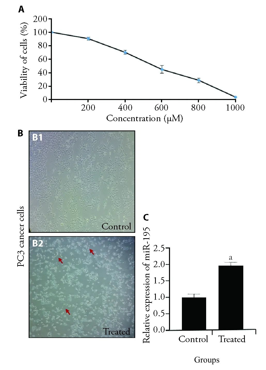
Figure 1 Upregulation of miR-195 in PC3 cancer cells by GTE
3.2.GTE suppressed EMT by upregulation of Ecadherin and down regulation of vimentin in PC3 prostate cancer cells
During EMT,epithelial cells lose cell-cell adhesion and enable to invade.On the other hand,expression of epithelial markers such as E-cadherin reduced and expression of mesenchymal markers such as vimentin,elevated (Figure 2A).28-30To find the possible effect of GTE on PC3 cancer cells migration,the wound healing assay was used (Figure 2B).RNAs from PC3 cells were next extracted and cDNA were synthesized.Finally,we performed qRT-PCR and the relative expressions of Ecadherin and vimentin were detected (Figure 2C,2D).Moreover,PC3 cells under treatment of GTE were lysed and total lysate of cells applied to western blot (Figure 2E).Our results showed that EMT was suppressed in PC3 cancer cells by green tea and thereby,the expression of E-cadherin was reduced and vimentin was up regulated in both gene and protein levels.
3.3.Anti-tumor effect of GTE on PC3 cancer cells by using a 3D cell culture model
A 3D cell culture system is more reflective ofin vivocell behaviors.By using 3D cell culture system,spheroids will form.Spheroids are a population of cells grown in suspension or embedded in a 3D system.Hanging drop technique is one of the general methods to form spheroids.31As already explained in materials and methods,PC3 cells were cultured,trypsinized and counted.Ten drops containing 20×103cells were pipetted into the bottom of the tissue culture dish lid and PBS was placed in its bottom (Figure 3A).After spheroid formation (around 3 d),we analyzed size and number of spheroids,which represents the lower size and number in the treated (cells+medium containing GTE) compared to the control (cells+medium) (Figure 3B,3C).In two days after spheroid formation,the expression of Ecadherin,vimentin and miR-195were examined through qRT-PCR method (Figure 3D-3F),which confirm the results related to 2D cell culture system.

Figure 2 Suppression of EMT by GTE in PC3 prostate cancer cells
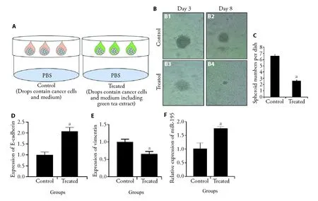
Figure 3 Anti-tumor effect of GTE on PC3 cancer cells by using a 3D cell culture model
4.DISCUSSION
PCa is one of the most causes of cancer-associated death among men worldwide.Although available treatments that have delayed progression of PCa,metastatic prostate cancer remains incurable.32,33It was fully established that EMT is a critical player in cancer progression and metastasis.In EMT,morphological and molecular characteristics of epithelial cell changes into mesenchymal cell such as enhanced migration ability and resistance to apoptosis.Therefore,identification of novel and more efficient biomarkers particularly for suppression of EMT is extremely required.miRNAs have been reported to be potential biomarkers for diagnosis and treatment of cancers due to their management of cellular dysregulations and deregulated expression in a variety of human cancer types.In the current study,we aimed to evaluate thein vitroantitumor activities of green tea in PC3 cancer cells.In this regard,we employed 2D and 3D cell culture models to perform our experiments.We found that GTE suppressed EMT and upregulated miR-195 expression in PC3 cancer cells after treatment with GTE in 2D and 3D cell culture models.
Green tea is produced fromCamellia sinensisand contains high content polyphenols.Anti-tumor activities of green tea and purified green tea catechins were demonstrated previously.11-13,25Several reports also showed the inhibitory effects purified green tea catechins on the cancer cells by the dysregulation of miRNAs.21-24It was known that miR-195 acts as a tumor suppressor and it could serve as a biomarker and therapeutic target in PCa.In this regards,it was revealed that miR-195 can regulate the invasiveness of PCa cells and metastasis of PCa xenografts by regulating breast canceroverexpressed gene 1 (BCOX1).34In addition,inhibition of the EMT process of PCa cells through its target gene FGF2 was shown.35Wuet al17found that in PCa cell lines,miR-195 shows an inhibitory effect on cell migration by targeting Fra-1.In the another study,Caiet al18reported that miR-195 can be implicated into the progression of PCa by regulating ribosomal protein S6 kinase B1 (RPS6KB1) signaling.Notably,it was found that miR-195 could directly inhibit the expression of proline rich 11 (PRR11) and the upregulation of miR-195 or knockdown PRR11 could suppress PCa angiogenesis and proliferationin vivoandin vitro.36These results suggested several potential targets of miR-195 in PC3 cancer cells.Also,it is still unknown the possible targets of miR-195 in PC3 cancer cells during treatment with GTE and thereby more experiments to clarify the related mechanisms were suggested.Notably,the anti-tumor effects of green tea extract and EGCG have been investigated in cancer stem cells and it seems that identifying of potential targets of green tea extract in cancer stem cells will be interested.37
In conclusion,miRNAs play a broad range of roles in cellular processes,such as proliferation,metastasis,invasion,apoptosis.It seems that miRNAs are an excellent promising platform in therapeutic approaches.Green tea with high content of polyphenols is a one of the most consumed beverage in the world and health beneficial activities of green tea were demonstrated.However,the therapeutic influences of green tea on various cancer cellsviamiRNAs are not completely understood.This study has provided new evidences of therapeutic effects of GTE in PC3 prostate cancer cells by the upregulation of miR-195 expression resulting EMT inhibition.More studies will be required to identify greater recognition of the miRNAs roles in carcinogenesis.
 Journal of Traditional Chinese Medicine2022年5期
- Journal of Traditional Chinese Medicine的其它文章
- Effectiveness and safety of tripterygium glycosides tablet (雷公藤多苷片) for lupus nephritis: a systematic review and Meta-analysis
- Qilan preparation (芪蓝颗粒) inhibits proliferation and induces apoptosis by down-regulating microRNA-21 in human Tca8113 tongue squamous cell carcinoma cells
- Tenglong Buzhong granules (藤龙补中颗粒) inhibits the growth of SW620 human colon cancer in vivo
- Yajieshaba prevents lipopolysaccharide-induced intestinal barrier injury via anti-inflammatory and anti-apoptosis
- Antihepatofibrotic effect of Guizhifuling pill (桂枝茯苓丸) on carbon tetrachloride-induced liver fibrosis in mice
- Huangqi decoction (黄芪汤) attenuates renal interstitial fibrosis via transforming growth factor-β1/mitogen-activated protein kinase signaling pathways in 5/6 nephrectomy mice