
Liqi Huoxue dripping pill protects against myocardial ischemia-reperfusion injury via the PI3K/Akt/GSK-3β signaling pathway in rats
2023-02-20 07:16JiaYiZhanYaoZhangXieZhongHanMaoXiangYunChenYaoFengLi
Traditional Medicine Research 2023年4期
Jia-Yi Zhan ,Yao Zhang ,Xie Zhong ,Han Mao ,Xiang-Yun Chen* ,Yao-Feng Li*
1School of Basic Medicine,Guizhou University of Traditional Chinese Medicine,Guiyang 550025,China.
Abstract Background:Liqi Huoxue dripping pill (LQHXDP),a traditional Chinese drug for coronary heart disease,has a protective effect on the heart of rats with myocardial ischemia-reperfusion injury (MIRI) in previous studies;however,its mechanism of action remains unclear.The purpose of this study was to investigate the protective mechanism of LQHXDP on MIRI in rats and its relationship with the PI3K/Akt signaling pathway.Methods:In this study,Sprague-Dawley rats were pre-infused with LQHXDP(175 mg/kg/d)for 10 days.PI3K inhibitor LY294002 (0.3 mg/kg) was intravenously injected 15 minutes before ischemia.The rat model of MIRI was established by ligating the left anterior descending coronary artery.Subsequently,cardiac hemodynamics,serum myocardial injury markers,inflammatory factors,myocardial infarct size,antioxidant indexes,myocardial histopathology,and phosphorylation levels of key proteins of PI3K/Akt signaling pathway were assessed in rats. Results: LQHXDP was found to improve cardiac hemodynamic indexes,reduce serum creatine kinase MB isoenzyme activity and cardiac troponin and heart-type fatty acid binding protein levels,lower serum interleukin-1 beta,interleukin-6 and tumour necrosis factor α levels,reduce the myocardial infarct size and enhance the antioxidant capacity of myocardial tissue in MIRI rats.Pathological analysis revealed that LQHXDP attenuated the extent of myocardial injury and protected mitochondria from damage in MIRI rats.Immunoblot analysis revealed that LQHXDP increased the expression levels of p-Akt and p-GSK-3β in MIRI rat cardiomyocytes.PI3K inhibitor LY294002 could impair these effects of LQHXDP. Conclusion: LQHXDP attenuated myocardial injury,attenuated oxidative stress injury and reduced inflammatory response in MIRI rats,and its protective effects were mediated by activating of PI3K/Akt/GSK-3β signaling pathway.
Keywords: Liqi Huoxue dripping pill;myocardial ischemia-reperfusion injury;myocardial injury;PI3K/Akt/GSK-3β signaling pathway
Highlights
This study was to investigate the protective mechanism of Liqi Huoxue dripping pill on myocardial ischemia-reperfusion injury in rats and its relationship with the PI3K/Akt signaling pathway.This study found that Liqi Huoxue dripping pill attenuated myocardial injury,attenuated oxidative stress injury and reduced inflammatory response in myocardial ischemia-reperfusion injury rats,and its protective effects were mediated by activating of PI3K/Akt/GSK-3β signaling pathway.
Medical history of objective
Liqi Huoxue dripping pill,a traditional Chinese medicine for coronary heart disease and angina pectoris,was developed by Professor De-Wen Qiu in the 1970s by combining the Miao people’s medication habits with modern traditional Chinese medicine theories,and its components areCinnamomum migaoH.W.Li,Allii MacrostemonisBulbus,Chuanxiong Rhizoma,and l-Borneolum(Chinese patent application number CN03135734.2;publication number CN1265828C).
Background
According to the World Health Organization,cardiovascular diseases have become a major threat to human health,among which ischemic heart disease has become the biggest killer.For patients with acute myocardial infarctions,early restoration of blood supply using reperfusion therapy is an effective treatment for severe ischemic myocardial injury [1].However,myocardial reperfusion can also cause a series of adverse reactions,such as severe arrhythmias,myocardial systolic dysfunction,and myocardial no-reflow,which further leads to an increase in the incidence and mortality of postoperative complications,and causes a more severe injury than ischemia,namely myocardial ischemia-reperfusion injury (MIRI) [2,3].Reperfusion injury can reduce the beneficial effects of vascular recanalization,so how to reduce the injury caused by reperfusion is an urgent issue.
Previous studies have shown that the etiopathogenesis of MIRI is complex,and is related to many pathways and pathophysiological processes such as oxidative stress responses,mitochondrial function injury,calcium overload,inflammatory responses,and apoptosis during myocardial reperfusion [4-6].The main therapeutic drugs commonly used in clinical practice are angiotensin II receptor antagonists,nitroglycerol,opioid receptor agonists,and statins[7-11].Numerous studies have shown that certain exogenous bioactive substances can replace endogenous protective factors and provide strong myocardial protection by activating PI3K/Akt signal pathway.The PI3K/Akt signal pathway is a key intracellular signaling pathway that is involved in ischemic pre-adaptation,thereby stimulating in vivo protective mechanisms,and its downstream effector molecules have functions such as promoting cell survival,antioxidant,and inhibiting inflammatory responses.In addition,activation of the PI3K/Akt signal pathway has been shown to be closely related to the protection of myocardial ischemia-reperfusion injury [12].
Traditional Chinese medicine (TCM) compound therapy has the synergistic effect of the combination of multi-component,and multi-target and system regulation.In recent years,the unique advantages of TCM in the prevention and treatment of MIRI have become increasingly prominent [13-15].Liqi Huoxue dripping pill(LQHXDP) is a compound preparation made according to TCM theory and a miao folk prescription from Guizhou Province.It is composed ofCinnamomum migaoH.W.Li,Allii MacrostemonisBulbus,Chuanxiong Rhizoma,and l-Borneolum,which have significant clinical effects on coronary heart disease and angina pectoris [16,17].Our previous studies showed that LQHXDP had a protective effect against the heart of MIRI rats,which was related to antioxidant activities,inhibition of inflammatory responses,and inhibition of myocardial cell apoptosis[18-20].To further explore the underlying mechanism of LQHXDP in MIRI rats,this study investigated the correlation between LQHXDP protection against MIRI and the activity of PI3K/Akt pathway.
Materials and methods
Reagents
LQHXDP(210206)was purchased from Guizhou Yibai Pharmaceutical Co.,Ltd.(Guizhou,China).Creatine kinase MB isoenzyme (CK-MB)assay kit (H197-1),cardiac troponin (cTnI) assay kit (H149-2),the superoxide dismutase (SOD) assay kit (A001-3),the catalase (CAT)assay kit (A007-2),the malondialdehyde (MDA) assay kit (A003-1),and the bicinchoninic acid total protein assay kit (A045-4) were procured from Nanjing Jiancheng Bioengineering Institute (Nanjing,China);heart-type fatty acid binding protein (H-FABP) ELISA kit(ZN2868) was procured from Beijing Biolab Technology Co.,Ltd.(Beijing,China);interleukin-1 beta (IL-1β) ELISA kit (abs553801),interleukin-6 (IL-6) ELISA kit (abs530003),tumour necrosis factor α(TNF-α) ELISA kit (abs530007) were purchased from Absin biotechnology Co.,Ltd.(Shanghai,China).2,3,5-Triphenyl-2H-Tetrazolium chloride (T8170),hematoxylin-Eosin stain kit (G1120),glutaraldehyde,2.5% (electron microscope grade)(P1126) were procured from Beijing Solarbio Science &Technology Co.,Ltd.(Beijing,China).GSK-3β antibody (AF5016) and phosphorylated glycogen synthase kinase 3 beta (p-GSK-3β) (Ser9)antibody(AF2016)were purchased from Affinity Biosciences Co.,Ltd.(Changzhou,China);protein kinase B rabbit monoclonal antibody(AF1789),phosphorylated protein kinase B (p-Akt) (Ser473) rabbit monoclonal antibody (AF1546) and β-actin rabbit monoclonal antibody (AF5003) were procured from Beyotime Biotechnology Co.,Ltd.(Shanghai,China).The PI3K specific inhibitor,LY294002(L9908),was procured from Sigma-Aldrich (St.Louis,MO,USA).
Animal experiments
Male Sprague-Dawley (SD) rats,weighing 180 ± 20 g,were purchased from Tianqin Biotechnology Co.,Ltd.(Changsha,China)(license number: SCXK (Xiang) 2019-0013).All animal experiments were performed in accordance with the Regulations on the Administration of Affairs Concerning Experimental Animals (China)and were approved by the Medical Ethics Committee at Guizhou University of Traditional Chinese Medicine (No.20210098).160 SD rats were selected and randomly divided into five groups,with 32 rats in each group,including Sham (only the left anterior descending coronary artery was threaded without ligation) group,I/R group,I/R+LQHXDP group,I/R+LQHXDP+LY294002 group,and I/R +LY294002 group.The rats in I/R+LQHXDP group and I/R +LQHXDP+LY294002 group were pre-gavaged with LQHXDP (175 mg/kg/d) for 10 days [19].The Sham group,I/R group and I/R +LY294002 group were given equal amounts of sterile deionized water.The rats in I/R+LQHXDP+LY294002 group and I/R+LY294002 group were intravenously injected with LY294002(0.3 mg/kg)15 min before ischemia[21].
Rats in each group were fasted for 12 hours before surgery.At 45 min after the last administration,they were fixed using 25% urethane anesthesia.Endotracheal intubation was performed and the animal ventilator was connected (frequency: 80-90 times/min;tidal volume:2 mL/100 g;exhalation/inhalation ratio: 3:2).The lead line of the limb was connected to a BL-420s biological and functional experimental system (TME Technology Co.,Ltd.,Chengdu,China),and lead II electrocardiograms were recorded.After skin preparation and disinfecting the left chest,left thoracotomy was performed to expose the heart.The left anterior descending coronary arteries of the I/R group were ligated with No.5 silk thread.Threading but no ligation was performed for the Sham group.The success of the ligation was determined by cyanosis or blue-purple staining of the anterior left ventricular wall,and by the elevated QRS amplitude and ST-segment elevation on the simultaneous electrocardiogram.After 0.5 h of ischemia,the silk thread was loosened and reperfused for 2 h to establish a rat MIRI model [21].
Hemodynamic monitoring
The right common carotid artery of SD rats was separated,a vein indwelling needle was prepared,and 1%heparin was injected into the right common carotid artery.The vein indwelling needle was linked with a pressure sensor,which was connected to the BL-420s biological and functional experimental system.The catheter was slowly pushed through the aortic valve into the left ventricle.Cardiac hemodynamic indices were measured,including the left ventricular systolic pressure(LVSP),left ventricular end-diastolic pressure(LVEDP),maximum rate of left ventricular pressure rise (+dp/dtmax),and the maximum rate of left ventricular pressure fall (-dp/dtmax).
Biochemical testing
CK-MB,cTnI,H-FABP,IL-1β,IL-6 and TNF-α in serum were analyzed by enzyme-linked immunosorbent assay using commercial kits.The left ventricular myocardial tissue was accurately weighed and added to 9 times the volume of saline in the ratio of weight(g): volume(mL)=1:9,and this mixture was mechanically homogenized in an ice-water bath and the supernatant was extracted by centrifugation at 2,500 rpm for 10 min.The supernatant was used for total protein,SOD,CAT and MDA determination,and these assays were performed with commercially available kits.
Determination of myocardial infarct size
At the end of the reperfusion period,rat hearts were excised and kept at-20 °C for 30 minutes,then hearts were cut into transverse sections and thawed at room temperature.The sections were incubated in buffer (phosphate buffer,pH 8.5) 1% 2,3,5-Triphenyl-2H-Tetrazolium chloride for 20 minutes.The non-infarcted area was stained brick red,while the infarcted area showed a grayish-white color.The sections were blotted dry with filter paper.The infarcted and non-infarcted areas were carefully separated and the weight of the total myocardium and infarcted area was weighed.Finally,infarct size was calculated as the ratio of infarct weight to total myocardium weight [22].
Histological analysis by hematoxylin-Eosin staining
Left ventricular myocardial tissue was fixed with 4% formaldehyde,dehydrated,embedded in paraffin,and serially sectioned with a microtome.H&E staining was used to observe the histopathological changes of myocardial tissue.
Ultrastructural analysis by transmission electron microscope
After reperfusion,the left ventricular myocardial tissue was rapidly excised and fixed with 2.5% glutaraldehyde overnight at 4 °C.The tissue was then fixed in 1% osmium tetroxide fixative for 2 h,graded acetone dehydration,epoxy resin Epon812 infiltration,embedding,and optical positioning of semithin sections to prepare ultrathin sections.The sections were double stained with uranyl acetate-lead citrate and then observed in a transmission electron microscope(Hitachi H-600 type,Tokyo,Japan).
Western blot analysis
The left ventricular myocardium was ground in liquid nitrogen and prepared in sodium dodecyl sulfate lysate solution containing protease phosphatase inhibitor for 1 h.The tissue lysate was crushed on ice using ultrasonic pulses,and was placed without treatment for 30 min.The supernatant was collected (centrifugation at 12,000 rpm for 15 min at 4 °C).After measuring protein concentration with bicinchoninic acid method,an appropriate amount of sodium dodecyl sulfate polyacrylamide gel electrophoresis loading buffer was added.The sample was boiled,and stored at -20 °C.The sample proteins were resolved using sodium dodecyl sulfate polyacrylamide gel electrophoresis and transferred to a polyvinylidene fluoride membranes.Polyvinylidene fluoride membranes were placed in blocking solution for 1.5 h,washed with phosphate buffered saline and tween-20 solution,then incubated overnight at 4 °C with the following primary antibodies: p-Akt (Ser473) (1:1000),Akt (1:1000),p-GSK-3β (Ser9) (1:1000),GSK-3β (1:1000),β-actin (1:1000).The membranes were then washed and incubated with appropriate horseradish peroxidase-conjugated goat anti-rabbit secondary antibody (1:2,000) for 1 h at room temperature.Antigen-antibody immunoreactivity was visualized using the BeyoECL Plus reagent.
Statistical analysis
The experimental data were analyzed by Prism 8 software(GraphPad,San Diego,CA,USA).A one-way analysis was used to compare multiple groups.The measurement data are expressed as the mean ±standard deviation.Pvalues less than 0.05 indicated a statistically significant difference.
Results
Effects of LQHXDP on cardiac hemodynamics in MIRI rats
As shown in Figure 1,compared to the Sham group,the LVSP,the maximum rate of left ventricular pressure rise (+dp/dtmax),the maximum rate of left ventricular pressure fall (-dp/dtmax) values of the I/R group were significantly decreased,and the LVEDP was significantly increased.Compared with the I/R group,the LVSP,+dp/dtmax,and-dp/dtmax values of the I/R+LQHXDP group were significantly increased and LVEDP was significantly decreased.The LVSP of rats in the I/R+LY group was lower than that of the I/R group,and the remaining indices were not significantly different.Compared to the I/R+LQHXDP group,the levels of LVSP,+dp/dtmax,and -dp/dtmax in the I/R+LQHXDP+LY group were significantly decreased,and LVEDP was increased.These above results indicated that LY294002 attenuated the improvement effect of LQHXDP on the left ventricular function in MIRI rats.

Figure 1 Effects of LQHXDP on cardiac hemodynamics in rats with myocardial ischemia-reperfusion injury. A.The levels of LVSP of rats in each group (n=8);B.the levels of LVEDP of rats in each group (n=8);C.the levels of +dp/dtmax of rats in each group(n=8);D.the level of-dp/dtmax of rats in each group(n=8).**P <0.01 versus the sham group;#P <0.05,##P <0.01 versus the I/R group;ΔP <0.05,ΔΔP <0.01 versus the I/R+LQHXDP group.I/R,myocardial ischemia-reperfusion injury;LVSP,left ventricular systolic pressure;LQHXDP,Liqi Huoxue dripping pill;LY,LY 294002;LVSP,the left ventricular systolic pressure;LVEDP,the left ventricular end-diastolic pressure;+dp/dtmax,the maximum rate of left ventricular pressure rise;-dp/dtmax,the maximum rate of left ventricular pressure fall.
Effects of LQHXDP on serum CK-MB activity and cTnI and H-FABP levels in MIRI rats
To examine the effect of LQHXDP on ischemia-reperfusion myocardial injury,serum CK-MB activity,cTnI and H-FABP levels were measured in rats at the end of reperfusion.As shown in Figure 2,compared with the Sham group,the serum CK-MB activity,cTnI and H-FABP levels of the rats in the I/R group were significantly increased.Compared with the I/R group,the serum CK-MB activity,cTnI and H-FABP levels of rats in the I/R+LQHXDP group were significantly decreased,the serum CK-MB activity and cTnI and H-FABP levels of rats in the I/R+LQHXDP+LY group were decreased,but the difference was not significant;and the serum CK-MB activity,cTnI and H-FABP levels of rats in the I/R+LY group were not significantly different.Compared with the I/R+LQHXDP group,the serum CK-MB activity and cTnI and H-FABP levels were significantly increased in the I/R+LQHXDP+LY and I/R+LY groups.
Effects of LQHXDP on serum IL-1β,IL-6 and TNF-α in MIRI rats
As shown in Figure 3,the serum levels of IL-1β,IL-6 and TNF-α levels were significantly higher in the I/R group compared with the Sham group.Compared with the I/R group,the serum IL-1β,IL-6 and TNF-α levels were significantly lower in the I/R+LQHXDP group,while the serum levels of IL-1β,IL-6 and TNF-α in the I/R+LQHXDP+LY and I/R+LY groups were not significantly different.Compared with the I/R+LQHXDP group,the serum levels of IL-1β,IL-6 and TNF-α in the I/R+LQHXDP+LY and I/R+LY groups were significantly increased.
Effects of LQHXDP on myocardial infarction,SOD activity,CAT activity and MDA content in MIRI rats
There was no myocardial infarction size of rats in the Sham group,while the percentage of myocardial infarctions of rats in the model group was (36.64 ± 6.12) %.Pretreatment with LQHXDP significantly reduced the myocardial infarction in MIRI rats.Whereas there was no significant difference between the I/R+LQHXDP+LY group and the I/R group.Compared with I/R+LQHXDP group,the myocardial infarct size of rats in the I/R+LQHXDP+LY group was significantly increased(Figure 4,Figure 5A).The results indicated that the PI3K inhibitor LY294002 decreased the ameliorative effect of LQHXDP on myocardial infarction in MIRI rats.
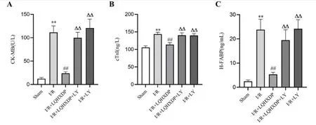
Figure 2 Effects of LQHXDP on serum CK-MB,cTnI and H-FABP in rats with myocardial ischemia-reperfusion injury.A.Serum CK-MB level of rats in each group (n=8);B.Serum cTnI level of rats in each group (n=8);C.Serum H-FABP level of rats in each group (n=8).**P<0.01 versus the sham group;##P <0.01 versus the I/R group;ΔΔP <0.01 versus the I/R+LQHXDP group.LQHXDP,Liqi Huoxue dripping pill;CK-MB,creatine kinase mb isoenzyme;cTnI,cardiac troponin I;H-FABP,heart-type Fatty acid binding protein;I/R,myocardial ischemia-reperfusion injury;LY,LY294002.

Figure 3 Effects of LQHXDP on serum IL-1β,IL-6 and TNF-α in rats with myocardial ischemia-reperfusion injury. A.Serum IL-1β level of rats in each group (n=8);B.Serum IL-6 level of rats in each group(n=8);C.Serum TNF-α level of rats in each group(n=8).**P<0.01 versus the sham group;##P <0.01 versus the I/R group;ΔΔP <0.01 versus the I/R+LQHXDP group.LQHXDP,Liqi Huoxue dripping pill;IL-1β,interleukin-1 beta;IL-6,interleukin-6;TNF-α,tumour necrosis factor α;I/R,myocardial ischemia-reperfusion injury;LY,LY294002.
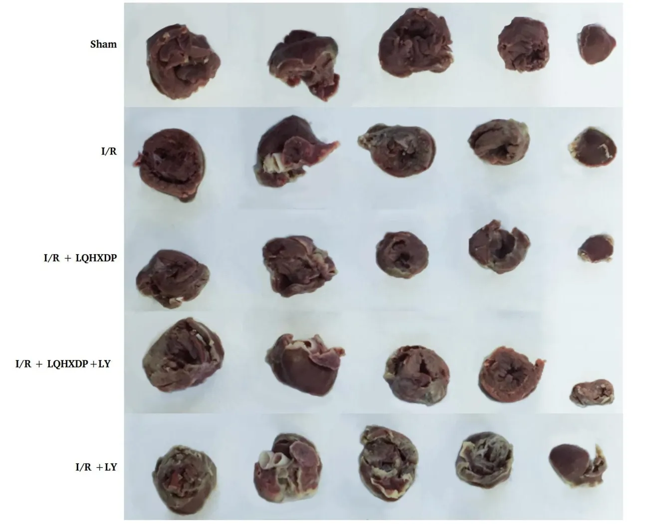
Figure 4 Effects of LQHXDP on myocardial infarct size in rats with myocardial ischemia-reperfusion injury. Myocardial infarct size was measured by TTC staining.Red-staining areas indicate normal tissue and unstained pale areas indicate infarct tissue.LQHXDP,Liqi Huoxue dripping pill;TTC,2,3,5-Triphenyl-2H-Tetrazolium Chloride.
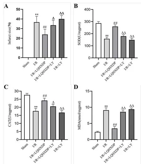
Figure 5 Effects of LQHXDP on myocardial infarct size and myocardial tissue SOD,CAT activity and MDA content in rats with myocardial ischemia-reperfusion injury. A.The myocardial infarct size of rats in each group (n=8);B.SOD activity of myocardial tissue of rats in each group (n=8);C.CAT activity of myocardial tissue of rats in each group (n=8);D.MDA content of myocardial tissue of rats in each group (n=8).**P <0.01 vs.the sham group;##P <0.01 vs.the I/R group;ΔP <0.05,ΔΔP <0.01 vs.the I/R+LQHXDP group.LQHXDP,Liqi Huoxue dripping pill;I/R,myocardial ischemia-reperfusion injury;LY,LY 294002;SOD,superoxide dismutase;CAT,catalase;MDA,malondialdehyde.
To analyze the antioxidant capacity of LQHXDP,we measured SOD,CAT activity and MDA content in myocardial tissue of rats in various group.As shown in Figure 5B-5D,compared to the Sham group,the activities of SOD and CAT were significantly decreased,and the content of MDA was significantly increased in the I/R group.Compared to the I/R group,the activities of SOD and CAT were significantly increased and MDA content was significantly decreased in the I/R+LQHXDP group.Compared to rats in the I/R group,Administration of LY294002 alone had no significant effect on the antioxidant capacity of myocardial tissue.Compared to the I/R +LQHXDP group,the addition of LY294002 decreased the effects of LQHXDP on SOD activity,CAT activity,and MDA content in the myocardial tissues of I/R rats.
Histological effects of LQHXDP on the myocardium of MIRI rats
The rats in Sham group had normal myocardial structure,while the rats in I/R group had disorganized myocardial cell morphology and structure,myocardial fibers appeared broken,myocardial cells had obvious membrane damage,edema and myonecrosis,and more inflammatory cell infiltration was seen in the interstitium.Compared with the I/R group,the myocardial fibers in the I/R+LQHXDP group were more neatly arranged,the myocardial cell membrane damage was greatly relieved,the myocardial cell structure was more normal,and the inflammatory cell infiltration was almost invisible.Compared with the I/R group,the myocardial histomorphology of rats in the I/R+LQHXDP+LY group and the I/R+LY group had no significant improvement (Figure 6).
Ultrastructural effects of LQHXDP on the myocardium of MIRI rats
In Sham group,myocardial fibers were neatly arranged,nuclei and mitochondria were normal,mitochondrial cristae were clear,and no abnormalities were observed between myocardium.In I/R group,myocardial cell structure was destroyed,some myocardial fibers were lysed and broken,interstitium was enlarged,some mitochondria were fixed and shrunken,some mitochondria were swollen or outer membrane was broken,mitochondrial cristae were irregular,broken into flocculent or missing.In the I/R+LQHXDP+LY group,some myocardial fibers were lysed and broken,myofiber gaps were widened,mitochondria were unevenly distributed,and some mitochondria were swollen or ruptured.There was no significant difference in ultrastructure between the I/R+LY group and I/R group (Figure 7).
Effects of LQHXDP on the protein expression of p-Akt and p-GSK-3β
Akt is an important protein in the PI3K/Akt signaling pathway.Akt phosphorylation is a key indicator of PI3K/Akt pathway activation,and GSK-3β is an important downstream factor regulated by Akt.The phosphorylation levels of Akt and GSK-3β in myocardial tissue of rats in various group were therefore measured.As shown in Figure 8,compared to the Sham group,the expressions of p-Akt/Akt and p-GSK-3β/GSK-3β in myocardial tissues of rats in the I/R group were significantly upregulated.Compared to the I/R group,the expressions of p-Akt/Akt and p-GSK-3 β/GSK-3β in myocardial tissues of rats in the LQHXDP group were also significantly upregulated.However,the expression of p-Akt and p-GSK-3β were down-regulated following co-treatment with LY294002 and LQHXDP.

Figure 6 Hematoxylin-Eosin staining of cardiac muscular tissue in the left ventricle of rats in each group (×200 magnification). I/R,myocardial ischemia-reperfusion injury;LQHXDP,Liqi Huoxue dripping pill;LY,LY294002.

Figure 7 Transmission electron microscopy images of cardiomyocytes in the left ventricle of rats in each group (×6,000 magnification).I/R,myocardial ischemia-reperfusion injury;LQHXDP,Liqi Huoxue dripping pill;LY,LY294002.

Figure 8 Western blotting analysis of p-Akt,Akt,p-GSK-3β and GSK-3β protein expression in the left ventricle of rats in each group.A.The expressions of p-Akt,Akt,p-GSK-3β and GSK-3β were evaluated by western blotting;B.the expression level of p-Akt/Akt;C.the expression level of p-GSK-3β/GSK-3β.β-actin is shown as internal reference protein.**P <0.01 vs.the Sham group;##P <0.01 vs.the I/R group;ΔΔP <0.01 vs.I/R+LQHXDP group.LQHXDP,Liqi Huoxue dripping pill;I/R,myocardial ischemia-reperfusion injury;LY,LY 294002;p-Akt,phosphorylated protein kinase B;Akt,protein kinase B;p-GSK-3β,phosphorylated glycogen synthase kinase 3 beta;GSK-3β,glycogen synthase kinase 3 beta.
Discussion
Myocardial ischemia-reperfusion injury is one of the major problems in the field of cardiovascular interventions.This study explored the protective mechanism of LQHXDP against myocardial ischemia-reperfusion injury in terms of hemodynamics,myocardial injury marker analysis,antioxidant index and histopathology.The results showed that LQHXDP attenuated MIRI by activating PI3K/Akt pathway,which is expected to provide a new theoretical basis for the myocardial protective effect of LQHXDP.
After MIRI occurs,cell structure is damaged,mitochondrial permeability is altered,oxygen free radicals are increased,and endothelial cell membranes are damaged,resulting in a significant increase in myocardial enzymatic markers released from cardiomyocytes into the blood.Cardiac enzyme CK-MB is one of the specific serum markers of myocardial injury.cTnI detection is the main indicator of myocardial ischemic necrosis or cell membrane permeability.Fatty acid binding protein is a carrier of free fatty acids specifically present in the cytoplasm of cardiomyocytes.H-FABP peaks within 1 h of reperfusion and is one of the indicators of perioperative myocardial injury [23].Therefore,CK-MB,cTnI and H-FABP can be used as sensitive indicators for detecting myocardial injury [24].In this study,LQHXDP was found to reduce serum CK-MB activity and cTnI and H-FABP levels in MIRI rats,indicating that LQHXDP could protect against myocardial ischemia-reperfusion injury in rats.In contrast,treatment with PI3K inhibitor LY294002 attenuated the protective effect of LQHXDP on myocardial ischemia-reperfusion injury in rats,indicating that the protective effect of LQHXDP on cardiomyocytes was related to the PI3K/Akt signaling pathway.
Due to myocardial damage after reperfusion,the systolic and diastolic functions of the myocardium are diminished.LVEDP,LVSP,+dp/dtmax and -dp/dtmax,the cardiac hemodynamic indices measured in this experimental study,reflect the myocardial systolic and diastolic capacities of the left ventricle.The results of this experiment showed that LQHXDP increased the LVEDP,+dp/dtmax and -dp/dtmax values and decreased the LVSP value,suggesting that LQHXDP can improve the cardiac systolic and diastolic capacities in MIRI rats.Observation of myocardial pathological morphology and calculation of myocardial infarct extent are the most visual methods of myocardial injury.In this study,we found that myocardial infarct extent was increased in MIRI rats,and myocardial fibers were disorganized,broken or lysed.Mitochondria were swollen or ruptured,and mitochondrial cristae were swollen or vacuolated.LQHXDP pretreatment significantly reduced the extent of myocardial infarction in MIRI rats,improved the ultrastructure of myocardial cells,reduced myocardial fiber rupture,reduced interstitial widening,and protected the normal morphological structure of mitochondria.Furthermore,LQHXDP reduced the serum levels of IL-1β,IL-6,TNF-α and attenuated inflammatory damage.However,PI3K inhibitor LY294002 treatment diminished the protective effect of LQHXDP on cardiomyocytes in MIRI rats.
Reactive oxygen species (ROS) are important causes of myocardial injury during MIRI [25].Excessive ROS attack lipids in cell membranes and cause peroxidation damage,resulting in the production of the lipid oxidation of the MDA end product,which indirectly reflects the degree of oxidative damage [26].SOD and CAT are protective enzymes acting against oxidative damage in cardiomyocytes.SOD can convert ROS to H2O2which is then reduced to H2O by CAT,thereby removing excess ROS in cells [27-29].LQHXDP enhanced the SOD and CAT activities in myocardial tissue,reduced the content of MDA,and alleviated the degree of myocardial infarction.It is therefore suggested that LQHXDP alleviated oxidative stress injury induced by MIRI and protected against myocardial injury.Notably,the expressions of p-Akt and p-GSK-3β were enhanced in rats of the I/R group,and the phosphorylation levels of Akt and GSK-3β were further increased after LQHXDP pretreatment.However,when pre-treated with the PI3K inhibitor LY294002,the expressions of p-Akt and p-GSK-3β in myocardial tissue were significantly reduced,which also weakened the protective effect of LQHXDP on MIRI,attenuated the improvement of cardiac function,decreased the ability of anti-oxidants,and reduced the improvement of myocardial infarction.The above results suggested that the PI3K/Akt/GSK-3β pathway was involved in the protective effect of LQHXDP against MIRI.The PI3K/Akt/GSK-3β pathway is an important membrane receptor signaling pathway,which can regulate apoptosis,autophagy,oxidative stress,inflammation,energy metabolism and other functions by regulating different downstream targets [30,31].Akt is a serine/threonine protein kinase that is a key downstream target of PI3K.Activated Akt can phosphorylate GSK-3β,which is a key regulator of cell proliferation,differentiation,and apoptosis [32,33].Phosphorylation of GSK-3β inhibits mitochondrial permeability transition pore opening and triggers a series of cascade that attenuates the process of apoptosis induced by mitochondria [34,35].At the same time,this pathway belongs to the reperfusion injury salvage kinase pathway,which plays an important role in the endogenous protection strategy of MIRI[12,36].It can promote myocardial tissue to use compensatory mechanisms to limit adverse reactions such as oxidative stress,inflammation,and apoptosis during I/R [37,38].Studies have shown that PI3K/Akt/GSK-3β pathway is a key signaling pathway that inhibits abnormal cardiomyocyte apoptosis and alleviates myocardial injury after I/R [33,39].The present study revealed that LQHXDP further enhanced the phosphorylation of Akt and GSK-3β in cardiomyocytes,implying that LQHXDP activates endogenous protective mechanism in cardiomyocytes after reperfusion.
Conclusion
In conclusion,LQHXDP has a protective effect on cardiomyocytes in MIRI rats.LQHXDP ameliorates myocardial tissue injury,reduces inflammatory cell infiltration and enhances the scavenging of oxidative free radicals in MIRI rats by activating the PI3K/Akt/GSK-3β pathway.This study provides a theoretical reference for further revealing the cardioprotective effect of LQHXDP.
 Traditional Medicine Research2023年4期
- Traditional Medicine Research的其它文章
- A review on cocrystal of active ingredients in traditional Chinese medicine
- Phoenix dactylifera:traditional uses,chemical constituents,nutritional benefit and therapeutic effects
- Advantages and prospects of traditional Chinese medicine in treating COVID-19
- The role of selected medicinal plants from Iranian traditional medicine for the treatment of fatigue in metabolic syndrome
- The effect of head and facial massage on sleep condition after coronary artery bypass graft surgery