
Effects of acupuncture and moxibustion on PINK1/Parkin signaling pathway in substantia nigra of Thy1-αSyn transgenic mice with Parkinson disease
2023-12-25 09:28XIAOYouzhi萧有智LIKunshan李昆珊CHENZiyi陈子怡SHENLin沈琳CHENYiyi陈奕奕LUJiajing陆佳婧XIEJing谢静LIJunxiong李俊雄WANGWenjia王文佳LILingjie李灵杰QIAOYu乔宇LIJing李璟
XIAO Youzhi (萧有智), LI Kunshan (李昆珊), CHEN Ziyi (陈子怡), SHEN Lin (沈琳), CHEN Yiyi (陈奕奕),LU Jiajing (陆佳婧), XIE Jing (谢静), LI Junxiong (李俊雄), WANG Wenjia (王文佳), LI Lingjie (李灵杰), QIAO Yu (乔宇),LI Jing (李璟)
1 Yueyang Hospital of Integrated Traditional Chinese and Western Medicine, Shanghai University of Traditional Chinese Medicine,Shanghai 200437, China
2 Shanghai Xinqidian Rehabilitation Hospital, Shanghai 201107, China
3 Shanghai Research Institute of Acupuncture and Meridian, Shanghai 200030, China
Abstract
Keywords: Acupuncture Therapy; Moxibustion Therapy; Acupuncture-moxibustion Therapy; Parkinson Disease; Alpha-Synuclein; Mitochondrial Autophagy; Mice
Parkinson disease (PD) is a progressive neurodegeneration with main pathological manifestations of progressive dopaminergic neuron loss in brain substantia nigra, decreased dopamine (DA) level in the striatum[1], and abnormal aggregation of alphasynuclein (αSyn)[2].Under the influence of heredity,aging, and environment, the pathogenesis of PD is related to mitochondrial dysfunction, oxidative stress,and abnormal ubiquitination protein system[3-4].Phosphatase and PTEN-induced putative kinase 1(PINK1)/Parkin signaling pathway mainly maintains the mitochondrial mass[5].Research showed, specific PINK1 gene-knockout mice showed PD-like symptoms lasting for one year, and the mechanism was related to the role of PINK1 in mitochondrial antigen presentation[6].PINK1 localizes in the outer membrane and induces the autophagy of mitochondria when mitochondria are damaged; PINK1 receives the mitochondrial damage signal and phosphorylates and activates the downstream Parkin protein to generate ubiquitin chain recognized by lysosomes.Intracellular autophagy vesicles wrap the damaged mitochondria and combine with lysosomes to degrade damaged mitochondria[7-8].Damaged mitochondria accumulate in cells and may cause nerve cell dysfunction due to mitochondrial autophagy dysfunction, suggesting that the pathogenesis of PD is closely related to mitochondrial autophagy mediated by PINK1/Parkin signaling pathway.
The clinical treatment of PD mainly consists of levodopa drugs and DA receptor agonists, which have little improvement in non-motor symptoms.After several years of treatment, the curative effect decreases,showing movement fluctuation and dyskinesia problems[9].Acupuncture, one of the traditional therapies of traditional Chinese medicine, effectively controls PD progression and reduces the dosage and adverse reactions of Western medications[10].Acupuncture and moxibustion are recommended in the consensus opinion of supplementary and alternative therapies for PD in 2018[11].Our previous research confirmed that acupuncture regulated autophagy and promoted the clearance of brain αSyn in PD mice[12],improved the diversity of intestinal microorganisms,regulated the abundance of intestinal flora, and improved the motor function of PD mice[13].In this study, Thy1-αSyn transgenic mice were used as experimental objects to observe the effects of acupuncture and moxibustion on the expression levels of PINK1/Parkin pathway-related molecules in the substantia nigra of Thy1-αSyn mice, and to explore the mechanism of acupuncture and moxibustion in treating PD.
1 Materials and Methods
1.1 Laboratory animals and grouping
Twenty-four specific-pathogen-free Thy1-αSyn transgenic mice[14]with a body mass of (22±2) g were used.The mice were bred by C57BL/6J-Tg (Th-SNCA)5Eric/J transgenic mice provided by Jackson Laboratory and C57BL/6J provided by Jiangsu Jicui Yaokang Biotechnology Co., Ltd., China.The gene carrying αSyn mutation was identified by polymerase chain reaction(PCR) gel electrophoresis.Mice were raised in the barrier environment of the Experimental Animal Center of Yueyang Hospital of Integrated Traditional Chinese and Western Medicine, Shanghai University of Traditional Chinese Medicine, with day-night rhythm alternation of 12 h/12 h, room temperature of 18-22 ℃,indoor humidity of 50%-70%, and free drinking water.The license number of the experimental animal was SYXK (Hu) 2018-0040.Twenty-four Thy1-αSyn transgenic (+) mice were randomly divided into a model group, an acupuncture group, an acupuncture +moxibustion group, and a Western medicine group,with 6 mice in each group.Six wild-type male mice identified as transgenic (-) by PCR in the same litter were used as the wild-type group.The treatment of experimental animals was in accordance with the Guiding Opinions on the Treatment of Experimental Animals issued by the Ministry of Science and Technology of the People’s Republic of China.This study was reviewed by the Animal Ethics Committee of Yueyang Hospital of Integrated Traditional Chinese and Western Medicine, Shanghai University of Traditional Chinese Medicine (Ethical Approval No.YYLAC-2019-060-1).
1.2 Drugs and reagents
Rapamycin (Cat.No.LC R-500, LC Laboratory, USA);chloroform (Cat.No.2006-06-08, Shanghai Reagent No.1 Factory, China); isopropanol (Cat.No.AR80109218,Sinopharm Chemical Reagent Co., Ltd., China); absolute ethanol (Cat.No.AR10009218, Sinopharm Chemical Reagent Co., Ltd., China); mRNA extraction Trizol (Cat.No.15596018, Invitrogen Corporation, USA).
Fatigue rod rotating instrument (Chengdu Taimeng Software Co., Ltd., China); 5810R desktop refrigeration high-speed centrifuge (Eppendorf Company, Germany);F6/10-6G ultrafine electric homogenizer (Fluko,Germany); Western blotting (WB) Bio-Rad electrophoresis apparatus (Bio-Rad, USA); WB electrophoresis film transfer instrument (Dalian Jingmai Technology Co., Ltd., China); gel imaging system(Bio-Rad, USA); timing horizontal shaker (Crystal, USA).
1.3 Intervention method
Acupuncture group: The mice were fixed on a special mouse rack.Hwato brand sterile disposable filiform acupuncture needles of 0.25 mm in diameter and 13 mm in length (Suzhou Medical Appliance Factory,China) were used.The needle was obliquely inserted 3 mm at Baihui (GV20) and 3 mm perpendicularly at Yanglingquan (GB34), twisting once every 5 min for 15 s.The needles were retained for 15 min, once a day,for 7 d.The points were positioned referring to Experimental Acupuncture Science[15].
Acupuncture + moxibustion group: The abdominal fur of mice was shaved.The fixation method, point selection, and stimulation method were the same as those in the acupuncture group.At the same time, a special thin moxa stick of 0.5 cm in diameter and 12 cm in length was ignited, and moxibustion was performed for 15 min/time 2 cm away from Guanyuan (CV4), once a day for 7 d.The local skin temperature of points was kept at (43±1) ℃.
Western medicine group: Thy1-αSyn transgenic PD mice were intraperitoneally injected with rapamycin at a dose of 10 mg/(kg·bw), once a day for 7 d.
The wild-type group and the model group received the same grasping and fixation as the acupuncture group and the acupuncture + moxibustion group without other interventions.
1.4 Behavioral detection
After intervention, the mice were placed on a rotary rod with a radius of about 1 cm, and the orientation of the mouse head was perpendicular to the long axis of the rotary rod of the instrument.The holding time of mice in each group was recorded at rotation speeds of 10, 15, 20, 25, and 30 r/min and was read 300 s when exceeding 300 s.The overall rod performance (ORP)score was calculated using the trapezoidal method and measured twice at intervals of 5 min.The average value was used as the final score.
1.5 Sample collection
Samples were collected after the behavioral experiment.The mice were anesthetized by intraperitoneal injection of 2% pentobarbital sodium at a dose of 50 mg/(kg·bw), which was confirmed by clamping the extremities of the mice with hemostatic forceps.The mice were sacrificed by cervical dislocation.The Petri dish was kept on the ice.Opened the skull,quickly peeled off the whole brain, and placed it in a brain mold.The left and right brains were separated along the middle suture.The substantia nigra of the midbrain was located by referring to the online map of Allen Brain Atlas (http://mouse.brain-map.org/static/atlas),and the substantia nigra of the brain was exposed.The left and right substantia nigra tissues were then separated.One piece was fixed in EP tubes with 4%paraformaldehyde, and the other piece was properly trimmed with a blade to clear.Put it into a cryopreservation tube containing liquid nitrogen, which was then transferred into a refrigerator at -80 ℃ for storage and later testing.
1.6 Immunohistochemical method
Paraffin sections were dewaxed, hydrated, antigen repaired, blocked, and incubated with the primary antibody, and then put into a refrigerator at 4 ℃overnight.On the second day, the second antibody was added for incubation.Washed with PBS and colored with DAB solution.Dehydrated and sealed after re-dyeing.Observed under the optical microscope with fixed brightness by randomly selecting 3 visual fields for photographing each slice.Analyzed and calculated the target positive areas (Area) and integrated optical density (IOD) of each photo using the Image-Pro Plus software.Calculated the average optical density (AOD)of the positive targets by the equation: AOD = IOD ÷Area.The calculated average value of the three visual fields on each slice was used as the positive target AOD of the slice and was used to observe the changes in tyrosine hydroxylase (TH) positive neurons in the brain.
1.7 Immunofluorescence chemical method
Paraffin sections were dewaxed, hydrated, antigen repaired, sealed, and then incubated with the primary antibody at 4 ℃ overnight.PBS rinsing was performed 3 times for 3 min/time.Incubated with the second antibody for 1 h in the dark and rinsed with PBST 3 times for 5 min/time.Re-dyed with DAPI after drying and washed with PBST 3 times for 5 min/time.Mounting was performed after drying with antiquenching mounting reagent in the dark.Computer images were obtained in a darkroom with the Olympus cellSens Standard image acquisition system.Each tissue slice was photographed under a fixed excitation intensity to detect the αSyn expression level in the substantia nigra of mesencephalon.
1.8 WB assay
A proper amount of RIPA lysate was added into the substantia nigra homogenate of the mouse midbrain.After full lysis, the supernatant was centrifuged at a high speed.The BCA protein quantitative method was used to determine the protein concentration.Denatured and loaded each sample according to the concentration.Blocked in blocking solution with 5%skim milk at room temperature for 1 h after electrophoresis and film transfer.Incubated in the primary antibody on the shaking table at 4 ℃ overnight.After washing of the membrane, the second antibody was added and incubated at room temperature for 1 h.Calculated the gray value in a Bio-Rad system by adding the developer after washing the membrane.The expression levels of PINK1, Parkin, and microtubuleassociated protein 1 light chain 3 (LC3)-Ⅱ/LC3-Ⅰ,autophagy protein sequencosome-1/protein 62(SQSTM-1/p62), and ubquitin-specific protease 30(USP30) in the mouse substantia nigra were detected.
1.9 Real-time quantitative PCR (RT-qPCR)
Mouse substantia nigra tissues were ground, lysed with Trizol reagent, and centrifuged to extract total RNA.Prepared reaction solution according to the instructions of reverse transcription kit for cDNA synthetization and stored at -20 ℃ for later use.A total of 20 μL of the reaction system was configured, and the reaction conditions were: 95 ℃ for 30 s with 1 cycle; 95 ℃ for 5 s, 60 ℃ for 30 s with 40 cycles in total.The mRNA expression levels of PINK1, Parkin, LC3B, p62, and USP30 in the substantia nigra were calculated by the 2-ΔΔCtmethod.The probes used are shown in Table 1.

Table 1 Primers for mRNA detection
1.10 Statistical processing
The SPSS version 24.0 software was used for statistical analysis.Normal distribution data were expressed as mean ± standard deviation (), while non-normal distribution data were expressed as median(lower quartile, upper quartile) [M (QL, QU)].The measurement data were tested for normality and homogeneity of variance first.One-way analysis of variance was used for data comparison between groups with the normal distribution and the homogeneity of variance.The least significant difference method was further used for pairwise comparisons if the difference between groups was statistically significant;nonparametric test (Kruskal-Wallis H rank-sum test) was used for comparisons between groups if the data did not conform to normal distribution or heterogeneity of variance; Dunnett’s T3 test was used to compare the two groups if the difference was statistically significant.All tests were conducted by two-tailed tests, and P<0.05 indicated that the difference was statistically significant.
2 Results
2.1 ORP score
Compared with the wild-type mice in the same litter,the ORP score of transgenic mice in the model group was significantly lower (P<0.01).Compared with the model group, the ORP score of the acupuncture group and the acupuncture + moxibustion group increased significantly (P<0.05), while that of the Western medicine group increased without statistically significant difference (P>0.05).Compared with the Western medicine group, the ORP score of the acupuncture group and the acupuncture + moxibustion group increased slightly without a statistically significant difference (P>0.05).See Table 2.
Table 2 Comparison of the ORP score among groups
Note: ORP=Overall rod performance; compared with the wildtype group, 1) P<0.01; compared with the model group, 2) P<0.05.
Wild-type 6 19 478.33±4 057.94 Model 6 10 644.17±1 653.601)Acupuncture 6 15 590.83±3 825.132)Acupuncture + moxibustion 6 16 307.50±4 231.012)Western medicine 6 15 244.17±5 069.54
2.2 Detection of PD pathological markers in mouse substantia nigra of each group
2.2.1 TH-positive neurons
The TH-positive neurons of the substantia nigra in wild-type mice are abundant, and the shape is complete.The TH-positive neurons were relatively fewer with morphological degeneration and deletion of the substantia nigra in the model group, suggesting that the PD mouse model was successfully established.The number of TH-positive neurons in mouse substantia nigra of the acupuncture group, the acupuncture +moxibustion group, and the Western medicine group increased compared with that of the model group,suggesting that acupuncture, acupuncture +moxibustion, and Western medicine treatment increase the TH-positive neurons in PD mice.See Figure 1 (the red arrows indicate the target positive neurons).
2.2.2 αSyn protein expression
The αSyn protein expression in the substantia nigra of wild-type mice was not significant with small shapes but was significant and aggregated in the model group.After intervention, the αSyn protein expression of the mouse substantia nigra in the acupuncture group, the acupuncture + moxibustion group, and the Western medicine group decreased compared with that in the model group.Compared with the wild-type group, the αSyn protein expression level in the model group was higher than that in the wild-type group with statistical significance (P<0.05); compared with the model group,the αSyn protein expression level in the acupuncture +moxibustion group decreased (P<0.05).See Figure 2 and Figure 3 (the red arrows indicate the target proteins).

Figure 2 Expression of αSyn protein in mouse substantia nigra of each group (n=6, immunofluorescence, ×200)

Figure 3 Comparison of αSyn protein expression in mouse substantia nigra among groups (n=6)
Group n ORP score/point
2.3 Expression levels of PINK1/Parkin pathway molecules in mouse substantia nigra of each group
Compared with the wild-type group, the mRNA and protein expression levels of PINK1 in the model group decreased significantly (P<0.05); the Parkin protein expression level in the model group decreased (P<0.05),and the Parkin mRNA expression level in the model group decreased without a significant difference(P>0.05).Compared with the model group, the Parkin and PINK1 protein expression levels in the acupuncture group, the acupuncture + moxibustion group and the Western medicine group increased significantly (P<0.05);the expression level of Parkin mRNA in the acupuncture group and the acupuncture + moxibustion group increased significantly (P<0.05); the expression level of PINK1 mRNA in the acupuncture group, the acupuncture + moxibustion group, and the Western medicine group increased without statistical significance (P>0.05).See Figure 4.
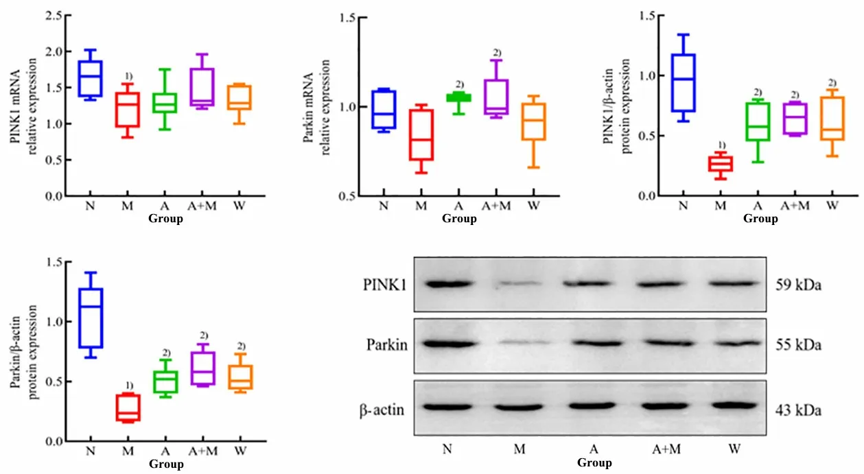
Figure 4 The mRNA and protein expression levels of PINK1/Parkin (n=6)
2.4 Expression levels of mitochondrial autophagyrelated molecules in substantia nigra of PD mice
Compared with the wild-type group, the USP30 mRNA expression level was significantly higher (P<0.05),the USP30 protein expression level was significantly higher (P<0.01), and the p62 protein expression level decreased significantly (P<0.01), while there was no significant difference in the expression level of LC3-Ⅱ/LC3-Ⅰ protein or the LC3B and p62 mRNAs in the substantia nigra of the model group (P>0.05).Compared with the substantia nigra in the model group,the expression level of USP30 mRNA in the acupuncture+ moxibustion group decreased (P<0.05), while the expression level of USP30 mRNA in the acupuncture group and the Western medicine group had no significant difference (P>0.05); the expression level of USP30 protein in the acupuncture group, the acupuncture + moxibustion group, and the Western medicine group decreased significantly (P<0.01); the expression levels of LC3B and p62 mRNAs in the acupuncture group and the acupuncture + moxibustion group had no significant difference (P>0.05), but the expression level of LC3-Ⅱ/LC3-Ⅰprotein in the acupuncture + moxibustion group and the Western medicine group had a significant difference (P<0.05).See Figure 5.

Figure 5 Protein and mRNA expression levels of mitochondrial autophagy-related molecules (n=6)
3 Discussion
PD belongs to the category of “flutter” and “quiver”in traditional Chinese medicine.Contemporary physicians believe that PD originates in the brain.In recent years, research on PD treatment with acupuncture and moxibustion has attracted more and more attention.Randomized controlled clinical trials have shown that acupuncture significantly improves gait disturbance in PD patients[16-17], relieves PD-related fatigue[18-19], and reduces anxiety symptoms[20].Meta-analysis suggests that acupuncture[21-22]and moxibustion[23]are effective in improving motor symptoms in PD compared with Western medicine alone, and have outstanding advantages in reducing Western medicine consumption and adverse drug reactions and treating non-motor symptoms[24-25].Acupuncture combined with moxibustion is often used to treat PD in clinics[24], but the corresponding mechanism is still unclear and needs to be evidenced by further investigations.
The occurrence of PD motor symptoms is often accompanied by abnormal accumulation of pathological αSyn in the substantia nigra[25].Excessive αSyn damages the information transmission of dopaminergic nerves,leading to insufficient DA release in the early stage of dopaminergic neuron axonal mutation in the substantia nigra[26].The interaction of oligomer αSyn with metal ions induces the production of reactive oxygen species(ROS).The increased ROS leads to the accumulation of oxidative DA, resulting in the dysfunction of mitochondria and lysosomes.The subsequent process is blocked when the abnormal αSyn signal is cleared[6,27].Here, we focused on the mitochondrial autophagy of PD and identified that the mitochondrial autophagyrelated signals of transgenic PD model mice had changed compared with those of the same litter wild-type mice, which indicated that the loss of dopaminergic neurons in the substantia nigra was related to mitochondrial autophagy dysfunction caused by excessive αSyn accumulation.
Restoring mitochondrial autophagy is a new way to treat PD.With the age of PD patients, the clearance ability of mitochondrial autophagy cannot bear the increasing oxidative stress and neurotoxin, leading to nerve cell dysfunction[28].The mitochondrial autophagy pathway mediated by PINK1/Parkin pathway is the key point of the above research.PINK1/Parkin signal is characterized by PINK1 located in the outer membrane of the damaged mitochondria, recruiting and activating Parkin through the phosphorylation of its ubiquitin-like domain[29].Activated Parkin labels damaged target mitochondria with ubiquitin, which is recognized by downstream mitochondrial autophagy-related proteins.Mutation of PRKN, the gene encoding Parkin protein,will lead to the loss of selective dopaminergic cells in the substantia nigra, which may be related to the decrease in mitochondrial function caused by mitochondrial clearance defects[30].
LC3, p62, and USP30 are downstream proteins activated by PINK1/Parkin.LC3 protein has two forms:LC3-Ⅰ and LC3-Ⅱ.The up-regulated expression levels of LC3-Ⅱ/LC3-Ⅰ protein and LC3 gene generally indicate an increased autophagy level.The p62 is a selective autophagic motor protein[31], which is a substrate linked to LC3[32].Decreased autophagy will lead to the accumulation of insoluble p62[33].In the mitochondrial autophagy, LC3 and p62 proteins function in the upstream and downstream processes,respectively.Examining only one of them is not enough to explain the dynamic process of autophagy flow.Joint detection of LC3-Ⅱ/LC3-Ⅰ and p62 is necessary to evaluate the autophagy activity level.USP30 is a deubiquitinase, which antagonizes the activity of autophagy ubiquitin chain mediated by Parkin protein by regulating the intracellular mitochondrial morphology[34-35].Overexpression of USP30 in neurons will weaken the induced mitochondrial depolarization to clear mitochondria by causing autophagy[36].The relative expression levels of Parkin and USP30 indirectly reflect the mitochondrial mass status.
Baihui (GV20) and Yanglingquan (GB34) are the most commonly used points in clinical trials and animal experimental research on PD[37].Many animal experiments on PD in our previous research were based on two points[38].Considering the stability of animal experimental models, the points in our previous animal experiments were used.Baihui (GV20) is compatible with Yanglingquan (GB34) to reconcile the rise and fall of Yin and Yang via the upper and lower compatibility.On this basis, the acupuncture + moxibustion group treated the mice with moxibustion to consolidate the foundation by warming the spleen and stomach.Rapamycin, a classical autophagy inducer, has shown neuroprotective effects in various brain degenerative diseases.Rapamycin alleviates the damage of dopaminergic neurons and improves the behavioral abnormalities of PD mice by enhancing autophagy activity of substantia nigra and reducing the oxidative stress level[39].Here, we identified that compared with the model group, the behavior score of PD mice in the acupuncture group and the acupuncture + moxibustion group was higher, while that was up-regulated, the TH-positive neuron expression level increased and αSyn signal decreased in the Western medicine group.Simultaneously, compared with the model group, the TH-positive neuron increased, and the αSyn signal decreased under the light microscope in the acupuncture group, the acupuncture + moxibustion group, and the Western medicine group, indicating that acupuncture and acupuncture + moxibustion interventions have similar effects to the Western medicine on improving the behavior and pathological changes in the Thy1-αSyn transgenic PD model mice.We further confirmed that the expression levels of p62,PINK1, and Parkin proteins and PINK1 mRNA decreased significantly, while the expression levels of USP30 protein and mRNA increased significantly in the PD model mice.However, the expression levels of LC3-Ⅱ/LC3-Ⅰ, p62, PINK1, and Parkin increased, and the USP30 protein expression level decreased after acupuncture, acupuncture + moxibustion, or Western medicine treatment, suggesting that acupuncture and acupuncture + moxibustion have similar effects to Western medicine, and can protect neurons of PD mice by promoting mitochondrial autophagy mediated by PINK1/Parkin, thus improving the motor performance of PD mice.
In conclusion, our current study suggests that acupuncture and moxibustion effectively eliminate abnormal accumulation of αSyn by regulating PINK1/Parkin-mediated mitochondrial autophagy signaling pathway in Thy1-αSyn transgenic PD model mice, which may be one of the mechanisms of acupuncture and moxibustion improving behavioral manifestations and pathological changes in PD model mice.Meanwhile, there are still some shortcomings in this study.E.g., the similarities and differences between acupuncture and acupuncture + moxibustion in regulating PD mitochondrial autophagy are not clear.In the future, it is necessary to further study the regulatory mechanism of acupuncture on PD autophagy based on the results of electron microscope.
Conflict of Interest
The authors declare that there is no potential conflict of interest in this article.
Acknowledgments
This work was supported by the Project of Scientific Research Project of Shanghai Municipal Health Commission (上海市卫生健康委员会科研课题,No.20204Y0271); Construction of Traditional Chinese Medicine Specialized Disease Alliance in East China and at the Municipal Level [华东片区及市级中医专科专病联盟建设, No.ZY(2021-2023)-0302]; Traditional Chinese Medicine High-level Key Discipline Construction Project of National Administration of Traditional Chinese Medicine(国家中医药管理局高水平中医药重点学科建设项目,No.zyyzdxk-2023068)].
Statement of Human and Animal Rights
This study was reviewed by the Animal Ethics Committee of Yueyang Hospital of Integrated Traditional Chinese and Western Medicine, Shanghai University of Traditional Chinese Medicine (Ethical Approval No.YYLAC-2019-060-1).The treatment of animals in this experiment conformed to the ethical criteria.
Received: 28 October 2022/Accepted: 18 May 2023
猜你喜欢
海南开放大学学报(2021年4期)2022-01-24
佛山科学技术学院学报(自然科学版)(2021年6期)2021-12-07
——生态学
黄山学院学报(2021年5期)2021-11-06
佛山科学技术学院学报(自然科学版)(2021年4期)2021-08-31
佛山科学技术学院学报(自然科学版)(2021年3期)2021-06-15
江西社会科学(2016年4期)2016-12-01
中国工程咨询(2016年4期)2016-02-14
中国石油大学学报(自然科学版)(2015年2期)2015-11-10
 Journal of Acupuncture and Tuina Science2023年6期
- Journal of Acupuncture and Tuina Science的其它文章
- Effects of electroacupuncture at Baihui (GV20) and Yintang (GV29) on endoplasmic reticulum stress in depressive rats caused by chronic unpredictable mild stress
- Acupuncture compound anesthesia for traditional thyroidectomy: a systematic review and meta-analysis
- Effects of abdominal Tuina on behavioral function and 5-hydroxytryptamine 1A receptor/synapsin-1 in hippocampal CA1 region of rats with hypoxic-ischemic brain injuries
- Effects of auricular point sticking on labor pain and anxiety
- Clinical study of Tuina combined with functional training to improve the clinical symptoms and balance function in patients with meniscus injury
- Study on the mechanism of P2X receptors involved in electroacupuncture treatment of neuropathicpain in dorsal root ganglion and spinal cord