
Brain mechanism of acupuncture for children with anisometropic amblyopia: a resting functional magnetic resonance imaging study based on voxel-mirrored homotopic connectivity
2024-02-23 10:23JueWangJingJiaYanSunChongBingMaYuZhuChenAnGuoLiuXingKeYan
Jue Wang, Jing Jia, Yan Sun, Chong-Bing Ma, Yu-Zhu Chen, An-Guo Liu, Xing-Ke Yan
1College of Acupuncture-Moxibustion and Tuina, Gansu University of Chinese Medicine, Lanzhou 730101, Gansu Province, China
2Department of Ophthalmology, Lanzhou Purui Ophthalmology Hospital, Lanzhou 730050, Gansu Province, China
Abstract
● KEYWORDS: resting functional magnetic resonance imaging; voxel-mirror homotopy connection; anisometropic amblyopia; acupuncture
INTRODUCTION
Amblyopia refers to the best corrected visual acuity in one or both eyes that is lower than the visual acuity of the corresponding age group due to monocular strabismus,refractive error, high refractive error, and form deprivation during visual development, or the visual acuity of both eyes differs by two lines or more, and the eye with the lower visual acuity is considered to have amblyopia[1].According to previous research, the prevalence of amblyopia in preschool children in China is 1.09%, of which anisometropia is the main cause of monocular amblyopia, accounting for approximately 40% of the cases of amblyopia in children[2].In addition to decreased monocular vision, anisometropic amblyopia is often accompanied by stereo vision defects[3], and the structure and function of visual cortex and central nervous system are accompanied by damage[4-5].It causes attention deficits among sick children[6], affecting their physical and mental health.Presently, in addition to traditional drug, covering, and glasses therapies and surgical treatment[7], a new video game therapy is used to treat amblyopia[8].However, this requires specific treatment environments or poor compliance of patients[9].Many studies have reported the benefits of acupuncture as an effective treatment for amblyopia.Acupuncture and moxibustion were shown to improve the prognosis of children with anisometropic amblyopia and provide continuous curative effect for refractive correction[10-11].According to the pathogenesis of amblyopia, which is eyesight weakness due toqideficiency, our research group has developed an acupuncture method to regulateqiand clear the eyes by selecting Jingming(BL1), Cuanzhu (BL2), Guangming (GB37), and Fengchi(GB20) acupoints, and conducted experimental studies based on the macroscopic perspective of synaptic structure[12-14],which have shown good clinical effects[15].
Because the lesions of amblyopia are in the visual cortex rather than in the retina and resting state-functional magnetic resonance imaging (rs-fMRI) can noninvasively study the brain activity, rs-fMRI technology is widely used to study the brain function changes of amblyopia patients[16-18].Voxelmirror homotopic connectivity (VMHC) is a method proposed by Starket al[19]in 2008 to study the difference in functional connectivity among the voxels in the symmetrical system of the bilateral hemispheres of the brain and to evaluate its coordination.Given its multi-parameter, non-radioactive,and accurate location, the VMHC method has been widely used in the analysis of a variety of ophthalmic diseases, such as primary open angle glaucoma, acute open eye injury,congenital nystagmus, and unilateral blindness[20-22].Based on our team’s previous clinical works, this study further uses rs-fMRI technology and VMHC to reveal and explore the brain mechanism of acupuncture as treatment for amblyopia,providing basis for the in-depth study and application of acupuncture effects in patients with amblyopia.
SUBJECTS AND METHODS
Ethical ApprovalThe study obtained approval from the Ethical Review Committee of Gansu Rehabilitation Center Hospital, and the researches explained in detail the study purpose to the children and their families, and asked their families to sign the informed consent form voluntarily.The China Clinical Trial Registration number is Chi CTR2000029384.
Design and ProceduresThe present investigation is a randomized controlled double-arm trial with repeated measurements from November 2018 to December 2020.In this trial, the treatments with glasses, red flash, grating, and visual stimulation were each performed for 5min each time.As a routine treatment, the Jingming (BL1), Cuanzhu (BL2),Guangming (GB37), and Fengchi (GB20) acupoints were taken on both sides in the acupuncture group.
Baseline DataIn the present study, 80 children with anisometropic amblyopia who visited the strabismus and amblyopia clinic of Gansu Rehabilitation Center Hospital from November 2018 to December 2020 were enrolled.All patients aged 6-12y had monocular amblyopia (including 41 and 39 cases of left and right eye amblyopia).
Diagnostic CriteriaAccording to the consensus of experts on amblyopia diagnosis (2011) issued by the Strabismus and Pediatric Ophthalmology Group of the ophthalmology branch of the Chinese Medical Association[23], amblyopia is a condition in which the best corrected visual acuity is lower than the lower limit of the best corrected visual acuity of the corresponding age group, or a difference in visual acuity greater than ≥2 lines between the two eyes, without any obvious organic pathology and caused mainly by functional factors.
Inclusion CriteriaPatients aged 6-12y, regardless of sex,with anisometropic monocular amblyopia in accordance with the diagnostic criteria were enrolled.The data collection was complete, and the treatment could be completed in accordance with the doctor’s advice and cooperated with the relevant indicators.
Exclusion CriteriaPatients who also received other treatments (including any drug therapy or other external therapy) that may have affected the efficacy of the intervention during the study were excluded.We also excluded those with severe systemic diseases and amblyopia caused by other diseases and those with conventional magnetic resonance scans revealing other organic or space-occupying lesions in the brain.Finally, children who were afraid of closed environments and had contraindications to magnetic resonance imaging (MRI)examination were also excluded from the analysis.
Exclusion and Shedding CriteriaWe excluded the children who were unable to follow the prescribed treatment of this study, those who were unable to complete the study interventions, and those whose curative effects could not be evaluated.
The shedding criteria were as follows: the patients who discontinued the treatment on their own due to adverse events, poor treatment efficacy, or adverse reactions; patients treated for <2wk; and those with serious complications and complications requiring urgent treatments.
The excluded cases were not analyzed for efficacy, whereas those who were documented and who received at least one treatment were analyzed for adverse effects.The patients who dropped out of the treatment due to adverse reactions and ineffectiveness were given appropriate measures according to their specific situation.
Research Instruments and MaterialsWe utilized the Kefu International Standard Vision Table (KF-DX17,Changsha Kefu Technology Co., Ltd.), fully automatic computer optometry (NIDEK-TONOREFII, Niedek Co.,Ltd., Japan), pattern visual-evoked potential (P-VEP) visual electrophysiology instrument (APS-60X, Chongqing Conghua Ruiming Technology Co., Ltd.), high field strength 3.0 T whole body magnetic resonance imager (MAGNETOM Skyra, Siemens Healthcare, Erlangen, Germany), high intensity mute anti-noise earplugs (3M Company, USA),red flicker amblyopia therapeutic apparatus (HS-II, Xi’an Ailo Electronic Technology Co., Ltd.), visual stimulation therapeutic instrument (CAM-1, Beijing Tongming Ophthalmic instrument Development Co., Ltd.), Huatuo brand filiform needle (0.30×25 mm or 0.30×40 mm, Suzhou Medical Products Factory Co., Ltd.), MOLSON eyeglass frame (Xiamen Yarui Optics Co., Ltd.), Apollo ultra-thin glasses (Guangzhou Su Mingda Optical Co., Ltd.), and GEDUN blindfold (Zhejiang Jinhua Simingtang enterprise store).
Research and Design
Clinical observationEighty children were randomly divided into the control (n=40) and acupuncture (n=40) groups.Visual acuity chart and computer optometry were used to evaluate the visual acuity and curative effect before and after treatment.The P-VEP instrument was used to detect the latency and amplitude of P100 wave in the amblyopic eyes.
Mechanism researchNine children with left amblyopia were randomly selected from each of the children in the control and acupuncture groups, and the brain function was evaluated by rs-fMRI.After the images were preprocessed, the VMHC algorithm was used to analyze the functional connections between the bilateral hemispheres.
Treatment schemeAfter giving spectacle masking treatment,the control group received three treatments with red flash(intensity II, frequency 4.0-10.0 Hz), grating, and visual stimulation (Cambridge Stimulator, CAM) with each eye for 5min, once every other day, three times per week, for a total of 4wk.For the acupuncture group, the position during acupuncture was in accordance with the relevant positioning standards in the latest version of the World Health Organization Standard Acupuncture Meridian and Point Positioning (Western Pacific Region)[24]; the Jingming (BL1), Cuanzhu (BL2),Guangming (GB37), and Fengchi (GB20) acupoints on both sides were selected.When performing acupuncturing at BL1 and BL2 around the eye, patients were instructed to close their eyes.BL1 was gently stabbed along the edge of the eye socket by 0.2 to 0.5 inches, BL2 was horizontally stabbed by 0.2 to 0.5 inches, the GB20 was stabbed 0.5-0.8 inches diagonally toward the nose tip, and GB37 was stabbed 0.5-0.8 inches directly.With sensation ofqi, mild reinforcing-reducing manipulation was achieved by lifting and twisting the needles.In the abovementioned procedure, the needle was retained for 30min each time, and then the periocular acupoint was pressed with sterilized dry cotton ball for 1-2min.The children were treated once every other day, three times a week, for 4wk.During the treatment, the children did not take any other drugs.rs-fMRI Scanning and Data AcquisitionThe two groups underwent rs-fMRI during admission and within 3d after treatment at MRI Room II, Department of Radiology, Gansu Provincial People’s Hospital.During the test, the child was required to be in a resting state with the eyes closed.First, routine T2-weighted image and routine T2 body fluid inversion recovery sequence scans were performed to determine whether intracranial organic lesions were present.Then, anatomical image scanning was performed with the following scanning parameters: repetition time=2530ms,echo time=2.35ms, reversal time=1100ms, turning angle=7°,field of view=256 mm ×256 mm, matrix=256×256, layer thickness=1.33 mm, spacing 50%, number of excitations(NEX)=1, scanning time 5min and 23s.We also conducted blood oxygen level-dependent gradient echo-echo plane imaging sequence scanning with the following parameters:repetition time=2000ms, echo time=30ms, flip angle=90°,visual field=224 mm ×224 mm, matrix=64×64, layer thickness=3.5 mm, spacing 20%, layer number=33, excitation times=1, 420 time points every time, and scanning time 14min and 8s.
Statistical AnalysisStatistical analyses of clinical data were performed using the SPSS 23.0 software.If the measurement data accorded with the normal distribution, it was expressed by mean±standard deviation.In the comparison between two groups,t-test of two independent samples was used if the variance was uniform, andt-test was used if the variance was uneven.Paired samplet-test was used for comparison within the group.P<0.05 was set to indicate statistically significant difference.For the statistical analysis of the VMHC data, the pretreatment results were analyzed using the DPARSF4.3 of the Data Processing & Analysis for Brain Imaging (DAPBI)software kit[25].The time series of each voxel in the brain hemisphere, which was preprocessed and registered to the standard Montreal Neurological Institute (MNI) template, was extracted, and the Pearson correlation coefficient between the time series and time series of mirror voxels in the contralateral cerebral hemisphere was calculated.Thezvalue was obtainedby Fisher’sr-to-ztransform to improve the normality of the data.By using the statistical module of the DPABI software,the functional connection data of VMHC in the control and acupuncture groups were tested by performing a single samplettest, and the VMHC diagrams of the single samplettest in control and acupuncture groups before and after treatment were obtained.The pairedt-test of the two groups before and after treatment was performed, and a comparison between the control and acupuncture group before and after treatment was conducted.Two independent samplet-tests were used.Age, sex, and educational level were taken as covariables,and multiple comparative correction of Gaussian random field was performed.After correction, the areas of single voxel withP-value of<0.05 and cluster sizeP-value of<0.05 were defined as brain regions with significant differences.
Table 1 Comparison of the baseline data between the control and acupuncture groups

Table 2 Comparison of the overall efficacy between the control and acupuncture groups
Table 3 Comparison of the latency and amplitude of the P-VEP P100 wave
RESULTS
General SituationAltogether, 80 patients were included in the present study, and one patient was excluded from the group because the treatment plan could not be carried out on these patients.One patient in the acupuncture group dropped out because personal reasons.The clinical data of 78 children were statistically analyzed to observe the clinical curative effects(39 cases in the control group and 39 cases in the acupuncture group).There was no significant difference in the age, case of left and right eye amblyopia, course of disease, and years of education between the two groups (Table 1).
Comparison of the Overall EfficacyThe efficacy evaluation was based on the domestic Consensus of Experts in 2011 on the Diagnosis of Amblyopia, and the proposed criteria for the efficacy evaluation of amblyopia are as follows: basic recovery,the patient’s vision recovered to ≥0.8, or the corrected vision was equal to or higher than the lower limit of the normal vision of healthy children of the same age; valid, the improvement of visual acuity of more than two lines, but the corrected visual acuity was lower than the lower limit of the normal visual acuity value of healthy children of the same age group (0.5 for those aged 3-5y; 0.7 for those aged ≥6y); and invalid, the vision deteriorated, remained the same, or improved by only one line.There was no significant difference in the corrected visual acuity before treatment between the two groups (P>0.05).After treatment, in the control group in 8 cases (20.51%) were basically cured, 23 cases (58.98%) showed treatment efficacy,and 8 cases (20.51%) showed ineffective treatment outcomes.The overall efficacy rate was 79.49%.In the acupuncture group, 15 cases (38.46%) were basically cured, 22 cases(56.41%) showed treatment efficacy, and 2 cases (5.13%)showed ineffective treatment outcomes.The overall efficacy rate was 94.87%.The difference in the rates between the two groups was statistically significant (P=0.047; Table 2).
Comparison of Latency and Amplitude of P-VEP P100 WaveThere was no significant difference in the latency and amplitude of P100 wave between the two groups before treatment (P>0.05).Compared with the data before treatment,the latency of P100 wave was shortened and the amplitude was increased in both groups after treatment (P<0.05).After treatment, the improvement in the latency and amplitude of P100 wave was significantly better in the acupuncture group than in the control group (P<0.05; Table 3).
Results of Mechanism ResearchCompared with the value before treatment, the VMHC value of the left temporal lobe/middle temporal gyrus/superior temporal gyrus increased in the control group after treatment.The value of VMHC in the bilateral anterior cerebellar lobe increased in the acupuncture group after treatment (Table 4, Figures 1 and 2).After treatment, the VMHC values of bilateral temporal lobe,superior temporal gyrus and middle temporal gyrus increased in the acupuncture group compared with the control group(Figure 3; Table 5).
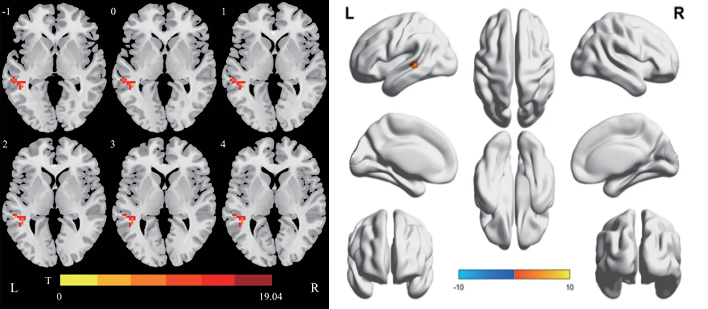
Figure 1 Difference in the VMHC value before and after treatment in the control group Compared with the values before treatment, the red area indicated that the VMHC value was increased after a comprehensive treatment, and the blue area indicated that the VMHC value was decreased.VMHC: Voxel-mirror homotopic connection.
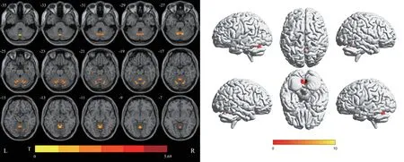
Figure 2 Difference in the VMHC value before and after treatment in the acupuncture group Compared with the values before treatment, the red area indicated that the VMHC value was increased after a comprehensive treatment, and the blue area indicated that the VMHC value was decreased.VMHC: Voxel-mirror homotopic connection.
DISCUSSION
There is no record of the term “amblyopia” in traditional Chinese medicine, but it can be referred to as “blurred vision”according to its clinical symptoms.According to the traditionalChinese medicine theory, the cause of amblyopia is mainly related to congenital deficiency, acquired loss of support,or related abnormal visceral function.Combined with the traditional Chinese medicine theory and results of our previous clinical works, we proposed that amblyopia is located in the eye, which is related toqideficiency of the heart, brain, liver,spleen, and kidney.Its basic pathogenesis is “unclear vision due to deficiency of the eye spiritqi” and the treatment is mainly “regulatingqiand unblocking meridians to bright vision”.After clinical verification, a good curative effect has been achieved.

Table 4 VMHC analysis of the different brain regions

Table 5 VMHC analysis of the brain region
Vision detection is one of the most important, intuitive, and convenient evaluation methods in the study of amblyopia.A P-VEP is a kind of visual evoked potential, which is the bioelectric signal of the cerebral cortex in response to visual stimulation, which mainly reflects the visual conduction pathway and visual cortex function[26].In the present study,compared with the data before treatment, the visual acuity and P-VEP of the two groups were improved after treatment, and the improvement in the acupuncture group was better than that of the control group, indicating that the clinical effect of acupuncture combined with routine treatment on children with amblyopia is better than that of routine treatment, and it is an effective treatment for children with anisometropic amblyopia.Amblyopia in children are often accompanied by abnormal functional activities of brain-related brain regions, whereas rs-fMRI can directly show the functional changes of brain tissues[27-28].Given that there are a large number of functional networks between cerebral hemispheres, VMHC can reflect the changes in information communication and integration disorders between the cerebral hemispheres, which makes it possible to detect the activity patterns of the cerebral neural circuits and the interaction between the cerebral hemispheres[29-30].VMHC can be used as a method for determining the clinical manifestation and curative effect of nervous system diseases, and it can also be used to evaluate the changes in the brain functional connections related to the patients’ behavior and cognition.Therefore, VMHC was used to analyze the rs-fMRI data.
In the present study, the brain regions with increased VMHC values in the control group after treatment were the temporal lobe, middle temporal gyrus and superior temporal gyrus.It is generally believed that the function of the temporal lobe is mainly to process auditory information.When the temporal lobe lesion damages the deep visual radiation fibers and visual bundles, the blind area in the same upper quadrant of the binocular contralateral visual field may appear.Studies using fMRI and diffusion tensor imaging have shown that, as the main white matter bundle connecting the occipital lobe and the temporal lobe, the inferior longitudinal tract of the temporal lobe is involved not only in visual processing tasks, but also in visual processing tasks, language, semantic function, mood regulation, reading, and neuropsychiatric status[31].Therefore,in the control group, the VMHC values of the temporal lobe,middle temporal gyrus and superior temporal gyrus increased after treatment, suggesting that routine treatments may improve the visual acuity by enhancing the function of middle and superior temporal gyrus through visual stimulation, such as using red flashes and a raster.At the same time, it may affect an individual’s emotional and auditory processing abilities.
In the acupuncture group, the brain area with an increased VMHC value was the anterior cerebellar lobe.The non-motor function of the cerebellum includes executive, language,and emotion regulation functions as well as visual spatial processing.Some scholars have utilized rs-fMRI to determine whether amblyopic patients have functional changes in the brain areas along this pathway,i.e., from the cerebellum,inferior parietal lobule to V1[27-32].A similar study found that the functional connection between the right anterior cerebellar lobe and primary visual cortex is decreased in patients with glaucoma-associated optic nerve damage, and even the higher visual cortex has similar manifestations, suggesting that there is a decline in visual integration in these patients[33].It is suggested that the decrease in the function of the anterior cerebellar lobe may be caused by the decrease in information input in the primary or higher visual cortex.Therefore, in the acupuncture group, the VMHC value in the anterior cerebellar lobe increased after treatment.It is also suggested that acupuncture may enhance the functional coordination between the bilateral cerebellum and improve the visual function caused by the enhancement of the functional network and fiber connection between the cerebellum and the cerebellum, and the improvement in visual ability will also enhance somatic motor functioning.In turn, it reactivates the functional connection of the cerebellum, forming a positive feedback mechanism.However, this mechanism needs to be confirmed in further research and analysis.
Compared with the acupuncture and control groups after treatment by VMHC analysis, the abnormal response of the brain area was observed in the temporal lobe.The temporal lobe is mainly responsible for auditory functioning, but it is also related to color vision and vision formation, participates in the visual pathway of figure vision and shape cognition of objects, and regulates the processing of stereo vision information[34].The temporal lobe is also related to the primary visual cortex.The changes in visual information transmission and integration caused by the changes in visual function are related to the synchronous changes in nerve signals in visual processing areas, such as the temporal lobe[35].The specific mechanism may be the transsensory remodeling of brain function after visual impairment,i.e., a change in the activity of a sensory system, which can cause the remodeling of other cortical and subcortical structures and nerve fiber connections[36].The high synchronization of spontaneous activity of the bilateral cerebral neurons,i.e., homotopy, is the basic characteristic of maintaining the function of the left and right hemispheres[37].In a previous study on homotopy function between the cerebral hemispheres, the superior temporal lobe and middle gyrus were related to multiple cognitive functions[38].
The results of the present study showed that the VMHC value of the bilateral superior and middle temporal lobes increased,and it was speculated that patients with amblyopia might have temporal lobe dysfunction, which could be improved by acupuncture.The previous study of the members of the present project found that acupuncture could activate the function of transsensory pathway reorganization (such as hearing) in monocular deprived rats, and they further speculated that BL1 and BL2 were selected as head and face acupoints in the acupuncture method of “regulatingqiand unblocking meridians to bright eyes”.The improvement in visual function due to acupuncture therapy on the scalp and facial acupoints in the treatment of amblyopia may be related to the role of temporal lobe after the reorganization of transsensory channels.To sum up, conventional therapy, such as red flash, grating, and other stimulations, may improve the visual function of children with amblyopia by changing the abnormal response of the temporal lobe, although the possible mechanism of acupuncture combined with conventional therapy in the treatment of amblyopia is the activation of the anterior cerebellar lobe.Through a comparative analysis, the regulation of acupuncture on the functional activity of the cerebellar anterior lobe may be an effective mechanism of acupuncture in the treatment of anisometropic amblyopia,which is obviously different from that of the routine treatment of amblyopia.
However, the present study also has some limitations due to objective factors.First, this study included a small sample size, and did not establish a normal control group or subgroup analysis (e.g., different age groups, types of amblyopia).Second, brain remodeling was found to be closely related to the reorganization of auditory and other sensory pathways, but only nine children were included in each group for the study.Hence, follow-up studies should include more patients, set up a normal control group, and conduct an in-depth study on the reorganization of other sensory pathways in the brain region to explore the correlation between imaging indicators and clinical variables and further increase the accuracy of the study results.In conclusion, acupuncture has a stronger overall effect in the treatment of anisometropic amblyopia, and it may improve the functions of body movement and emotional processing; the process involves a default-mode network and sensory crosschannel reorganization mechanism, reflecting the multi-level and multi-channel characteristics of the effect of acupuncture therapy.This provides a train of thought and preliminary basis for the study on the brain function mechanism of acupuncture intervention for amblyopia and other visual diseases.
ACKNOWLEDGEMENTS
Authors’ contributions:Wang J conceptualized and designed the study, drafted the initial manuscript.Wang J, Jia J, Sun Y, Ma CB, and Chen YZ collected the data and carried out the initial analyses.Liu AG and Yan XK critically reviewed the manuscript for important intellectual content.All authors approved the final manuscript as submitted and agree to be accountable for all aspects of the work.
Foundations:Supported by National Natural Science Foundation of China (No.82160935; No.82260965);Traditional Chinese Medicine Discipline “Qi Huang Ying Cai”Tutor Special Fund Doctoral Program (No.ZYXKBD-202208);Higher Education Innovation Fund Project of Gansu Province(No.2021A-087); Natural Science Foundation of Gansu Province (No.22JR5RA583); Traditional Chinese Medicine Discipline “Qi Huang Ying Cai” Tutor Special Fund Master’s Supervisor Program (No.ZYXKSD-202220); Youth Research Fund Project of Gansu University of Chinese Medicine (No.ZQ2017-9); Gansu Province 2023 Provincial Key Talent Project (No.2).
Conflicts of Interest:Wang J,None;Jia J,None;Sun Y,None;Ma CB,None;Chen YZ,None;Liu AG,None;Yan XK,None.
 International Journal of Ophthalmology2024年2期
- International Journal of Ophthalmology的其它文章
- Using choroidal thickness to detect myopic macular degeneration
- lmpact of multifocal gas-permeable lens designs on short-term choroidal response, axial length, and retinal defocus profile
- Baerveldt glaucoma implant with Supramid© ripcord stent in neovascular glaucoma: a case series
- Efficacy and safety of Usights UC100 illuminated microcatheter in microcatheter-assisted trabeculotomy
- Quantifying peripapillary vessel density and retinal nerve fibre layer in type 1 diabetic children without clinically detectable retinopathy using OCTA
- Nomogram to predict severe retinopathy of prematurity in Southeast China