
Shape change in viable eggs of the collembolan Folsomia candida provides insight into the role of Wolbachia endosymbionts
2010-12-25 01:03NinaHaferNathanPike
Zoological Research 2010年6期
Nina Hafer, Nathan Pike
(Department of Zoology, University of Oxford, Tinbergen Building, South Parks Road, Oxford, OX1 3PS, UK)
Shape change in viable eggs of the collembolanFolsomia candidaprovides insight into the role ofWolbachiaendosymbionts
Nina Hafer, Nathan Pike*
(Department of Zoology, University of Oxford, Tinbergen Building, South Parks Road, Oxford, OX1 3PS, UK)
The endosymbiotic bacteria of the genusWolbachiathat infect the collembolan speciesFolsomia candidaare responsible for facilitating parthenogenetic reproduction in their hosts. This study made empirical observations of the development of eggs ofF. candidawhich contained normal populations ofWolbachiaand of eggs which were cured ofWolbachiaby treatment with the antibiotic rifampicin. A marked increase in egg size accompanied by a significant change in shape from spherical to discoid occurred in viable eggs three to four days after laying. These changes did not occur in the universally inviable eggs which came from the antibiotic treatment or in the 7% of untreated eggs which were naturally inviable. We infer thatWolbachiaplays a critical role in zygotic or embryonic development during or before the first three days after laying and we draw on existing knowledge in speculating on the developmental mechanisms thatWolbachiamay influence.
parthenogenesis, embryonic development, Collembola, hatching rate
The genusWolbachiarepresents a diverse taxonomic group of α-protobacteria that are endosymbionts of insects, mites, crustaceans and nematodes (Werren et al, 1995; Stouthamer et al, 1999). These endosymbionts are known to influence reproduction in a number of profound ways including (i) creation of incompatibility of crosses between infected males and uninfected females (Werren, 1997), (ii) phenotypic male feminisation (Rousset et al, 1992), (iii) male killing (Stevens et al, 2001), and (iv) thelytokous parthenogenesis (Stouthamer et al, 1993).
Folsomia candida(Collembola: Isotomidae) is a species of springtail that has a worldwide distribution and is widely used as a standard organism in toxicological assays and as a biological marker of soil pollution (International Standards Organization, 1999; Fountain & Hopkin, 2005). Unlike closely related species,F. candidareproduces by parthenogenesis (Hopkin, 1997; Fountain & Hopkin, 2005) andWolbachiais an obligate facilitator of this parthenogenesis (Pike & Kingcombe, 2009; Timmermans & Ellers, 2009).F. candidais diplodiploid(Kiauta, 1970) and it is thus remarkable in being the first non-haplodiploid organism for whichWolbachia-induced parthenogenesis has been recorded. The supergroup E strain ofWolbachiathat is hosted byF. candidais unique relative to the strains known from non-collembolan taxa (Vandekerckhove et al, 1999). AlthoughWolbachiahas a critical role to play in the life history ofF. candida, infections can be cured through relatively heavy and sustained doses of the antibiotic rifampicin (Pike & Kingcombe, 2009).
The current study made experimental observations of normal and anomalous development of eggs ofF. candidawith and withoutWolbachiaendosymbionts. The key aim was to use these observations to make inferences about the developmental period during whichWolbachiafacilitates parthenogenesis and to subsequently unite these findings with existing knowledge of embryonic development to shed light on the cellular processes with whichWolbachiamay be involved.
1 Materials and Methods
1.1 Study organism and rearing conditions
Eggs were obtained from individuals from a clonal population ofF. candida(that was established from a single parthenogenetic female). Collembolans were kept individually and observed daily at the same time such that laying and developmental times for individual clutches of eggs could be accurately recorded. Collembolans designated to the control group were fedad libitumon a paste of powdered brewer’s yeast and water. Other collembolans were assigned to a treatment group which was exposed to the antibiotic rifampicin byad libitumprovision of a yeast paste in which rifampicin powder (Sigma-Aldrich) comprised 2.7% of the dry weight. Eggs from the rifampicin treatment group were not observed until two generations had passed and elimination of endosymbioticWolbachiabacteria had been confirmed using the technique set out in Pike & Kingcombe (2009). All individuals were kept in sealed containers on a substrate of 10% charcoal, 90% plaster of Paris at a constant temperature of (21 ± 0.2)°C and a relative humidity of 100%.
1.2 Experimental observations
The same observation protocols were applied to the control group and to the rifampicin treatment group. Eggs were observed on the day on which they were laid and on every day thereafter until either hatching occurred or 15 days had elapsed. High-resolution photographs of eggs were taken with the assistance of a light microscope on the day of laying and again four days after laying. These photographs were subsequently used to obtain a measurement of the longest axis of the egg. The accuracy of this measuring technique was verified: repeat measurements made on the same egg differed from the mean value by just (0.95% ± 0.56)% (mean ± 95% C.I.).
Analysis-of-variance procedures were used to investigate differences among experimental groups after conformity with assumptions of normality and homoscedasticity had been verified. Analyses were conducted using version 2.10.1 of the R statistical computing package (R Development Core Team, 2009). Mean values ± 95% confidence intervals along with sample sizes or degrees of freedom are reported in all cases.
2 Results
A marked change of shape occurred in viable eggs between three and four days after laying. Viable eggs (which corresponded to 93.2 ± 1.0% of eggs laid by the control group,n= 3736) were essentially spherical in shape on the day of laying (at which time the long axis measured 127.33 ± 1.06 µm,n= 85) and they remained spherical until three to four days after laying. Four days after laying, the viable eggs flattened along one axis to take on a discoid shape in which the longest axis measured 158.97 ± 1.62 µm (n= 85). This shape change which is equivalent to an increase in the longest axis of(25.66 ± 1.30)% over the first three to four days is significant (F1,168= 1033,P< 0.001). The change of shape is illustrated in Fig. 1.
Eggs were not viable if the shape change just described did not occur. Eggs produced by rifampicintreated collembolans were inviable in all cases. As no difference in size or shape was detected when comparing the universally inviable eggs from the rifampicin-treated group with the inviable eggs from the control group, data on inviable eggs from both groups were pooled. The average size at laying of inviable eggs was 125.95 ± 1.60 µm (n= 54). The size difference between inviable and viable eggs on the day of laying was thus not significant (F1,137= 2.18,P= 0.14). Four days after laying, the longest axis of the inviable eggs was 130.05 ± 1.63 µm (n= 54) and inviable eggs were thus clearly smaller than viable eggs from the same period (F1,137= 554,P<0.001).
Shape change at four days after laying was a very good predictor of egg viability (albeit not a perfect one). Seventy-nine of the 85 eggs which changed shape (~93%) hatched to produce nymphs. Eggs that changed shape are vastly more likely to be viable than eggs that did not change shape (logistic regression: Wald’sz= 165,P< 0.001).
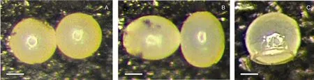
Fig. 1. Shape change in eggs of F. candida
3 Discussion
The observed change of shape in eggs is clearly a sign of normal embryonic development. Similarly, the absence of this change by three to four days after laying appears to be a perfect predictor of failed embryonic development. The current results accord with earlier studies which demonstrated that antibiotic elimination ofWolbachiaendosymbionts fromF. candidaresults in the exclusive production of inviable eggs (Pike & Kingcombe, 2009; Timmermans & Ellers, 2009). The current results provide two further pieces of information: (i) eggs which are inviable due to natural or unknown causes are visibly indistinguishable from eggs that are inviable as a result of antibiotic curing, and (ii) antibiotic curing disrupts development at some pointpriorto the third day after laying.
Most freshly laid eggs are at the metaphase I stage of meiosis and the first mitotic stages commence between three and ten hours after laying (Riparbelli et al, 2006). The majority of development in the first three days after laying is likely to be associated with cleavage and formation of the blastoderm and germ band. The marked shape change in eggs at three to four days after laying corresponds to one of the most fundamental and rapid periods of embryonic development inF. candida(Gao et al, 2006). Visible embryonic development and apparent tissue differentiation commence only after 72 hours have elapsed since laying. The mandibles, antennae, and legs differentiate between three to four days after laying. It is not until four to five days after laying that the furca (the forked springing organ) begins to form and the appendages begin to elongate and segment. Our findings suggest that the bulk of the growth in the egg and virtually all of the change in shape occurs during this developmental period which lasts between one and two days. Our own casual observations nevertheless concur with the observations of Gao et al (2006) that eggs continue to grow at a reduced but detectable rate right up until hatching.
While the natural causes of egg inviability remain uncertain, we note that the shape change we report presents a fast and convenient method of sorting the living eggs from the dead. Good evidence exists that the egg inviability associated with the rifampicin treatment group is a direct result of the absence ofWolbachiainfection rather than as a side-effect related to toxicity of the antibiotic: Timmermans & Ellers (2009) observed that collembolans that had been cured ofWolbachiainfection through heat treatment in early life also produce inviable eggs. We can thus use the information obtained in the current study to inform speculation on the developmental mechanisms ofF. candidain whichWolbachiaplays an obligate role.
Until recently, it was thought that theWolbachiathat induce parthenogenesis may be functionally restricted to organisms that have a haplodiploid mechanism of sex determination (Stouthamer, 1997). The basis for this suspicion is that haplodiploids need only to achieve diploidisation of the unfertilised egg to be parthenogenetic. Diplodiploid organisms likeF. candidamust achieve the additional feat of inducing eggs to develop in the absence of the sperm that usually perform this task. It is now clear thatF. candidahas overcome both of these obstacles to parthenogenesis and thatWolbachiaendosymbionts have been co-opted into an obligate role early in the parthenogenetic developmental process (Pike & Kingcombe, 2009; Timmermans & Ellers, 2009).
A comparative study of parthenogeneticF. candidaand its obligately sexual sibling speciesFolsomia fimetariaby Riparbelli et al (2006) highlighted a key difference in early development: the first mitotic division in the zygote ofF. fimetariarelies on a centrosome which is derived from the basal body of the fertilising sperm while the first mitotic division in the unfertilised egg ofF. candidais facilitated by remarkablede novoformation of cytoplasmic microtubules that take on a unique spindle structure relative to normal centrosomes.Wolbachiaare known to cluster near the centrosome during cell division and, in doing so, they adhere to the evolutionary imperative to be passed on by inclusion in new cells and, most critically, in the cells of the host’s germ line (Kose & Karr, 1995). This close spatial association betweenWolbachiaand centrosomes is certainly true in the context ofF. candida(Riparbelli et al, 2006). Given the observation that theWolbachiaendosymbionts are firmly in the right places at the right times, one might hypothesise thatWolbachiais involved in enabling the formation of centrosome-like structures that replace the true centrosomes that would be provided by fertilising sperm in other collembolan species.
An alternative but not exclusive hypothesis for the role ofWolbachiarelates to the fact that meiosis inF. candidais clearly aberrant in that the oocyte undergoes only a single maturation division (Palévody, 1973). It is thus feasible thatWolbachiacould facilitate parthenogenesis within the oocytes at meiosis II by preventing separation of sister chromatids and thereby effecting diploidisation.
Two obvious candidate mechanisms by whichWolbachiacould facilitate successful parthenogenetic development of itsF. candidahosts have been set out. Whether either of these occur in reality remains to be elucidated by further study. However, we can assert with confidence that the obligatory facilitation of parthenogenesis happens some time before the shape change that occurs in eggs three to four days after laying.
Acknowledgement:We are very grateful to Hui Hua of the Department of Zoology, University of Oxford for translating key parts of the scientific literature from Chinese to English.
Fountain MT, Hopkin SP. 2005.Folsomia candida(Collembola): A“standard” soil arthropod[J].Annu Rev Entomol,50: 201-222.
Gao Y, Bu Y, Luan YX, Yin WY. 2006. Preliminary observation of the embryonic development ofFolsomia candida(Collembola: Isotomidae)[J].Zool Res,27: 519-524. (in Chinese)
Hopkin ST. 1997. Biology of the Springtails[M]// Insecta: Collembola. Oxford: Oxford University Press.
International Standards Organization. 1999. Soil Quality-inhibition of Reproduction of Collembola (Folsomia candida) by Soil Pollutants[C]. Report No. ISO 11267:199(E). Geneva,16.
Kiauta B. 1970. Review of the germ cell chromosome cytology of Collembola, with a list of chromosome numbers and data on two species new to cytology[J].Genen en Phaenen,13: 89-99.
Kose H, Karr T. 1995. Organization ofWolbachia pipientisin theDrosophilafertilized egg and embryo revealed by an anti-Wolbachiamonoclonal antibody[J].Mech Dev, 51: 275-288.
Palévody C. 1973. Etude cytologique de la parthénogenèse chezFolsomia candida(Collembole, Isotomide) [J].Comptes Rendus de l'Académie des Sciences,277: 2501-2504.
Pike N, Kingcombe R. 2009. Antibiotic treatment leads to the elimination ofWolbachiaendosymbionts and sterility in the diplodiploid collembolanFolsomia candida[J].BMC Biol,7: 54.
R Development Core Team. 2009. R: A language and environment for statistical computing[DB/OL]. Vienna: R Foundation for Statistical Computing, http://www.r-project.org/.
Riparbelli MG, Giordano R, Callaini G. 2006. Centrosome inheritance in the parthenogenetic egg of the collembolanFolsomia candida[J].Cell Tissue Res,326: 861-872.
Rousset F, Bouchon D, Pintureau B, Juchault P, Solignac M. 1992. Wolbachia endosymbionts responsible for various alterations of sexuality in arthropods[J].Proc Biol Sci,250: 91-98.
Stevens L, Giordano R, Fialho RF. 2001. Male-killing, nematode infections, bacteriophage infection, and virulence of cytoplasmic bacteria in the genusWolbachia[J].Annu Rev Ecol Syst,32: 519-545.
Stouthamer R. 1997. Wolbachia-induced parthenogenesis[M]// O'Neill SL, Hoffmann AA & Werren JH. Influential Passengers: Inherited Microorganisms and Invertebrate Reproduction. Oxford: Oxford University Press, 102-124.
Stouthamer R, Breeuwer JA, Hurst GD. 1999.Wolbachia pipientis: microbial manipulator of arthropod reproduction[J].Annu Rev Microbiol,53: 71-102.
Stouthamer R, Breeuwer JAJ, Luck RF, Werren JH. 1993. Molecular identification of microorganisms associated with parthenogenesis[J].Nature,361: 66-68.
Timmermans M, Ellers J. 2009.Wolbachiaendosymbiont is essential for egg hatching in a parthenogenetic arthropod[J].Evol Ecol,23: 931-942.
Vandekerckhove TTM, Watteyne S, Willems A, Swings JG, Mertens J, Gillis M. 1999. Phylogenetic analysis of the 16SrRNA of the cytoplasmic bacteriumWolbachiafrom the novel hostFolsomia candida(Hexapoda, Collembola) and its implications for wolbachial taxonomy[J].FEMS Microbiol Letters,180: 279-286.
Werren JH. 1997. Biology ofWolbachia[J].Annu Rev Entomol,42: 587-609.
Werren JH, Zhang W, Guo RL. 1995. Evolution and phylogeny ofWolbachia: reproductive parasites of arthropods[J].Proc Biol Sci,261: 55-71.
从白符虫兆(弹尾纲)活性卵的形状变化探讨沃尔巴克氏体的共生作用
Nina Hafer, Nathan Pike*
(Department of Zoology, University of Oxford, Tinbergen Building, South Parks Road, Oxford, OX1 3PS, UK)
Wolbachia属共生菌的侵染是引起跳虫—— 白符虫兆孤雌生殖的原因。对带有正常沃尔巴克氏体菌群的白符虫兆卵和通过利福平处理剔除沃尔巴克氏体菌群的白符虫兆卵的胚胎发育进行实验观察。白符虫兆的活性卵产出3到4天后, 卵体大小显著性地增大, 并伴随卵体形状从球形到圆饼形的变化。这些变化在利福平处理的或者是7%自然失活的非活性卵中都没有出现。推测沃尔巴克氏体在白符虫兆卵产出后的3天之内或者3天之前的受精卵发育或胚胎发育中发挥着重要作用; 同时根据目前已有的研究结果推断沃尔巴克氏体对白符虫兆卵发育可能的影响机制。
孤雌生殖; 胚胎发育; 弹尾纲; 孵化率
Q969.140.4; Q954.4
A
0254-5853(2010)06-0623-04
2010-06-28; 接受日期:2010-11-24
10.3724/SP.J.1141.2010.06623
date: 2010-06-28; Accepted date: 2010-11-24
This work was partially funded by a grant awarded to NP by the Nuffield Foundation
*Corresponding author (通讯作者), Tel: + 44 1865271119, Fax: + 44 1865310447, E-mail: nathan.pike.1998@pem.cam.ac.uk
猜你喜欢
实用手外科杂志(2022年2期)2022-08-31
当代水产(2022年6期)2022-06-29
西藏研究(2021年1期)2021-06-09
实用手外科杂志(2018年1期)2018-07-03
西藏研究(2017年5期)2018-01-30
现代园艺(2017年19期)2018-01-19
中国继续医学教育(2015年1期)2016-01-06
中国卫生标准管理(2015年15期)2015-01-26
中国现代药物应用(2011年14期)2011-02-10
中国社区医师(2009年10期)2009-06-11

- Zoological Research的其它文章
- Digestive enzyme and alkaline phosphatase activities during the early stages of Silurus soldatovi development
- Isolation and characterization of Hainantoxin-II, a new neurotoxic peptide from the Chinese bird spider (Haplopelma hainanum)
- Morphological changes of silver and bighead carp in the Yangtze River over the past 50 years
- Positive influence of traditional culture and socioeconomic activity on conservation: A case study from the black-and-white snub-nosed monkey (Rhinopithecus bieti) in Tibet
- Bats and marsupials as indicators of endemism in the Yungas forest of Argentina
- A new record of Dasyatid fish in China: Dasyatis laosensis