
3.0T高分辨磁共振评价脑缺血患者症状侧与非症状侧颈动脉粥样硬化性斑块的差异
2014-04-21 00:45刘丹青李依霞蔡幼铨蔡剑鸣
解放军医学院学报 2014年7期
韩 旭,刘丹青,崔 豹,马 露,李依霞,蔡幼铨,蔡剑鸣
解放军总医院 放射诊断科,北京 100853
3.0T高分辨磁共振评价脑缺血患者症状侧与非症状侧颈动脉粥样硬化性斑块的差异
韩 旭,刘丹青,崔 豹,马 露,李依霞,蔡幼铨,蔡剑鸣
解放军总医院 放射诊断科,北京 100853
目的 采用3.0T高分辨磁共振成像评价脑缺血患者症状侧(患者一侧肢体出现脑缺血临床症状,对侧颈动脉则定义为症状侧)与非症状侧颈动脉粥样硬化斑块的差异。方法收集2012年6月- 2013年6月本院脑梗死或短暂性脑缺血患者102例,2周内进行双侧颈动脉高分辨磁共振成像,分析症状侧与非症状侧颈动脉粥样硬化斑块的差异。结果102例脑缺血患者中,脑梗死52例,短暂性脑缺血50例。症状侧与非症状侧颈动脉粥样硬化斑块所致颈动脉管腔狭窄程度比较无统计学差异(P>0.05),最大管壁厚度及斑块累及范围比较均有显著统计学差异(P=0.000);症状侧颈动脉粥样硬化斑块内易损成分(出血、富脂质核及纤维帽破裂)的发生率明显高于非症状侧,并且有显著统计学差异(Z=-6.525,P=0.000)。结论症状侧颈动脉粥样硬化斑块的严重性及易损性明显高于非症状侧斑块,症状侧斑块内易损成分发生率更高。
高分辨磁共振成像;颈动脉;粥样硬化斑块;脑缺血
缺血性脑血管病已成为我国居民主要致死、致残性疾病[1]。动脉粥样硬化是一种慢性炎症性疾病,可同时累及全身多个血管床[2]。颈动脉粥样硬化易损斑块主要表现为斑块内出血、大的富脂质核及纤维帽破裂,这些指标与缺血性脑血管病症状密切相关[3-4]。研究发现粥样硬化性斑块常同时出现在双侧颈动脉[5-6]。本研究旨在采用3.0T高分辨磁共振技术评价发生单侧肢体症状的脑缺血患者症状侧与非症状侧(患者一侧肢体出现脑缺血临床症状,对侧颈动脉定义为症状侧,同侧颈动脉定义为非症状侧)颈动脉粥样硬化斑块的差异,为预防脑血管事件提供研究资料。
对象和方法
1 研究对象 选取2012年6月- 2013年6月本院脑梗死或短暂性脑缺血患者102例,平均年龄(63.5±12.3)岁,其中男性68例;发生脑梗死52例,出现短暂性脑缺血症状(包括突发肢体无力、麻木等症状)50例。所有患者只出现一侧肢体症状并接受颅外颈动脉高分辨率磁共振成像。
2 磁共振检查方法 颅外颈动脉高分辨磁共振成像采用3.0T超导型MR扫描仪(GE Signa Excite,GE Medical System,USA)。其梯度场40 mT/m,梯度切换率150 mT/(m·s)。扫描线圈为4通道颈动脉相控阵表面线圈(Department of Radiology,University of Washington,USA)。扫描序列包括时间飞跃法(TOF)MRA序列、黑血T1加权(T1WI)以及增强T1WI序列、黑血质子加权(PDWI)和T2加权(T2WI)。扫描序列及参数见表1。
3 图像分析 由2位经验丰富的放射科医师对颈动脉MR图像进行定量及定性分析。定量分析包括管腔狭窄程度、最大管壁厚度及斑块累及范围。最大管腔狭窄程度按照北美症状性颈动脉内膜剥离术临床试验(North American Symptomatic Carotid Endarterectomy Trial,NASCET)的方法进行计算:最大狭窄度=(B-A)/B×100%,B为邻近相对正常的管腔直径(颈内动脉取远侧、颈总动脉及分叉处取近侧),A为最狭窄处剩余管腔直径[7]。定性分析包括斑块内出血(3D-TOF及T1加权序列都表现为高信号)、斑块内富脂质核(T1加权呈高信号,3D-TOF序列为等信号、T2加权及T1增强扫描序列上相应位置信号减低)、斑块表面纤维帽破裂(T1增强扫描序列中纤维帽信号连续性缺失)。按照颈动脉粥样硬化斑块内含有易损成分(出血、富脂质核及纤维帽破裂)的种类分为4个级别,“0”代表颈动脉斑块无易损成分,“1”代表含有1种易损成分,以此类推。
4 统计学方法 采用SPSS16.0统计软件,计量资料采用表示。定性指标(斑块内出血、富脂质核及纤维帽破裂)采用率表示。症状侧与非症状侧颈动脉粥样硬化性斑块所致管腔狭窄、最大管壁厚度及斑块累及范围的差异采用配对样本t检验(Paired-Samples T Test);斑块内易损成分含量差异采用等级资料秩和检验。
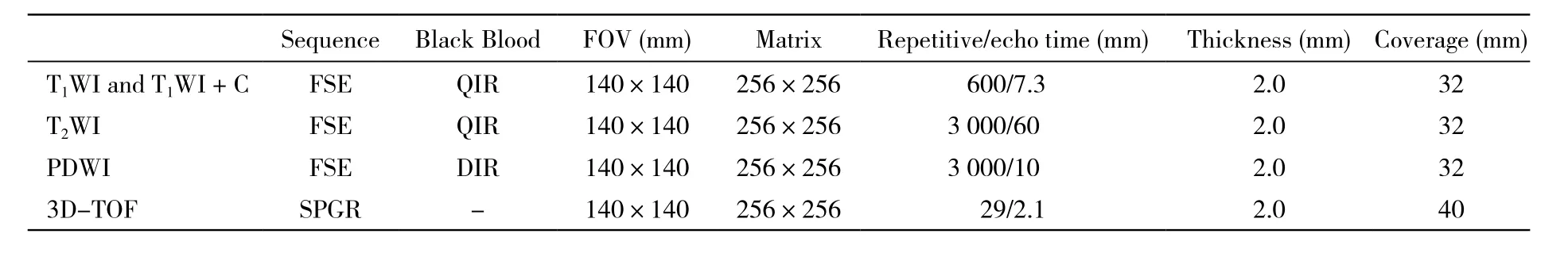
表1 颈动脉高分辨磁共振成像参数Tab. 1 Parameters of carotid high-resolution MR imaging
结 果
症状侧颈动脉斑块所致最大管壁厚度及斑块累及范围比非症状侧严重(P=0.000<0.05),而管腔狭窄无统计学差异(P=0.452>0.05)。症状侧颈动脉粥样硬化性斑块内易损成分的发生率及斑块级别明显高于非症状侧(Z=-6.525,P=0.000<0.05) (表2)。症状侧颈动脉斑块内同时含有出血及纤维帽破裂的概率明显高于非症状侧(46.1% vs 1.0%)。图1为一男性患者,主诉一过性左侧肢体麻木及无力。右侧颈动脉(症状侧)为3级斑块,内含大片出血、富脂质核及纤维帽破裂(图1D右侧、图1E箭头所示);左侧颈动脉(非症状侧)为1级斑块含富脂质核,T1增强序列上完整的纤维帽呈线样高信号(图1D左侧箭头示)。
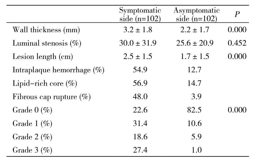
表2 症状侧与非症状侧颈动脉粥样硬化斑块差异Tab. 2 Differences of symptomatic and asymptomatic carotid AS plaques
讨 论
本研究发现症状侧颈动脉粥样硬化斑块内易损成分的发生率、斑块所致管壁厚度及累及范围均明显高于非症状侧颈动脉。症状侧高级别(3级)斑块的发生率也大于非症状侧。

图 1 颈动脉症状侧(右侧)斑块级别高于非症状侧(左侧)。 A、B、C及D分别为横轴位TOF、 T1WI、 T2WI及T1增强序列,E为右侧颈动脉矢状位T1WI序列Fig. 1 The plaque in carotid symptomatic side (right) showed higher grade than asymptomatic side (left). The A, B, C and D represented axial TOF, T1WI, T2WI and T1enhanced sequences, respectively. The E was right sagittal T1WI sequence
组织病理学研究证实,动脉粥样硬化性易损斑块主要表现为斑块内出血、大的富脂质核、薄纤维帽及纤维帽破裂等,颈动脉斑块内同时含有出血和纤维帽破裂的患者,脑血管疾病发生率明显升高[8-9]。本研究发现,症状侧颈动脉斑块内同时含有出血及纤维帽破裂的比率远高于非症状侧(46.1% vs 1.0%)。当临床发现一侧颈动脉斑块内同时含有出血及纤维帽破裂时,该侧颈动脉出现相关区域脑血管事件的可能性将明显增加。
颈动脉狭窄程度一直以来被认为是评价颈动脉粥样硬化斑块严重性的主要指标。近来研究表明,颈内动脉管腔狭窄(<50%)的症状性脑血管病患者中,超过20%的动脉血管存在易损斑块(斑块内出血和(或)纤维帽破裂)[9-10]。另有研究显示,颈动脉管腔无明确狭窄的症状性脑血管病患者中,仍有6% ~ 8%的患者发生颈动脉粥样硬化性易损斑块[11]。这是因为在动脉粥样硬化疾病发生、发展过程中,动脉血管存在正性重构效应,表现为向管腔外膨胀生长的趋势,而不引起管腔向心性狭窄[12-13]。
治疗颈动脉粥样硬化性斑块的方法主要有内科治疗、血管内介入及颈动脉内膜剥脱术。患者经内科治疗后,高分辨磁共振检查技术可以对其颅外颈动脉斑块的形态及内部成分的变化作出准确的判断,从而指导临床药物用量的调整[9,14]。NASCET临床试验结果显示,对症状性颈动脉中、重度狭窄患者行颈动脉内膜剥脱术,具有较好的疗效及远期预后[7]。磁共振检查技术不仅可以准确区分完全闭塞及接近于完全闭塞的血管腔,而且可以提供准确的斑块定位及定性信息,为介入或手术治疗提供准确的影像证据,同时可以用于患者的随访观察[15]。
综上所述,发生单侧肢体症状的脑缺血患者中,症状侧颈动脉粥样硬化斑块易损成分的发生率高于非症状侧斑块。磁共振高分辨成像对颈动脉粥样硬化斑块特性的判断,可以为临床医师提供不同的预防及诊疗思路。
1 Liu L, Wang D, Wong KS, et al. Stroke and stroke care in China:huge burden, significant workload, and a national priority[J]. Stroke, 2011, 42(12):3651-3654.
2 Wong KS, Li H, Chan YL, et al. Use of transcranial Doppler ultrasound to predict outcome in patients with intracranial large-artery occlusive disease[J]. Stroke, 2000, 31(11):2641-2647.
3 Ijäs P, Saksi J, Soinne L, et al. Haptoglobin 2 allele associates with unstable carotid plaque and major cardiovascular events[J]. Atherosclerosis, 2013, 230(2): 228-234.
4 Kurosaki Y, Yoshida K, Endo H, et al. Association between carotid atherosclerosis plaque with high signal intensity on T1-weighted imaging and subsequent ipsilateral ischemic events[J]. Neurosurgery, 2011, 68(1):62-67.
5 Adams GJ, Simoni DM, Bordelon CB, et al. Bilateral symmetry of human carotid artery atherosclerosis[J]. Stroke, 2002, 33(11):2575-2580.
6 Jeng JS, Tang SC, Liu HM. Epidemiology, diagnosis and management of intracranial atherosclerotic disease[J]. Expert Rev Cardiovasc Ther, 2010, 8(10):1423-1432.
7 Barnett HJ, Taylor DW, Eliasziw M, et al. Benefit of carotid endarterectomy in patients with symptomatic moderate or severe stenosis. North American Symptomatic Carotid Endarterectomy Trial Collaborators[J]. N Engl J Med, 1998, 339(20):1415-1425.
8 Takaya N, Yuan C, Chu B, et al. Association between carotid plaque characteristics and subsequent ischemic cerebrovascular events:a prospective assessment with MRI--initial results[J]. Stroke,2006, 37(3):818-823.
9 Zhao X, Underhill HR, Zhao Q, et al. Discriminating carotid atherosclerotic lesion severity by luminal stenosis and plaque burden:a comparison utilizing high-resolution magnetic resonance imaging at 3.0 Tesla[J]. Stroke, 2011, 42(2): 347-353.
10 Saam T, Underhill HR, Chu B, et al. Prevalence of American Heart Association type VI carotid atherosclerotic lesions identified by magnetic resonance imaging for different levels of stenosis as measured by duplex ultrasound[J]. J Am Coll Cardiol, 2008, 51(10):1014-1021.
11 Dong L, Underhill HR, Yu W, et al. Geometric and compositional appearance of atheroma in an angiographically normal carotid artery in patients with atherosclerosis[J]. AJNR Am J Neuroradiol, 2010,31(2):311-316.
12 Matsuo Y, Takumi T, Mathew V, et al. Plaque characteristics and arterial remodeling in coronary and peripheral arterial systems[J]. Atherosclerosis, 2012, 223(2):365-371.
13 Miura T, Matsukawa N, Sakurai K, et al. Plaque vulnerability in internal carotid arteries with positive remodeling[J]. Cerebrovasc Dis Extra, 2012, 1(1): 54-65.
14 Cai JM, Hatsukami TS, Ferguson MS, et al. Classification of human carotid atherosclerotic lesions with in vivo multicontrast magnetic resonance imaging[J]. Circulation, 2002, 106(11): 1368-1373.
15 王庆军,王勇,蔡剑鸣,等.斜矢状位黑血MRI增强扫描对颈动脉内膜剥脱手术的术前评估[J].南方医科大学学报,2011,31(3):385-391.
Differences of carotid atherosclerotic plaques between symptomatic and asymptomatic sides in cerebral ischemic patients detected using 3.0T high-resolution MR imaging
HAN Xu, LIU Dan-qing, CUI Bao, MA Lu, LI Yi-xia, CAI You-quan, CAI Jian-ming
Department of Radiology, Chinese PLA General Hospital, Beijing 100853, China
CAI Jian-ming. Email:caijm301@yahoo.com
ObjectiveTo investigate the differences of carotid atherosclerotic (AS) plaques between symptomatic and asymptomatic sides in cerebral ischemic patients detected using 3.0T high-resolution MR imaging.MethodsOne hundred and two patients with infarction or transient ischemic attack (TIA) in our hospital from June 2012 to June 2013 were recruited in our study. Each patient was received high-resolution MR examination of bilateral carotid arteries in two weeks. The differences of carotid atherosclerotic plaques between symptomatic and asymptomatic sides were investigated.ResultsOf the 102 cerebral ischemic patients, 52 had infarction and 50 developed TIA. Compared with the carotid atherosclerotic plaques between symptomatic and asymptomatic sides, the luminal stenosis showed no difference (P>0.05) while the maximum wall thickness and lesion length had signifcant statistic differences (P=0.000). The occurrence rates of vulnerable compositions such as hemorrhage, lipid-rich core and fbrous cap rupture in symptomatic sides were obviously higher than those in asymptomatic sides and showed signifcant statistic difference (Z=-6.525, P=0.000).ConclusionThe severity and vulnerability of carotid atherosclerotic plaque in symptomatic side are obviously higher than the asymptomatic side. The occurrence rates of vulnerable compositions in symptomatic sides are higher.
high-resolution magnetic resonance imaging; carotid artery; atherosclerotic plaque; cerebral ischemia
R 543.4
A
2095-5227(2014)07-0654-04
10.3969/j.issn.2095-5227.2014.07.002
时间:2014-04-14 17:31
http://www.cnki.net/kcms/detail/11.3275.R.20140414.1731.001.html
2014-02-21
全军“十二五”科研计划面上项目(CWS11J065)
Supported by Military Medical Research "12th Five-Year Plan"General Program of China(CWS11J065)
韩旭,男,在读硕士。研究方向:颈动脉粥样硬化斑块。Email:hxlryt@126.com
蔡剑鸣,男,博士,主任医师,博士生导师。Email:caijm301@yahoo.com
猜你喜欢
影像研究与医学应用(2022年23期)2023-01-11
中国卫生产业(2021年17期)2021-09-22
昆明医科大学学报(2020年11期)2020-12-28
中国医学装备(2019年11期)2019-11-27
天然产物研究与开发(2018年2期)2018-04-04
中西医结合心血管病电子杂志(2016年30期)2017-05-27
实用临床护理学杂志(电子版)(2017年26期)2017-04-01
罕少疾病杂志(2016年6期)2016-03-11
中西医结合心血管病杂志(电子版)(2016年30期)2016-01-20
中国康复理论与实践(2015年10期)2015-12-24
