
Effect of low intensity pulsed ultrasound on expression of TIMP-2 in serum and expression of mmp-13 in articular cartilage of rabbits with knee osteoarthritis
2015-10-31 03:25JingBoJiXueFuLiLeiLiuGuangZhongWangXinFengYan
Jing-Bo Ji, Xue-Fu Li, Lei Liu, Guang-Zhong Wang, Xin-Feng Yan
1Department of Bone and Joint, Affiliated Qianfoshan Hospital of Shandong University, Ji'nan Shandong, 250014
2Department of Orthopedics, Liaocheng No.3 Hospital, Liaocheng, Shandong, 252000
Effect of low intensity pulsed ultrasound on expression of TIMP-2 in serum and expression of mmp-13 in articular cartilage of rabbits with knee osteoarthritis
Jing-Bo Ji1,2, Xue-Fu Li2, Lei Liu2, Guang-Zhong Wang2, Xin-Feng Yan1*
1Department of Bone and Joint, Affiliated Qianfoshan Hospital of Shandong University, Ji'nan Shandong, 250014
2Department of Orthopedics, Liaocheng No.3 Hospital, Liaocheng, Shandong, 252000
ARTICLE INFO
Article history:
in revised form 20 October 2015
Accepted 3 November 2015
Available online December 2015
Low intensity pulsed ultrasound
Osteoarthritis
TIMP-2
MMP-13
Objective: To study the effect of low intensity pulsed ultrasound (LIPUS) on the expression of tissue inhibitor of metalloproteinase-2 (TIMP-2) in the serum and expression of matrix metallopeptidase 13 (MMP-13) in the articular cartilage cells of rabbits with knee osteoarthritis(OA). Methods: Inner patellar ligament defect method was used to establish the model of knee OA. Four weeks after the modeling, the arterial blood was drawn from the ear of each rabbit, while ELISA was employed to detect the expression of TIMP-2 in the serum. The chondrocytes were separated from animals in each group and then cultured in vitro. All rabbits were divided into control group, OA model group and OA + LIPUS group. Cells in the control and OA groups were not treated, while cells in the OA+ LIPUS group were treated with LIPUS(40 mW/cm2, 1 time/day). Cells were collected 7 d later and the RNA and total protein were extracted respectively. Real-time PCR and Western blotting were employed to analyze the expression of MMP-13 in chondrocytes at the mRNA and protein level, respectively. Results:The success rate of establishment of OA model was 83%. The results of ELISA showed that the content of TIMP-2 in the serum of animals with OA was 22.3%, lower than the one in the control group (P<0.05). Compared with the normal control group, the expression of TIMP-2 in the OA model group was significantly increased, while the expression of MMP-13 was significantly increased (P<0.05). After the stimulation of LIPUS, the expression of TIMP-2 and MMP-13 was close to the one in the normal control group. Conclusions: The inner patellar ligament defect method is a mature method to establish the rabbit OA model, with high success rate. The expression of serum TIMP-2 in the OA model group is significantly decreased. LIPUS can up-regulate TIMP-2 and down-regulate MMP-13.
Document heading doi:10.1016/j.apjtm.2015.11.003
1. Introduction
Osteoarthritis (OA) is also named degenerative osteoarthropathy,a common disease that frequently occurs in the middle-aged and elderly people. The pathological features of OA include the atrophyand degeneration of articular cartilage, osteophyte formation and bone and synovial hyperplasia on the edge and surface of joints. Most of patients would suffer from the limitation or loss of joint function finally, which greatly limits the activities of patients and affected their quality of life[1,2]. There are limited measures in the clinical treatment of osteoarthritis. The drug therapy is mainly to make use of anti-inflammatory and analgesic drugs to relieve or control the symptoms, but it cannot change the progression of diseases, only provide the temporary solutions to the acute pain episode. The injection of prednisolone (prednisolone acetate) orhydrocortisone acetate in the joints could control the symptoms[3]. The ultrasound-based physical therapy is to make use of the physical effect of clinical significance that is caused by the mechanical and elastic vibration to apply the miniature pressure to the tissues, which has been widely applied in the treatment of neurologic, muscular and bone diseases and traumas[4,5]. The frequency of low intensity pulsed ultrasound (LIPUS) is usually 1-3 MHz, with the strength less than 100 mW/cm2. LIPUS could stimulate the healing of fracture and thus shorten the healing time of fracture. Its mechanism is that LIPUS can promote the chondrogenesis and endochondral ossification and also the secretion of collagen Ⅱ and proteoglycan (especially the 6-chondroitin sulfate) by the transparent chondrocytes, as well as the secretion of many cytokines[6-8].
Matrix metallopeptidase (MMPs) play a key role in the physiological and pathological degradation of extracellular matrix(ECM). The previous researches also indicate that MMPs are of critical importance in the degradation of ECM of articular cartilage[9]. As the member of MMPs, MMP-13 can degrade the collagenⅡ and also help other subtypes of MMPs to degrade the collagenⅡ. Accordingly, the change of MMP-13 could reflect the change in the metabolism of collagen Ⅱ and proteoglycan in the cartilage matrix[10]. The excessive secretion of MMPs by the chondrocytes break the balance between the MMPs and the tissue inhibitor of metallo proteinases (TIMPs) and then cause the excessive degradation and degeneration of cartilage. Therefore, the balance adjustment of MMPs-TIMPs plays the critical role in the occurrence and development of OA[11,12].
2. Materials and methods
2.1. Materials and reagents
A total of 40 SPF New Zealand male rabbits with the weight of(1.0±0.5) kg and age of 1 month were purchased from Institute of Laboratory Animal Sciences, CAMS & PUMC. Rabbits were given the diet and water freely. The ventilation was good, with the natural lighting day and night. The culture temperature was maintained at 18-25 ℃.
The high glucose DMEM was purchased from GIBCO (America,item No. 12491-015); the fetal bovine serum from GIBCO (America,item No. 10082147); the total RNA extraction kit from QIAGEN(Germany, item No. 74134); the reverse transcription kit (iScript cDNA Synthesis Kit) from Bio-Rad (America, item No. 1708891);Real-time PCR fluorescent quantitative kit (SsoAdvanced SYBR Green Super mix) from Bio-Rad (America, item No. 1708882);ReadyPrep protein extraction kit from Bio-Rad (America); BCA protein quantitative kit from Vazyme Biotech (China, item No. E112-01); β-actin and MMP-13 monoclonal antibody from Santa Cruz Biotechnology (America, item No. sc-47778 and 30073); TIMP-2 monoclonal antibody from Abcam (Britain, item No. ab1828);horseradish peroxidase (HRP) labeled secondary antibody from Beijing Zhongshan Jinqiao Biotechnology; ECL Chemiluminescent Substrate Reagent Kit from Life Technologies (America, item No. WP20005); and PVDF film from Millipore (America).
The LIPUS instrument was Ito-HT2009-1; DNA/RNA analyzer was Qubit Fluorometer; CO2cell culture incubator was Thermo Scientific Series 8000; and the fluorescent quantitative PCR system was Bio-Rad -CFX96 Touch.
2.2. Methods
2.2.1. Modeling of knee OA rabbits
The inner patellar ligament defect method was used to establish the model of knee OA rabbits. After one week of adaptive feeding,the laboratory rabbits were given the intravenous injection of 3% sodium pentobarbital (1 mL/kg) at the edge of ear for the anesthesia. They were fixed on the laboratory table at the supine position. The hair around the knee joints of two hind limbs were sheared using the shearing scissors. The povidone-iodine was used for the disinfection of skin. The incision was performed in the tubercle between the patella and tibia along the medial margin of patellar ligament of inner knee joint to cause the inner patellar ligament defect of two hind limbs. It was washed with the sterile saline solution. After the surface of wound was stitched layer by layer, rabbits were put back to the cage. After the operation, the intramuscular injection of 200 000 units of penicillin was performed every day to avoid the infection. One week after the operation, the model animals were forced to take activities for 1 h every day. Rabbits in the control group did not receive the treatment, while other feeding conditions were the same as the model group.
2.2.2. Separation and culture of primary chondrocytes
Four weeks after the modeling, the animals were executed by the air embolism method. Knee joints were taken out under sterile conditions rapidly (the joint capsule should not be broken). After being washed with the normal saline, it was immersed in the normal saline containing 1% streptomycin and penicillin. The soft tissues around the knee joint were removed. After opening the joint capsule, the ligament and soft tissues around were also removed. The cartilage on the surface of knee joint was separated using the sterile scalpel blade. It was then put in the sterile Petri dish that contained the phosphate buffer solution (PBS). The cartilage was cut into about 1 mm3bone slices. It was washed with PBS twice. Afteradding 0.25% trypsin, it was digested at 4℃ for 4 h. Afterwards,the fetal bovine serum was added and the digested was stopped. It was centrifuged at 1 500 rmp for 5 min and then washed with high glucose DMEM twice. After being centrifuged, the supernatant was removed. The cells were resuspended and the suspension was seeded on 75 cm2tissue culture flask. 15% fetal bovine serum was added in the high glucose DMEM and it was then cultured at 5% CO2and 37 ℃. When cells were paved on the bottom of flask about 60%-70%, the subculture was performed by the ratio of 1:4.
2.2.3. Stimulation of LIPUS
LIPUS instrument in this study was HT2009-1, with the doubleprobe output (same power). Cells in the control and OA groups were not treated, while cells in the OA + LIPUS group were treated with LIPUS during the culture (40 mW/cm2, 1 time/day), lasting for 7 d.
2.2.4. ELISA
The blood samples were kept still at 37 ℃ for 1 h. After being centrifuged at 1 000 rmp and 4 ℃ for 20 min, the supernatant was drawn. Samples were labeled and then diluted. It set the standard wells, samples wells to be tested and blank wells. There were 7 standard wells. The standard samples with the different concentrations were added at one time. Then the serum samples to be tested were added. The ELISA plate was covered with the film and then incubated at 37 ℃ for 2 h. A total of 100 μL biotinylated primary antibodies were added in each well. It was than incubated at 37 ℃ for 1 h. The solution in the well was removed and each well was washed with 350 μL cleaning solution for 1-2 min. The plate was washed repeated 3 times. A total of 100 μL HRP-labeled secondary antibodies were added in each well. The ELISA plate was covered with the film and then incubated at 37 ℃ for 30 min. A total of 90 μL TMB substrates were added in each well. The ELISA plate was covered with the film and then colored at 37 ℃ and in a dark place for 15-25 min. When the first 3-4 of standard wells showed the obvious gradient blue, the reaction stopped. A total of 50 μL 2M H2SO4was then added. The plate reader was employed to measure the OD value of each well at 450 nm. Taking X-axis as the OD value and Y-axis as the log of concentration, the standard curve was drawn. According to the OD value of samples, the log of related concentration could be calculated from the standard curve;afterwards, the concentration value could be calculated according to the log of concentration; multiplying by the dilution factor, the actual concentration of samples could be obtained.
2.2.5. Western blotting
The collected cells were washed with PBS twice. After being centrifuged, the supernatant was removed. The precipitated cells were lysed with the protein extraction kit. After being put on the ice for 30 min, the probe-type ultrasound was used to produce the short impact with the appropriate frequency on the ice. The lysis mixture was centrifuged at 4 ℃ and 13 000 rpm/min for 20 min. The supernatant was transferred to the new centrifuge tube. BCA kit was employed to detect the protein concentration.
SDS-PAGE electrophoresis was performed on the protein samples. The gel was soaked in the transfer buffer for 10 min of equilibrium. It was installed with the transfer ‘sandwich', the transfer buffer was added and the electrodes were inserted, 100 V and 45-60 min. After the transfer, PVDF film was washed with TBS for 10-15 min. The film was placed in TBS/T blocking buffer containing 5% (w/v)skimmed milk powder and shaken at the room temperature for 1 h. Then the primary antibody with the appropriate degree of dilution was added [diluted with TBST containing 1% (w/v) skimmed milk powder]. It was incubated at the room temperature for 2 h and then the film was washed with TBST for 3 times. The film was incubated with the secondary antibody (1:10 000, HRP-labeled)that was diluted with TBST containing 0.05% (w/v) skimmed milk powder. It was incubated at the room temperature for 1 h and then the film was washed with TBST for 3 times. It was exposed and then photographed to save the experimental results. Quantity one v4.62 was used to measure the gray value of molecular band (trace tracking). The optical density curve could be drawn according to the optical density of different electrophoretic bands. The area under the curve of optical density was then calculated as the quantitative basis of electrophoretic bands (namely the semi-quantitative value of protein expression = optical density of protein band / related reference optical density). The statistical analysis was performed as well.
2.2.6. Real-time PCR
The collected cells were washed with PBS (RNase free). Then QIAGEN total RNA extraction kit was used to extract the total RNA and Qubit Fluorometer system to detect the concentration and purity of RNA. The total RNA was reversely transcripted to cDNA following the instruction manual of reverse transcription kit. The Real-time PCR was employed to detect the expression of related genes (Table 1, 2). The mRNA sequence of MMP-13 and β -actin genes could be referred to NCBI database and then the realtime PCR primers could be designed. All primers were synthesized by Shanghai Sangon Biotech, with the specific sequence shown in Table 3. The double △Ct method was adopted to calculate the relative expression of target gene: the mean of three parallel repeated experiments was treated as the Ct value of each sample, △Ct =Ct (Target gene) - Ct (reference), △△Ct=△Ct (sample) -△Ct(contro1). Therefore, the relative expression of target gene =2-△△Ctand the relative expression of control group was 20=1.

Table 1 Synthetic system of reverse transcription.
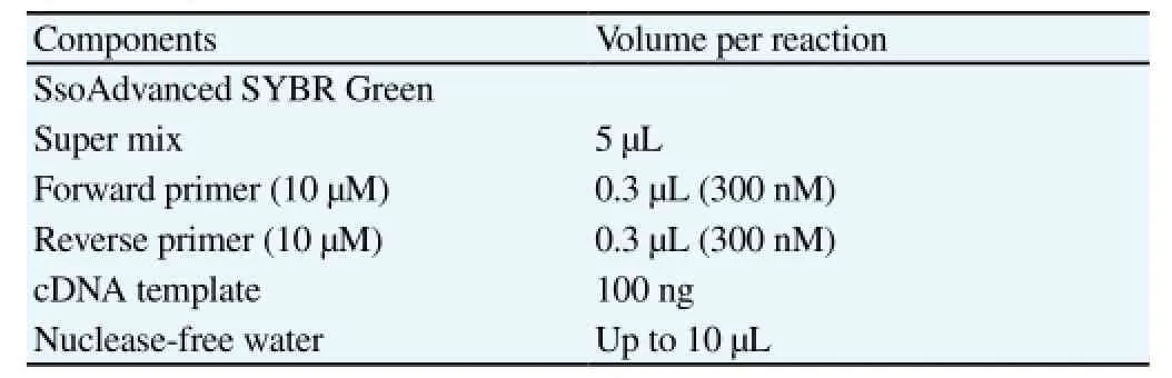
Table 2 Synthetic system of PCR.
2.3. Statistical analysis
The experimental data was treated with SPSS16.0. The measurement data was expressed by mean±SD. The student-t test was employed for the comparison between groups, with the significant level of α=0.05.
3. Results
3.1. Modeling of knee OA
Four weeks after the modeling, the animals were executed by the air embolism method. There were 10 rabbits in the normal control group, with no obvious effusion in the knee joint. The separated cartilage showed the transparent and smooth bone surface, without any defect or new organism. Mankin's score of cartilage structure was 0, which indicated that the form of knee joint before the modeling was normal. There were 30 rabbits in the model group,where 25 rabbits had the successful modeling (among 30 rabbits in the model group, 2 died because of the postoperative infection and 3 had no obvious OA feature). There was the effusion in the left knee joint of rabbits with the successful modeling. The separated cartilage showed the dim and grayish yellow surface, with the ulcers occurred on the coarse surface. According to Mankin's scores of cartilage structure, 4 rabbits got 4 points, 12 ones got 3 points and 9 ones got 2 points.
3.2. Expression of serum TIMP-2
The results showed that the expression of TIMP-2 was (24.86±6.45)ng/mL and (19.31±4.45) ng/mL in the normal control group and OA model group, respectively (Figure 1). According to the results,compared with the control group, the expression of TIMP-2 was significantly decreased in the OA model group (P<0.05), where the expression of TIMP-2 in the serum of OA rabbits was 22.3% lower than that of the control group.
3.3. Effect of LIPUS on expression of TIMP-2 and MMP-13 in rabbit chondrocytes
The results showed that (Figure 2), compared with the normal control group, the expression of TIMP-2 was significantly decreased and the expression of MMP-13 was significantly increased in the OA model group. After the stimulation of LIPUS, the expression of TIMP-2 and MMP-13 was close to the one in the normal control group (P<0.05).

Table 3 Primers used in real-time PCR.
4. Discussion
The apoptosis of chondrocytes and progressive degradation of ECM are main pathological features of OA. The degree of differentiation for chondrocytes is relatively high. As the main component of articular cartilage, its cellular activity is of critical importance to maintain the metabolism of cartilage and normal micro-environment of joint. The main component of ECM is aggrecan, which mainly maintains the elasticity and viscosity of transparent cartilage. The excessive hydrolysis of cartilage ECM by the protease was regarded as the early manifestation of destructive process of osteoarthritis[13,14]. MMPs belong to the zinc ion-dependent proteolytic enzyme family, as the most important proteolytic system for the degradation of ECM[15]. MMPs are mainly divided into the subfamilies of collagenases (MMP-1, MMP-8 and MMP-13), gelatinases (MMP-MMP-9) and stromelysins (MMP-3,MMP-7, MMP-10 and MMP-11). Where, MMP-13 can degrade the collagen II that is representative and has the highest content in the cartilage matrix. Besides, many other subtypes of MMPs will rely on it to play their role in the degradation of collagen II. Therefore, the change of MMP-13 is closely related to the metabolism of collagen II in the cartilage matrix. As the member of MMPs family, MMP-13 is the most effective degrading enzyme of collagen II. Especially,in the pathological situation, because of abundant MMPs secreted by chondrocytes, it could degrade collagen II and cause the loss of proteoglycan to result in the destroy and defect of cartilage and then the occurrence of osteoarthritis[16].
The animal experiment is the main method to study the pathogenesis of osteoarthritis. There are two animal models of OA at present: spontaneous model and induced model. The spontaneous model mainly refers to the OA animal model that the genetic mutation is reserved through the genetic breeding, while the induced model means that OA is induced by the operation or drugs. As the structure of rabbit's knee joint is relatively close to the human's, the biochemical indicators of rabbit OA model are relatively in accordance with the human OA[17], which is suitable for the experimental studies in the pathogenesis and medication of OA. Therefore, the inner patellar ligament defect method was employed in this study to build the model of rabbit's knee osteoarthritis. The primary chondrocytes were separated from the cartilage matrix,which could obtain the abundant chondrocytes with the high purity in such way[18]. The generation and amplification of primary cells are limited in vitro. With the increase in times of generation and amplification, the cellular activity will be gradually decreased. Thus the amplification was controlled at 3 times in this study to guarantee that the highest biological activity of chondrocytes that were suitable for the study.
TIMPs are the natural inhibitor of MMPs. The balance disorder of MMP-TIMP can result in the excessive degradation of articular cartilage ECM and then OA. Where, TIMP-2 is some kind of nonglycosylated protein, as the important metalloproteinase inhibitor, which can block the activity of all activated hydrolase of MMPs[19,20]. The inner patellar ligament defect method was employed to build the model of knee OA, where 10 rabbits were chosen in the control group without the treatment and 30 ones were modeled. Four weeks after the modeling, 25 New Zealand rabbits had the successful modeling (among 30 rabbits in the model group,2 died because of the postoperative infection and 3 had no obvious OA feature). Afterwards, the arterial blood was drawn from the ear of each rabbit. The TIMP-2 ELISA kit was employed to detect the expression of TIMP-2 in the serum. According to the results,compared with the control group, the expression of TIMP-2 was significantly decreased in the OA model group (P<0.05), where the expression of TIMP-2 in the serum of OA rabbits was 22.3% lower than that of the control group.
The application of ultrasound in the medicine could be traced back to 1942, in which Austrian doctor Dussik used it in the medical sector for the first time[21]. With the in-depth research on the mechanism of ultrasound and the progress of related applied technologies, the ultrasound has been widely applied in the clinical practice now. The previous researches reported that the sound wave in the form of pressure wave could apply the miniature pressure to the bone and its surrounding soft tissues[22,23]. The frequency of LIPUS is usually 1-3 MHz, with the strength less than 100 mW/ cm2. According to the researches in this year, LIPUS could stimulate the healing of fracture and thus shorten the healing time of fracture. Its mechanism is that LIPUS can promote the chondrogenesis and endochondral ossification and also the secretion of II collagen and proteoglycan (especially the 6-chondroitin sulfate) by the transparent chondrocytes, as well as the secretion of many cytokines[6-8]. Therefore, after building the animal model in this study, the primary cells were separated and the stimulation of LIPUS was applied at thecellular level. Real-time PCR and western blot were used to analyze the expression of TIMP-2 and MMP-13 at mRNA and protein level respectively. The results showed that, compared with the normal control group, the expression of TIMP-2 was significantly decreased and the expression of MMP-13 was significantly increased in the OA model group. After the stimulation of LIPUS, the expression of TIMP-2 and MMP-13 was close to the one in the normal control group (P<0.05). The results indicate that the balancing relationship between TIMP-2 and MMP-13 is of critical importance to maintain the micro-environment of joint, especially the activity of chondrocytes. The change in the specific signaling pathway caused by the stimulation of LIPUS will be the focus in the further studies.
Conflict of interest statement
We declare that we have no conflict of interest.
[1] Brown JP, Boulay LJ. Clinical experience with duloxetine in the management of chronic musculoskeletal pain. A focus on osteoarthritis of the knee. Ther Adv Musculoskelet Dis 2013; 5(6): 291-304.
[2] Kluzek S, Newton JL, Arden NK. Is osteoarthritis a metabolic disorder?Br Med Bull 2015; 80(6): 568-573.
[3] Thomas S, Vieira CS, Hass MA, Lopes LB. Stability, cutaneous delivery,and antioxidant potential of a lipoic acid and α-tocopherol codrug incorporated in microemulsions. J Pharm Sci 2014; 103(8): 2530-2538.
[4] Armijo-Olivo S, Fuentes J, Muir I, Gross DP. Usage patterns and beliefs about therapeutic ultrasound by Canadian physical therapists:An exploratory population-based cross-sectional survey. Physiother Can 2013; 65(3): 289-299.
[5] Griffin XL, Costello I, Costa ML. The role of low intensity pulsed ultrasound therapy in the management of acute fractures: a systematic review. J Trauma 2008; 65(6): 1446-1452.
[6] Khan Y, Laurencin CT. Fracture repair with ultrasound: clinical and cellbased evaluation. J Bone Joint Surg Am 2008; 90(Suppl 1): 138-144.
[7] Li S, Glynne-Jones P, Andriotis OG, Ching KY, Jonnalagadda US, Oreffo RO, et al. Application of an acoustofluidic perfusion bioreactor for cartilage tissue engineering. Lab Chip 2014; 14(23): 4475-4485.
[8] Korstjens CM, van der Rijt RH, Albers GH, Semeins CM, Klein-Nulend J. Low-intensity pulsed ultrasound affects human articular chondrocytes in vitro. Med Biol Eng Comput 2008; 46(12): 1263-1270.
[9] Xu Y, Zhang JA, Xu Y, Guo SL, Wang S, Wu D, et al. Antiphotoaging effect of conditioned medium of dedifferentiated adipocytes on skin in vivo and in vitro: a mechanistic study. Stem Cells Dev 2015; 24(9): 1096-1111.
[10] Attur M, Yang Q, Shimada K, Tachida Y, Nagase H, Mignatti P, et al. Elevated expression of periostin in human osteoarthritic cartilage and its potential role in matrix degradation via matrix metalloproteinase-13. FASEB J 2015; 23(Suppl 2): A135-A136.
[11] Giannopoulos G, Pavlakis K, Parasi A, Kavatzas N, Tiniakos D,Karakosta A, et al. The expression of matrix metalloproteinases-2 and -9 and their tissue inhibitor 2 in pancreatic ductal and ampullary carcinoma and their relation to angiogenesis and clinicopathological parameters. Anticancer Res 2008; 28(3B): 1875-1881.
[12] Groblewska M, Mroczko B, Gryko M, Pryczynicz A, Guzi ska-Ustymowicz K, K dra B, et al. Serum levels and tissue expression of matrix metalloproteinase 2 (MMP-2) and tissue inhibitor of metalloproteinases 2 (TIMP-2) in colorectal cancer patients. Tumour Biol 2014; 35(4): 3793-3802.
[13] Takaishi H, Kimura T, Dalal S, Okada Y, D'Armiento J. Joint diseases and matrix metalloproteinases: a role for MMP-13. Curr Pharm Biotechnol 2008; 9(1): 47-54.
[14] Xia P. Effect of low intensity pulsed ultrasound on chondrocytes of rabbit's knee osteoarthritis through integrin-FAK-MAPK chemical pathway. Master's Thesis of Nanjing Medical University; 2014.
[15] Yuan LJ, Niu CC, Lin SS, Yang CY, Chan YS, Chen WJ, et al. Effects of low-intensity pulsed ultrasound and hyperbaric oxygen on human osteoarthritic chondrocytes. J Orthop Surg Res 2014; 9: 5.
[16] Bramono DS, Richmond JC, Weitzel PP, Kaplan DL, Altman GH. Matrix metalloproteinases and their clinical applications in orthopaedics. Clin Orthop Relat Res 2004; (428): 272-285.
[17] Chen LX, Yu JK, Yu CL. Histomorphological comparison of articular cartilage of rats, rabbits and human. J Clin Reh Tissue Engineering Res 2007; 11(41): 8230-8233.
[18] Tian F, Cui XS, Zhang S. Improved stepwise digestion method for chondrocyte primary culture. J Clin Reh Tissue Engineering Res 2011;15(20): 3633-3635.
[19] Desmeules P, Trudel D, Turcotte S, Sirois J, Plante M, Grégoire J, et al. Prognostic significance of TIMP-2, MMP-2, and MMP-9 on high-grade serous ovarian carcinoma using digital image analysis. Hum Pathol 2015;46(5): 739-745.
[20] Chen X, Guo Z, Wang P, Xu M. Erythropoietin modulates imbalance of matrix metalloproteinase-2 and tissue inhibitor of metalloproteinase-2 in doxorubicin-induced cardiotoxicity. Heart Lung Circ 2014; 23(8): 772-777.
[21] Shampo MA, Kyle RA. Karl Theodore Dussik--pioneer in ultrasound. Mayo Clin Proc 1995; 70(12): 1136.
[22] Griffin XL, Costello I, Costa ML. The role of low intensity pulsed ultrasound therapy in the management of acute fractures: a systematic review. J Trauma 2008; 65(6): 1446-1452.
[23] Lin Y, Hu AH, Liu YW, Lv CW. Effects of low intensity pulsed ultrasound on the proliferation and differentiation of rabbit bone marrow stromal cells. J Clin Reh Tissue Engineering Res 2009; 13(36): 7043-7046.
15 September 2015
Xin-Feng Yan, Professor, PhD., Attending Physician,Department of Bone and Joint, Shandong Provincial Qianfoshan Hospital, Shandong University, 16766 Jingshi Road, Ji'nan, Shandong 250014, China.
Tel: 13869112527
E-mail: 13869112527@163.com
Foundation project: It was supported by Shandong Key Scientific and Technological Project Fund (No.: 2012GSF11845).
 Asian Pacific Journal of Tropical Medicine2015年12期
- Asian Pacific Journal of Tropical Medicine的其它文章
- Immunomodulatory effect of garlic oil extract on Schistosoma mansoni infected mice
- Larvicidal activity, inhibition effect on development, histopathological alteration and morphological aberration induced by seaweed extracts in Aedes aegypti (Diptera: Culicidae)
- Human ocular dirofilariasis due to Dirofilaria repens in Sri Lanka
- Childhood brucellosis: Review of 317 cases
- Effect of cyclophosphamide on fungal infection in SLE mice detected by fluorescent quantitative PCR
- Therapeutic effect of okra extract on gestational diabetes mellitus rats induced by streptozotocin