
Larvicidal activity, inhibition effect on development, histopathological alteration and morphological aberration induced by seaweed extracts in Aedes aegypti (Diptera: Culicidae)
2015-10-31 03:25KeXinYuChingLeeWongRohaniAhmadIbrahimJantan
Ke-Xin Yu, Ching-Lee Wong, Rohani Ahmad, Ibrahim Jantan*
1Drug and Herbal Research Centre, Faculty of Pharmacy, Universiti Kebangsaan Malaysia, 50300 Kuala Lumpur, Malaysia
2School of Biosciences, Taylor's University, Taylor's Lakeside Campus, 47500 Subang Jaya, Selangor, Malaysia
3Medical Entomology Unit, Infectious Disease Research Centre, Institute for Medical Research, 50588 Kuala Lumpur, Malaysia
Larvicidal activity, inhibition effect on development, histopathological alteration and morphological aberration induced by seaweed extracts in Aedes aegypti (Diptera: Culicidae)
Ke-Xin Yu1, Ching-Lee Wong2, Rohani Ahmad3, Ibrahim Jantan1*
1Drug and Herbal Research Centre, Faculty of Pharmacy, Universiti Kebangsaan Malaysia, 50300 Kuala Lumpur, Malaysia
2School of Biosciences, Taylor's University, Taylor's Lakeside Campus, 47500 Subang Jaya, Selangor, Malaysia
3Medical Entomology Unit, Infectious Disease Research Centre, Institute for Medical Research, 50588 Kuala Lumpur, Malaysia
ARTICLE INFO
Article history:
in revised form 20 October 2015
Accepted 3 November 2015
Available online 20 December 2015
Mosquito larvicidal activity
Dengue vector
Aedes aegypti
Bryopsis pennata
Sargassum binderi
Padina australis
Objective: To investigate the larvicidal activity, inhibition effect on development,histopathological alteration and morphological aberration induced by the extracts derived from seaweeds Bryopsis pennata (B. pennata), Sargassum binderi (S. binderi) and Padina australis in Aedes aegypti (Ae. aegypti) larvae and to characterize the phytochemical components of the three seaweeds. Methods: Larvicidal activity of the seaweeds towards the larvae of Ae. aegypti was determined according to WHO. The inhibition effect of seaweeds was assessed by determining the mortality, adult emergence rate, larval and pupa duration of the treated larvae. Histopathological effect on midgut epithelium of larvae and morphological aberration induced by the methanol extracts were examined. Phytochemical analysis was done to determine the presence of alkaloids, saponins, steroids and terpenoids in the seaweeds. Results: Chloroform partition of B. pennata extract exhibited the strongest larvicidal activity (LC50= 82.55 μg/mL),followed by methanol extract of B. pennata (LC50= 160.07 μg/mL) and chloroform partition of S. binderi extract (LC50= 192.43 μg/mL). The methanol extract of S. binderi exhibited the strongest effect on prolongation of larval period (1.5-fold longer as compared to control) and resulted in strongest inhibition effect in adult emergence (98.67 %). The histopathological study showed that larvae treated with seaweed extracts had cytopathological alteration of the midgut epithelium. The morphological observation revealed that the anal papillae and terminal spiracles of larvae were the common sites of aberrations. Conclusions: The study provided information on various effects of seaweed extracts on Ae. aegypti. Further investigation on identifying the active compounds and their mechanisms of action is recommended.
Document heading doi:10.1016/j.apjtm.2015.11.011
1. Introduction
Aedes aegypti (L.) (Diptera: Culicidae) (Ae. aegypti) is a mosquito vector for several important viral diseases of human and animals[1]. Of all the viral diseases carried by Ae. aegypti, dengue fever has been reported to increase dramatically around the world. WorldHealth Organization currently estimates over 40% of the world's population are at risk of dengue. Dengue fever cases reported across the Americas, South-east Asia and Western Pacific exceeded 2.3 million in 2010 and continued to increase[2]. However, vaccines and drugs for dengue treatment are not available till date. Disease preventive operation still solely depends on the anti-vector measures. Elimination of mosquito by killing mosquito larvae using larvicide is effective[3]. Larvicides that are commonly used in the mosquito control programme are chemical synthetic insecticides,namely organophosphates, organochlorines and carbamates. The extensive and widespread use of synthetic insecticides has causedsome concerns on the safety and toxicological impacts towards the environment, human and other organisms. The repetitive application of chemical insecticide results in the development of resistance in mosquitoes globally. Therefore, the search for new insect control agents from natural products which are target specific, biodegradable and of low environmental toxicity is crucial[4].
Seaweeds have been reported to possess primary and secondary metabolites with a wide range of novel biological activities[5]. One of the bioactivity that possessed by seaweed secondary metabolites is the mosquitocidal properties. Many reports have described the pronounced mosquitocidal properties of seaweed. Recently, Yu et al[6] described the mosquito larvicidal activity of 42 extracts and 13 compounds of seaweeds. For instance, halogenated sesquiterpene, elatol isolated from red seaweed Laurencia dendroidea has been reported to exhibit active insecticidal activity against Ae. aegypti larvae (LC50= 10.7 ppm)[7]. Besides the killing effect,the insecticidal compounds and extracts of seaweed are proven to influence the metabolism of insect in a wide range of diverse ways, such as through toxicity, mortality, growth and development,feeding behavior, utilization of food, oviposition and reproduction system[8,9]. This is evidence in the study of Elbanna and Hegazi[8],when they observed a longer larval duration for mosquito Culex pipiens compared to the control larvae, after the treatment of dried ground seaweeds namely Caulerpa prolifera, Caulerpa serrulata,Jania rubens, Nitophyllum punctatum, Cystoseira myrica and Padina pavonica (P. pavonica).
Bryopsis pennata (B. pennata) is a green seaweed with glossy dark green filamentous thallus and feather-like fronds found in tropical to temperate marine waters[10]. B. pennata exhibits antibacterial,antifungal[11] and antimicrobial activities[12]. Besides, B. pennata has been reported to exhibit cardiac effect by inducing inotropic effect towards ventricular muscle strips of toad and positive chronotropic action towards isolated right atria of rat by Freitas et al[13]. P. australis is a brown seaweed with thallus in leaf-like clusters and fan-shaped blades having chalky white alternating with light brown bands[14]. P. australis exhibits antibacterial activity against Grampositive and Gram-negative bacteria[15], antioxidant activity[16]and in vitro cytotoxic effect[17]. Sargassum binderi (S. binderi) is a bushy brown seaweed that has a differentiated thallus consists of basal holdfast and main axis with blades. S. binderi has been studied for its cytotoxic activity on brine shrimp Artemia salina[18]. In addition, Sargassum spp. have been used in Traditional Chinese Medicine for nearly 2000 years to treat diseases such as goiter,arteriosclerosis, skin diseases, high blood pressure, chronic bronchitis, sore throat, etc[19].
In view of the biopotential of seaweed, the present study aimed to determine the larvicidal activity and inhibition effect on development of B. pennata, S. binderi and P. australis against Ae. aegypti. Besides,histopathological alteration and morphological aberration induced by the extracts of the three seaweeds in Ae. aegypti larvae were evaluated.
2. Materials and methods
2.1. Seaweed material
Fresh seaweeds were collected from Teluk Kemang (Latitude 2º 26.29' N and Longitude 101º 51.42' E), Port Dickson, Malaysia. All samples were transported with ice back to the laboratory, washed and air-dried at (26±1) ºC. The samples were identified by using the standard taxonomic keys. All voucher specimens were deposited at the Herbarium of Universiti Kebangsaan Malaysia.
2.2. Preparation of extract
Dried samples were ground, sieved and macerated with methanol(60 g/L) for 72 h and stirred with the aid of a magnetic stirrer. The samples were extracted for 3 times. Then, the samples were filtered and concentrated by rotary evaporator at 50 ℃ to dryness[20]. The crude methanol extracts were liquid-liquid partitioned into hexane, chloroform and aqueous partitions[21]. The partitions were concentrated by rotary evaporator to dryness and kept in vials at 4 ℃.
2.3. Larvicidal assay
Laboratory strain of Ae. aegypti was obtained from the insectary of the Institute for Medical Research (IMR), Malaysia. The guidelines of Entomology Unit, Infectious Disease Research Centre, IMR for maintain and use of mosquitoes have been followed. All procedures performed in mosquito bioassays were in accordance with the ethical standards of IMR. The larvicidal assay was conducted according to World Health Organization[22]. Batches of 25 of fourth instar larvae were introduced to 200 mL paper cups filled with various concentrations of seaweed extract diluted from stock solution(in methanol and distilled water). Malathion and 0.25% v/v of methanol were used as positive and negative controls, respectively. The experiment was repeated five times with triplicates. The larval mortality was recorded after 24 h.
2.4. Morphological observation
Morphological changes of the treated larvae were studied and recorded and further compared to the control larvae after treatment of the methanol extracts at LC50for 24 h. For scanning electron microscope study, the larvae were washed with distilled water and treated with glutaraldehyde and osmium tetroxide prior to dehydration in graded ethanol and acetone series. Then, the samples were dried by using the critical point dryer, subsequently spurted with 45 nm gold, attached to the stubs and viewed under scanning electron microscope (JSM-7001F, JEOL, Tokyo, Japan)[23].
2.5. Histopathological observation
Histopathological changes of the midgut epithelial cells of treated larvae were observed. Larvae that were alive after 24 h of extract treatment at LC50were collected for examination. The larvae wererinsed with distilled water before fixation with bouins solution,followed by dehydration in graded ethanol and toluene series. Then,the larvae were embedded in paraffin, sectioned and stained with Haematoxylin and Eosin before the examination using compound microscope[24].
2.6. Sublethal effect on the growth and development
The surviving larvae from the larvicidal assay treated with methanol extract at concentration of LC50were transfered to distilled water and fed with partially cooked liver. The larvae were monitored daily to determine the mortality, period of pupation and adult emergence rate of treated larvae. Harley mean index was used for comparing the effect of different seaweed extracts towards the growth and survival rates of the treated larvae[25]. The index is calculated as follows. The experiment was repeated five times with triplicates.
2.7. Phytochemical analysis
Phytochemical screening was performed for the qualitative determination of phytochemical constituents of seaweeds. The presence of alkaloids was determined by formation of precipitation using Mayer's reagent[26]. Froth test was conducted to determine the presence of saponins. Liebermann-Burchard test using acetic anhydride and sulfuric acid was conducted to determine the presence of triterpenes or steroids[27]. Total phenolic content was estimated by using Folin-Ciocalteu method. Phloroglucinol was used as standard for the calibration curve. The results were expressed as mg phloroglucinol equivalent per gram (mg PGE/g)[28].
2.8. Data analysis
The LC50values were calculated using BioStat 2009(AnalystSoftInc., Alexandria, VA). The effects of different treatments were compared through one way analysis of variance (ANOVA),using Statistically Package for Social Sciences (SPSS version 15,Chicago, IL). P< 0.05 is considered statistically significant.
3. Results
3.1. Larvicidal assay
The larvicidal effect of methanol extract and its hexane, chloroform and aqueous partitions of the three seaweeds against Ae. aegypti is shown in Table 1. Among all the solutions tested, chloroform partition of B. pennata extract exhibited the strongest larvicidal activity (LC50= 82.55 μg/mL), followed by methanol extract of B. pennata (LC50= 160.07 μg/mL) and chloroform partition of S. binderi extract (LC50= 192.43 μg/mL).
3.2. Morphological observation
Morphological observation of the larvae treated with methanol extract of B. pennata, P. australis and S. binderi revealed a similar manifestation trend of toxicity. Seaweed extract treated mosquito larvae exhibited morphological aberrations, such as damaged anal papillae, distorted body, darken body and pale body. The commonly observed aberrations of the treated larvae were darkening and shrunken cuticle of anal papillae (Figure 1) and destructive structure at the stigmal plate on the siphon apex (Figure 2).
3.3. Histopathological observation
For the histopathological study, the effects of seaweed extracts towards the anterior and posterior midgut epithelial cells were examined. All treated larvae exhibited similar destruction in the midgut epithelial cells (Figure 3). The anterior midgut epithelium of control larvae exhibited flattened regular cells with pale clear cytoplasm and regular microvilli lining the apical surface, was closely attached to the basal lamina (Figure 3A). In contrast,anterior midgut epithelial cells treated by seaweed extract showed cytopathological alterations, such as the existence of vesicles in various sizes, destruction of microvilli and swollen cells (Figure 3B). On the other hand, the posterior midgut epithelial of control larvae was lined by large, irregular cells with large globular nuclei(Figure 3C). Seaweed extract-treated larvae showed detachment of the posterior midgut cells from the basal lamina, and formation of globular protrusion towards the lumen causes releasing of cellular content (Figure 3D).
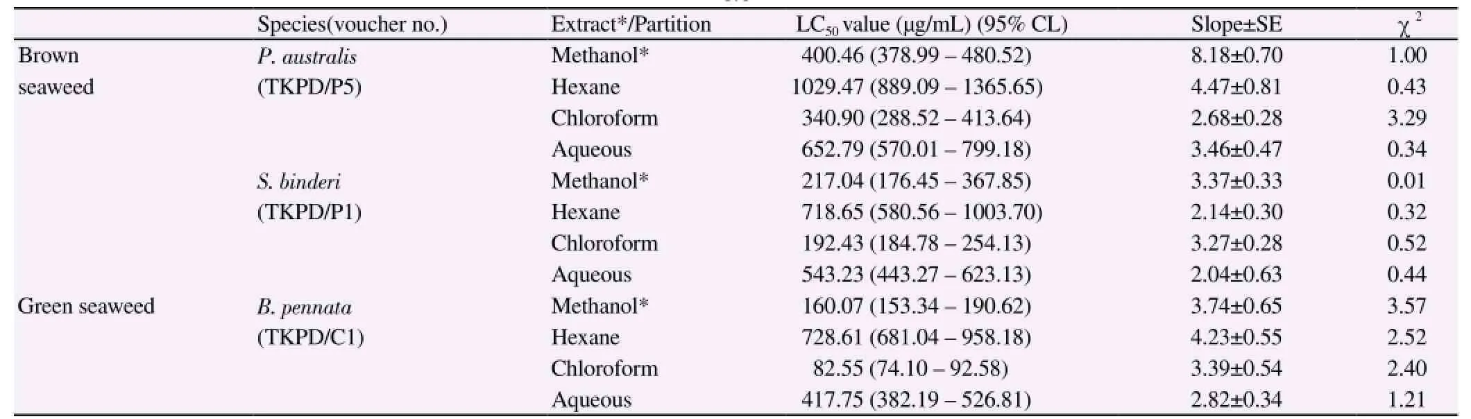
Table 1 Mosquito larvicidal activity of extracts and partition of seaweed towards Ae. aegypti.
3.4. Sublethal effect on the growth and development
The sublethal effects of methanol extracts of B. pennata, P. australis and S. binderi towards Ae. aegypti larvae were monitored until the death or emergence of all larvae (Table 2). Larvae treated with methanol extract of S. binderi showed the highest inhibition rate of adult emergence [(98.67 ± 0.21)%], longest larval period [(14.50 ± 0.71) d], longest median day of pupation (13 d) and lowest Harley Mean Index (0.58 ± 0.13) among all the treated larvae.
3.5. Phytochemical analysis
Phytochemical screening revealed the presence of alkaloids, saponins,steroids and terpenoids in all the seaweeds studied. Green seaweed B. pennata exhibited a significantly higher total polyphenol content[(20.37 ± 0.20) mg PGE/g extract] (P< 0.05) as compared to brown seaweeds, namely P. australis [(8.75 ± 0.54) mg PGE/g extract] and S. binderi [(10.90 ± 0.33) mg PGE/g extract].
4. Discussion
This is the first report of the mosquito larvicidal activity of S. binderi and P. australis. In our report, the larvicidal activity of chloroform partition of green seaweed B. pennata (LC50=82.55 μg/mL) is considered effective (LC50less than 100 mg/L)according to the classification of Thangam and Kathiresan[29], whilst the other 2 brown seaweeds tested - S. binderi and P. australis are considered as ineffective larvicide (LC50more than 200 μg/mL). However, other members of the Sargassum and Padina genus have been reported for their larvicidal activity towards mosquito and the activity reported is more effective compared to the activity in our report. For instance, Sargassum wightii has been reported to exhibit larvicidal action against the mosquito larvae of Ae. aegpyti and Culex quinquefasciatus[30]. Furthermore, ethyl acetate fraction of Sargassum swartzii has shown active larvicidal effect against mosquito larvae of Anopheles stephensi[31] and Anopheles sundaicus[32]. On the other hand,Padina tetrastromatica has also exhibited toxic effect against mosquito larvae of Ae. aegypti and Culex quinquefasciatus[30]. Wide difference in bioactivity between the individual species within the same genus can be due to geographical and ecological factors which affect the production of carbon-based bioactive secondary metabolites. Different compositions of chemical constituents in the seaweeds also may result in various degrees of bioactivity.
Besides causing death to mosquito larvae, the effect of intoxication also manifests through the aberration of structures. It is evident as Ae. aegypti larvae treated with extracts of natural products such as red seaweed Laurencia dendroidea and dried fruits of peppercorns have been reported to have darkening and shrunken cuticle of anal papillae after the treatment[7,33]. Similarly, the mosquito larvae treated with seaweed extract in our report were observed to exhibit the same manner of aberrations on the anal papillae. The deleterious effect on anal papillae interrupts the ion regulation of larvae and further causes the imbalance of homeostasis. Furthermore, the rupture of larval stigmal plate observed in the present report is suggested to cause destruction to the hydrophobe surface of stigma plate, causing water/medium to enter the tracheal trunk which harms the respiration system of the larvae. Similar action has been described in one of the study whereby kerosene entered the tracheal trunks from terminal spiracles and caused the finest capillaries to disappear from the tracheal system[34]. Interruption of the osmo-regulatory system (damaged anal papillae) and the spiracles of respiratory system (disrupted stigma plate) are suggested to contribute to the death of larvae.
The midgut of insect plays an important role in the secretion of digestive enzymes and absorption of nutrients[1]. Allelochemicals are proven to exert detrimental effect on the digestive epithelial cells and further decrease the survivability of the insect. For instance, mosquito larvae treated with plant extracts, namely Melia azedarach and Derris urucu have been reported to experience extensive damage on the midgut epithelium and peritrophic matrix[24,35]. These observations are in agreement with the findings of present study whereby the destruction and detachment of cells were observed from the midgut epithelium of the seaweed-treated larvae. The severe damage of midgut cells is suggested to disrupt the function of midgut, leading to the death of larvae.
Sublethal effects of the seaweed are proven to alter various stages of mosquito life cycle. In fact, delayed larval metamorphosis of the treated larvae is probably caused by the hormonal imbalance of post treatment. For instance, polyphenolic compounds of brown seaweed P. pavonica are suggested to prolong the life cycle intervals and pupation duration of mosquito larva Culex pipiens[8]. Reduction of total body protein and DNA content has also been observed in the red cotton bug Dysdercus cingulatus larvae treated with P. pavonica extract[9]. This is in line with the observation of the present study where methanol extract of B. pennata, P. australis and S. binderi exhibited their sublethal effects towards mosquito larvae Ae. aegypti, resulting in prolongation of larval duration and reduction of adult emergence.
Chemical composition of the seaweed plays important role in bioactivity. Previous studies have shown that brown seaweed P. australis contains fucoxanthin, aurantiamide acetate, mannitol D,palmitic acid and fatty acids[36]. Bioactive compounds derived from the Sargassum species such as meroterpenoids, phlorotanins, fucoidans,sterols and glycolipids have been reported to have a wide range of pharmacological properties[5]. Bryopsis species has been investigated for its pigment, sterol and fatty acid composition[37]. The phytochemical data obtained in the present study is in line with the previously reported data, where alkaloids, saponins, steroids and terpenoids are present in the seaweeds B. pennata, P. australis and S. binderi. This information serves as fundamental data for further investigation of the active constituents that are responsible for toxicity action in the assay. Comparative study of larvicidal extracts and compounds should be carried out to elucidate the possible mode of action in the future studies. The data obtained in this study provided information on the toxicity of 3 Malaysian seaweeds towards mosquito larvae Ae. aegypti. Chloroform fraction of green seaweed B. pennata showed the strongest larvicidal activity among all the samples tested, while methanol extract of brown seaweed S. binderi showed the strongest inhibition effect towards the development of larvae. Based on the results, it is evident that seaweed which is abundantly found in tropical country possesses potential as a mosquito larvicide. Further investigation aiming at ascertaining the active compounds and/or synergists, which are responsible for the larvicidal action, can be carried out to determine the use of seaweeds extracts and compounds in the implementation of efficient mosquito control strategies.
Conflict of interest statement
We declare that we have no conflict of interest.
Acknowledgments
Technical assistance and financial support from Faculty of Pharmacy of Universiti Kebangsaan Malaysia (Grant No.: Dana Modal Insan 13-00-09-018), Medical Entomology Unit of Institute for Medical Research, School of Biosciences of Taylor's University, and Faculty of Engineering and Science of Universiti Tunku Abdul Rahman are acknowledged. The authors are thankful to Miss Chiong Kai Shing for proofreading the manuscript.
[1] Christophers SR. Aedes aëgypti (L.) the yellow fever mosquito: its life history,bionomics and structure. New York: Cambridge University Press; 1960.
[2] World Health Organization. Dengue and severe dengue. Geneva: WHO; 2013.
[3] Kumar K, Singh PK, Tomar J, Baijal S. Dengue: epidemiology, prevention and pressing need for vaccine development. Asian Pac J Trop Med 2010;3(12): 997-1000.
[4] Ignacimuthu S, David BV. Ecofriendly insect pest management. Dehli: Elite Publishing House; 2009.
[5] Holdt SL, Kraan S. Bioactive compounds in seaweed: functional food applications and legislation. J Appl Phycol 2011; 23(3): 543-597.
[6] Yu KX, Jantan I, Ahmad R, Wong CL. The major bioactive components of seaweeds and their mosquitocidal potential. Parasitol Res 2014; 113(9): 3121-3141.
[7] Bianco EM, Pires L, Santos GK, Dutra KA, Reis TN, Vasconcelos ER, et al. Larvicidal activity of seaweeds from northeastern Brazil and of a halogenated sesquiterpene against the dengue mosquito Aedes aegypti. Ind Crop Prod 2013; 43: 270-275.
[8] Elbanna SM, Hegazi MM. Screening of some seaweeds species from South Sinai, Red Sea as potential bioinsecticides against mosquito larvae; Culex pipiens. Egypt Acad J Biol Sci 2011; 4(2): 21-30.
[9] Sahayaraj K, Kalidas S. Evaluation of nymphicidal and ovicidal effect of a seaweed, Padina pavonica (Linn.) (Phaeophyceae) on cotton pest, Dysdercus cingulatus (Fab.). Indian J Mar Sci 2011; 40(1): 125-129.
[10] Sharma OP. Algae. Series on diversity of microbes and cryptogams. New Dehli:Tata McGraw Hill; 2011.
[11] Usmanghani K, Shameel M, Sualeh M, Khan K, Mahmood Z. Antibacterial and antifungal activities of marine algae from Karachi seashore of Pakistan. Fitoterapia 1984; 55: 73-77.
[12] Puglisi MP, Engel S, Jensen PR, Fenical W. Antimicrobial activities of extracts from Indo-Pacific marine plants against marine pathogens and saprophytes. Mar Biol 2007; 150(4): 531-540.
[13] Freitas JC, Sakamoto MI, Caprara L. Cardiac effects induced by extract of the seaweed Bryopsis pennata (Chlorophyta, Caulerpales). Toxicon 1995;33(3): 301.
[14] Geraldino PJL, Liao LM, Boo SM. Morphological study of the marine algal genus Padina (Dictyotales, Phaeophyceae) from southern Philippines: 3 species new to Philippines. ALGAE-INCHON- 2005; 20(2): 99-112.
[15] Chiao-Wei C, Siew-Ling H, Ching-Lee W. Antibacterial activity of Sargassum polycystum C. Agardh and Padina australis Hauck (Phaeophyceae). Afric J Biotechnol 2013; 10(64): 14125-14131.
[16] Hongayo MC, Larino RC, Malingin DL. Antibacterial and antioxidant effects of brown alga Padina australis Hauck crude extract. IAMURE Int J Sci Clin Lab 2012; 2(1). DOI: http://dx.doi.org/10.7718/iamure.ijscl.v2i1.388.
[17] Jaswir I, Noviendri D, Salleh HM, Taher M, Miyashita K. Isolation of fucoxanthin and fatty acids analysis of Padina australis and cytotoxic effect of fucoxanthin on human lung cancer (H1299) cell lines. Afric J Biotechnol 2013; 10(81): 18855-18862.
[18] Ara J, Sultana V, Ehteshamul-Haque S, Qasim R, Ahmad VU. Cytotoxic activity of marine macro-algae on Artemia salina (Brine shrimp). Phytother Res 1999; 13(4): 304-307.
[19] Liu L, Heinrich M, Myers S, Dworjanyn SA. Towards a better understanding of medicinal uses of the brown seaweed Sargassum in Traditional Chinese Medicine: A phytochemical and pharmacological review. J Ethnopharmacol 2012; 142(3): 591-619.
[20] Sheikh TZB, Yong CL, Lian MS. In vitro antioxidant activity of the hexane and methanolic extracts of Sargassum baccularia and Cladophora patentiramea. J Appl Sci 2009; 9(13): 2490-2493.
[21] Jones WP, Kinghorn AD. Extraction of plant secondary metabolites. In:Sarker SD, Latif Z, Gray AI. (eds.) Natural products isolation. New York:Humana Press; 2005, p. 323-351.
[22] World Health Organization. Guidelines for laboratory and field testing of mosquito larvicides. Geneva: WHO; 2005.
[23] Neves Filho R, da Silva CA, da Silva C, Brustein VP, Dos Santos F. Improved microwave mediated synthesis of 3-(3-aryl-1, 2, 4-oxadiazol-5-yl) propionic acids and their larvicidal and fungal growth inhibitory properties. Chem Pharm Bull (Tokyo) 2009; 57(8): 819-825.
[24] Al-Mehmadi RM, Al-Khalaf AA. Larvicidal and histological effects of Melia azedarach extract on Culex quinquefasciatus Say larvae (Diptera: Culicidae). J King Saud University - Sci 2010; 22(2): 77-85.
[25] Harley KLS. A note on the influence of a range of plant chemicals on the growth and survival of Aedes aegypti L. larvae. Can J Zoolog 1967; 45(6):1297-1300.
[26] Culvenor CCJ, Fitzgerald JS. A field method for alkaloid screening of plants. J Pharm Sci 1963; 52(3): 303-304.
[27] Said IM, Din LB, Samsudin M, Zakaria WZ, Yusoff NI, Suki U, et al. A photochemical survey of Ulu Kincin, Pahang, Malaysia. Malaysia Nature J 1990; 43: 260-266.
[28] Zhang Q, Zhang J, Shen J, Silva A, Dennis DA, Barrow CJ. A simple 96-well microplate method for estimation of total polyphenol content in seaweeds. J Appl Phycol 2006; 18(3-5): 445-450.
[29] Thangam TS, Kathiresan K. Marine plants for mosquito control. In:Proceedings of the Second International Conference on Urban Pests; 1996.
[30] Manilal A, Thajuddin N, Selvin J, Idhayadhulla A, Kumar RS, Sujith S. In vitro mosquito larvicidal activity of marine algae against the human vectors, Culex quinquefasciatus (Say) and Aedes aegypti (Linnaeus) (Diptera:Culicidae). Int J Zoolog Res 2011; 7(3): 272-278.
[31] Khanavi M, Toulabi PB, Abai MR, Sadati N, Hadjiakhoondi F,Hadjiakhoondi A, et al. Larvicidal activity of marine algae, Sargassum swartzii and Chondria dasyphylla against malaria vector Anopheles stephensi. J Vector Borne Dis 2011; 48(4): 241-244.
[32] Kumar KP, Murugan K, Kovendan K, Kumar AN, Hwang JS, Barnard DR. Combined effect of seaweed (Sargassum wightii) and Bacillus thuringiensis var. israelensis on the coastal mosquito, Anopheles sundaicus, in Tamil Nadu,India. Science Asia 2012; 38(2012): 141-146.
[33] Kumar S, Warikoo R, Wahab N. Larvicidal potential of ethanolic extracts of dried fruits of three species of peppercorns against different instars of an Indian strain of dengue fever mosquito, Aedes aegypti L.(Diptera: Culicidae). Parasitol Res 2010; 107(4): 901-907.
[34] Wigglesworth V. A theory of tracheal respiration in insects. Proceedings of the Royal Society of London. Series B, Containing Papers of a Biological Character. 1930, p. 229-250.
[35] Gusmão DS, Páscoa V, Mathias L, Vieira IJC, Braz-Filho R, Lemos FJA. Derris (Lonchocarpus) urucu (Leguminosae) extract modifies the peritrophic matrix structure of Aedes aegypti (Diptera: Culicidae). Memórias do Instituto Oswaldo Cruz 2002; 97(3): 371-375.
[36] Kamenarska Z, Gasic M, Zlatovic M, Rasovic A, Sladic D, Kljajic Z, et al. Chemical composition of the brown alga Padina pavonia (L.) Gaill. from the Adriatic Sea. Bot Mar 2002; 45(4): 339-345.
[37] Siddiqui S, Usmanghani K, Shameel M. Sterol and fatty acid compositions of a marine alga Bryopsis pennata (Bryopsidophyceae, Chlorophyta). Pakistan J Pharm Sci 1994; 7(1): 73-82.
15 September 2015
Ibrahim Jantan, Drug and Herbal Research Centre, Faculty of Pharmacy, Universiti Kebangsaan Malaysia, 50300 Kuala Lumpur, Malaysia.
Tel: +603 92897315
Fax: +603 26983271
E-mail: profibj@gmail.com
Foundation project: This project was supported by Faculty of Pharmacy of Universiti Kebangsaan Malaysia (Grant No.: Dana Modal Insan 13-00-09-018).
 Asian Pacific Journal of Tropical Medicine2015年12期
- Asian Pacific Journal of Tropical Medicine的其它文章
- Immunomodulatory effect of garlic oil extract on Schistosoma mansoni infected mice
- Human ocular dirofilariasis due to Dirofilaria repens in Sri Lanka
- Childhood brucellosis: Review of 317 cases
- Effect of cyclophosphamide on fungal infection in SLE mice detected by fluorescent quantitative PCR
- Therapeutic effect of okra extract on gestational diabetes mellitus rats induced by streptozotocin
- Effect of low intensity pulsed ultrasound on expression of TIMP-2 in serum and expression of mmp-13 in articular cartilage of rabbits with knee osteoarthritis