Lanthanide(La,Tb,Dy)Complexes with Quinolinyloxy Acetamide Ligand:Crystal Structures and Fluorescence Properties
2016-04-05 08:11MAOPanDongYANLingLingWuWeiNaLIUMinQiZHOULiHuaFuSiLian
无机化学学报 2016年6期
MAO Pan-DongYAN Ling-Ling*,Wu Wei-Na*,LIU Min-QiZHOU Li-HuaFu Si-Lian
(1Department of Physics and Chemistry,Henan Polytechnic University,Jiaozuo,Henan 454000,China)
(2School of Materials Science and Engineering,Henan Polytechnic University,Jiaozuo,Henan 454000,China)
Lanthanide(La,Tb,Dy)Complexes with Quinolinyloxy Acetamide Ligand:Crystal Structures and Fluorescence Properties
MAO Pan-Dong1YAN Ling-Ling*,1Wu Wei-Na*,1LIU Min-Qi2ZHOU Li-Hua2Fu Si-Lian2
(1Department of Physics and Chemistry,Henan Polytechnic University,Jiaozuo,Henan 454000,China)
(2School of Materials Science and Engineering,Henan Polytechnic University,Jiaozuo,Henan 454000,China)
Three lanthanide(Ⅱ)complexes,[LaL2(NO3)3]·CH3CN(1),[Ln(L)(NO3)3(H2O)](Ln=Tb(2)and Dy(3)) based on L(L=N-phenyl-2-(5-chloro-quinolin-8-yloxy)acetamide)have been synthesized and characterized by elemental analyses,IR spectra and X-ray diffraction analyses.The results reveal that the La(Ⅱ)ion in complex 1 is surrounded by two tridentate amide ligands with NO2donor set and three bidentate nitrate anions,thus giving distorted icosahedron coordination geometry.By contrast,both complexes 2 and 3 are isostructral with those of the Pr,Nd,Sm,Eu,Gd and Er complexes bearing same ligand.In each complex,the center Ln(Ⅱ)ion with bicapped square antiprism coordination geometry is coordinated by one tridentate L,three bidentate nitrate anions and one water molecule.In addition,in solid state,complexes 2 and 3 could exhibit strong fluorescence emission in the visible region.CCDC:1438578,1;1438579,2;1438580,3.
amide type ligand;quinoline;lanthanide complex;fluorescence;crystal structure
The amide open chain ligands are a well suited type of antenna for lanthanide(Ⅱ)(Ln)ions,because they could shield the encapsulated lanthanide ion from interaction with the surroundings effectively,and thus to achieve strong fluorescent emission of metal ions[1-7].Up to now,a large amount of Eu(Ⅱ)and Tb(Ⅱ) complexes bearing such type of ligands have been widelyinvestigatedprimarilyduetotheirgoodluminescent properties[8-9].Recently,our previous work has demonstrated that the Sm(Ⅱ)and Eu(Ⅱ)complexes withN-phenyl-2-(5-chloro-quinolin-8-yloxl)acetamide exhibit the characteristic emission of the Eu(Ⅱ)and Sm(Ⅱ)ions,respectively[10-11].As a continuation of our research,we report here the structures of the ligands another three Ln(Ⅱ)complexes(Ln=La,Tb and Dy), together with their fluorescence properties in solid state.
1 Experimental
1.1 Materials and measurements
Solvents and starting materials for synthesis were purchasedcommerciallyandusedasreceived. Elemental analysis was carried out on an Elemental Vario EL analyzer.The IR spectra(ν=4 000~400 cm-1) were determined by the KBr pressed disc method on a Bruker V70 FTIR spectrophotometer.The UV spectra wererecordedonaPurkinjeGeneralTU-1800 spectrophotometer.Fluorescencespectrawere determinedonaVarianCARYEclipse spectrophotometer,andinthemeasurementsof emission and excitation spectra the pass width is 5 nm.
1.2 Preparations of complexes 1~3
The title Ln(Ⅱ)complexes weresynthesized according to the literature method[10-11].
1:Colorlessblocks.Anal.Calcd.for C36H29Cl2N8O13La(%):C,43.61;H,2.95;N,11.30. Found(%):C,43.33;H,3.09;N,11.42.FTIR(cm-1): ν(C=O)1 670,ν(C=N)1 582,ν(Ar-O-C)1 174,ν1(NO3)1 500,ν4(NO3)1 283.
2:Colorlessblocks.Anal.Calcd.for C17H15ClN5O12Tb(%):C,30.22;H,2.24;N,10.36. Found(%):C,30.20;H,2.16;N,10.43.FTIR(cm-1): ν(O-H)3403,ν(C=O)1 660,ν(C=N)1 571,ν(Ar-O-C) 1 172,ν1(NO3)1 505,ν4(NO3)1 293,ρ(O-H)879.
3:Colorlessblocks.Anal.Calcd.for C17H15ClN5O12Dy(%):C,30.06;H,2.23;N,10.31. Found(%):C,30.19;H,1.96;N,10.22.FTIR(cm-1): ν(O-H)3 402,ν(C=O)1 664,ν(C=N)1 573,ν(Ar-OC)1168,ν1(NO3)1 503,ν4(NO3)1 289,ρ(O-H)877. 1.3.1X-ray crystallography
The X-ray diffraction measurement for complexes 1~3 were performed on a Bruker SMART APEXⅡCCDdiffractometerequippedwithagraphite monochromatized Mo Kα radiation(λ=0.071 073 nm) by using φ-ω scan mode.Semi-empirical absorption correction was applied to the intensity data using the SADABS program[12].The structures were solved by direct methods and refined by fullmatrixleast-square on F2using the SHELXTL-97 program[13].All nonhydrogen atoms were refined anisotropically.The H atoms for water molecules are located from difference Fourier map and refined with restraints in bond length and thermal parameters.All the other H atoms were positioned geometrically and refined using a riding model.SQUEEZE procedure was applied to deal with the crystal solvent molecules of the complex 1.Details ofthecrystalparameters,datacollectionand refinements for complexes 1~3 are summarized in Table 1.
CCDC:1438578,1;1438579,2;1438580,3.
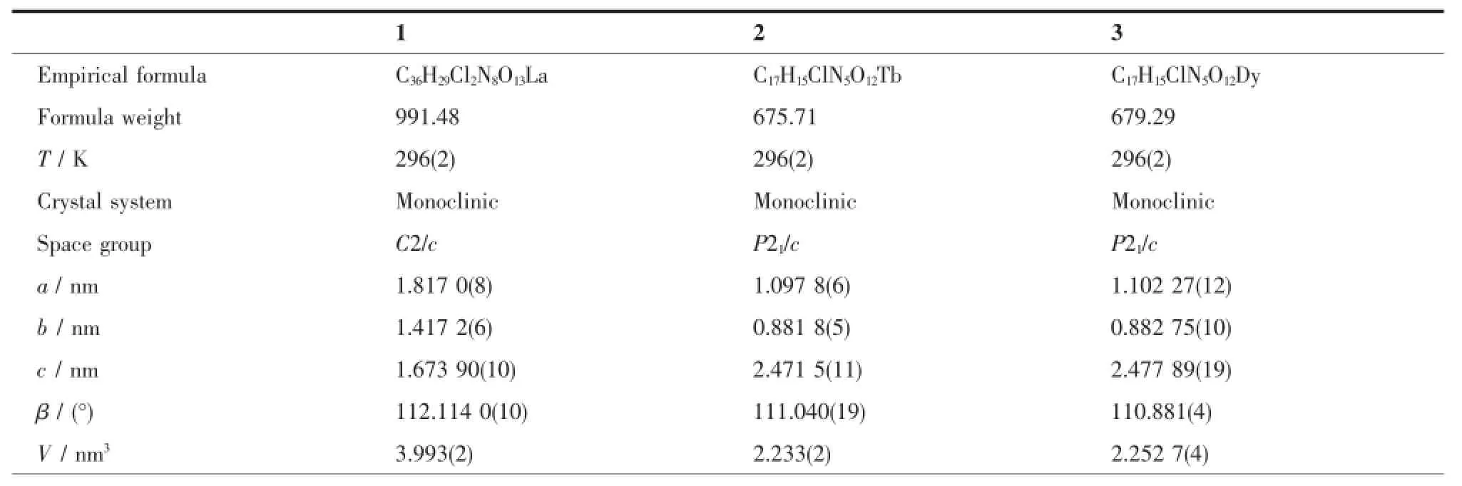
Table 1Selected crystallographic data for complexes 1~3
Continued Table 1
2 Results and discussion
2.1 Crystal structure of the complexes
Generally,N-phenyl-2-(5-chloro-quinolin-8-yloxy) acetamide ligand coordinates to Ln(Ⅱ)ions with molar ratio of ligand and metal being 1∶1[10-11].By contrast,it forms stable 2∶1 type complex with La(Ⅱ)ion.As shown in Fig.1a,La(Ⅱ)ion in complex 1 is surrounded by two tridentate amide ligands with NO2donor set andthreebidentatenitrateanions,thusgiving distorted icosahedron coordination geometry(Fig.1d). The La-O/N bond lengths are in the range of 0.254 8(4)~0.283 1(4)nm(Table 2),comparable to the La(Ⅱ) complexes with similar donor sets[6-7].The crystal structure of 1 is similar as that of[LaLa2(NO3)3]and [LaLb2(NO3)3]·H2O in our previous work[6-7](La=N,N-diphenyl-2-(quinolin-8-yloxy)acetamide,Lb=N-(naphthalene-1-yl)-2-(quinolin-8-yloxy)acetamide).In the crystal,pairs of intermolecular N-H…O hydrogen bonds between the amide N atoms and adjacent nitrate O atoms link the complexes into chains along c axis(Fig.1e).
Complexes 2(Fig.1b)and 3(Fig.1c)are isostructural and crystallize in the monoclinic,space group P21/c,same as the Pr(Ⅱ),Nd(Ⅱ),Sm(Ⅱ),Eu(Ⅱ),Gd(Ⅱ) and Er(Ⅱ)complexes with same ligand[10-11].The center Ln(Ⅱ)ion with bicapped square antiprism coordination geometry is coordinated by one tridentate L,three bidentate nitrate anions and one water molecule. Similarly,inthecrystalofeachcomplex, intermolecular O-H…O(O12-H12A…O4iiiand O12-H12B…O8iv)and N-H…O(N2-H2A...O3v)hydrogen bonds link the complex into a 2D supramolecular network(Fig.1f,Symmetry codes:iii-x+1,y+1/2,-z+1/2;iv-x+1,y-1/2,-z+1/2;v-x+1,-y,-z.

Table 2Selected bond lengths(nm)in complexes 1~3
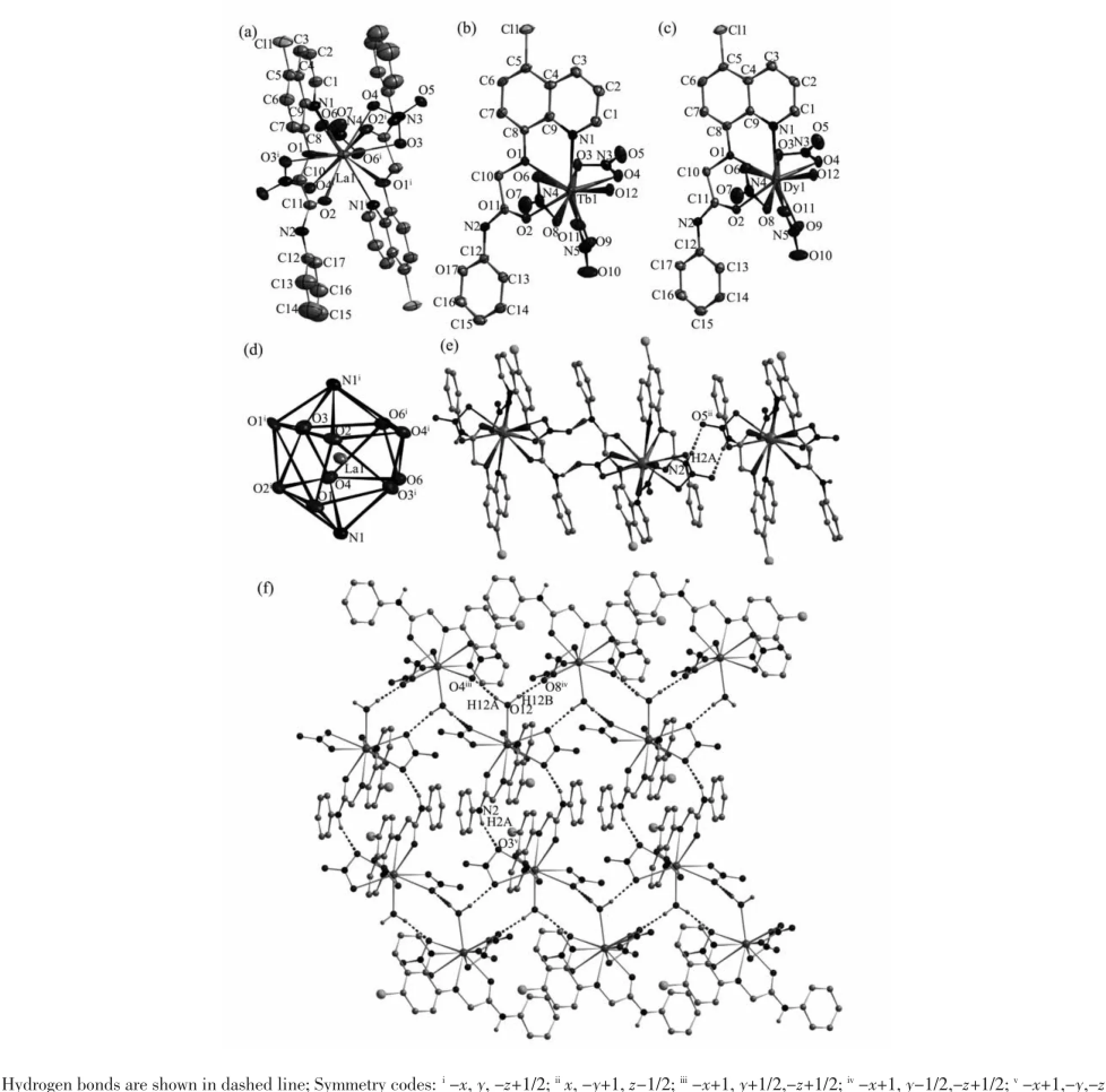
Fig.1Molecular structures of complexes 1~3(a~c)shown with 30%probability displacement ellipsoids;(d)Coordination geometry of the center La(Ⅱ)ion in complex 1,atoms shown with 30%probability displacement ellipsoids;(e)Extended chain-like structure along c axis in complex 1 formed by intermolecular N-H…O hydrogen bonds;(f)Extended 2D supramolecular structure in complex 2 formed by intermolecular hydrogen bonds.
Table 3Hydrogen bonds information in complexes 1~3
Continued Table 3
2.2 IR spectra
The spectral regions for all the complexes are moreorlesssimilarduetothesimilarityin coordination modes of the ligand with the metal center.The free ligand L exhibit three absorption bands at 1 682,1 598 and 1 241 cm-1,assigned to ν(C=O),ν(C=N)and ν(C-O-C),respectively[10]. However,in the complexes,such bands shift evidently to lower frequency,indicating that the oxygen atoms of the carbonyl group,quinoline nitrogen atoms and ethereal oxygen atoms take part in coordination to the central Ln(Ⅱ)ion[3-5].Additionally,the general pattern oftheIRspectroscopysupportsthenormal coordination of the bidentate nitrate group[6-7].It is in accordance with the result of the crystal structure study.
2.4 UV spectra
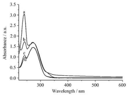
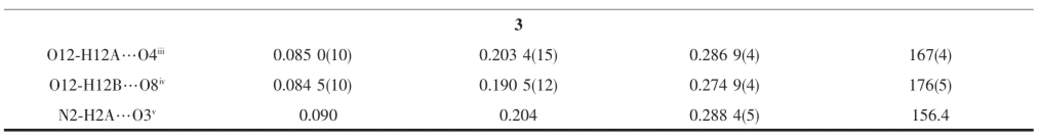
Fig.2UV spectra of the ligand L(a),1(b),2(c)and 3 (d)in the acetone solution at room temperature
The UV spectra of the ligand L and complexes 1~3 in acetone solution(1×10⁻⁵mol·L-1)were measured at room temperature(Fig.2).The spectra of L features two main bands located around 240(ε= 304 366 L·mol-1·cm-1)and 272 nm(ε=169 788 L·mol-1·cm-1).The bands could be assigned to characteristic π-π*transitions centered on benzene and quinoline units,respectively[6].Similar bands can be observed in the spectra of 1(241 nm,ε=174 235 L·mol-1· cm-1;273 nm,ε=168 132 L·mol-1·cm-1),2(241 nm, ε=107 529 L·mol-1·cm-1;273 nm,ε=146 195 L·mol-1·cm-1)and 3(242 nm,ε=98 529 L·mol-1·cm-1;273 nm,ε=145 803 L·mol-1·cm-1).However,the hyperchromicities indicate that the ligand L takes part in the coordination in all complexes.
Fig.3Fluorescence emission spectra of the ligand L(a), 1(b),2(c)and 3(d)in solid state at room temperature
2.4 Fluorescence spectra
The fluorescence spectra of the ligand L and complexes 1~3 have been studied in solid state at room temperature.The results show that the emission spectra of the ligand L and complex 1 exhibit similar emission peak at about 400 nm when excited at 310 nm.It also can be seen that the emission intensity of the complex 1 is much higher than that of L,which may be explained from two aspects:first,in the complex 1,the molar ratio of the ligand and La(Ⅱ) ion is 2∶1;second,the coordination of La(Ⅱ)ion may enhancetheπ→π*electrontransitionofthe acetamide ligand[6].By contrast,complexes 2 and 3 show quite different peak at about 500 nm.Thebehavior of Tb(Ⅱ)and Dy(Ⅱ)ions coordinated to the ligand is regarded as that of emissive species resulted from a CHEF effect(chelation enhancement of the fluorescence emission)[7].
[1]Yan Z Z,Hou N,Wang C M.Spectrochim.Acta A,2015, 137:1265-1269
[2]Song X Q,Xing D Y,Lei Y K,et al.Inorg.Chim.Acta, 2013,404:113-122
[3]Wu W N,Tang N,Yan L.Spectrochim.Acta A,2008,71: 1461-1465
[4]Wu W N,Cheng F X,Yan L,et al.J.Coord.Chem., 2008,61:2207-2215
[5]Wu W N,Tang N,Yan L.J.Fluoresc.,2008,18:101-107
[6]ZHANG Zhao-Po(张照坡),WANG Yuan(王元),WU Wei-Na (吴伟娜),et al.Chinese J.Inorg.Chem.(无机化学学报), 2013,29:2239-2244
[7]YE Xing-Pei(叶行培),CAI Hong-Xin(蔡红新),WU Wei-Na (吴伟娜),et al.Chinese J.Struct.Chem.(结构化学), 2014,33:1057-1062
[8]Binnemans K.Coord.Chem.Rev.,2015,295:1-45
[9]Bünzli J-C G.Coord.Chem.Rev.,2015,293-294:19-47
[10]CAI Hong-Xin(蔡红新).Thesis for the Master of Henan Polytechnic University(河南理工大学硕士论文).2014.
[11]MAO Pan-Dong(毛盼东),CHEN Liang(陈亮),WU Wei-Na (吴伟娜),et al.Chinese J.Inorg.Chem.(无机化学学报), 2016,32:336-342
[12]Sheldrick G M.SADABS,University of Göttingen,Germany, 1996.
[13]Sheldrick G M.SHELX-97,Program for the Solution and the Refinement of Crystal Structures,University of Göttingen, Germany,1997.
镧系元素(La、Tb、Dy)的喹啉氧基乙酰胺配合物的结构及荧光性质
毛盼东1闫玲玲*,1吴伟娜*,1刘珉琦2周利华2伏思连2
(1河南理工大学物理化学学院,焦作454000)
(2河南理工大学材料科学与工程学院,焦作454000)
合成并通过单晶衍射表征了3个稀土配合物[LaL2(NO3)3]·CH3CN(1),[Ln(L)(NO3)3(H2O)](Ln=Tb(2),Dy(3),L=N-苯基-2-(5-氯-8-喹啉氧基)乙酰胺)。在配合物1中,十二配位的La(Ⅱ)离子采取扭曲的二十面体配位构型,分别与来自2个酰胺配体L的4个氧原子和2个氮原子,及3个双齿配位硝酸根配位。配合物2和3的结构与拥有相同有机配体的Pr、Nd、Sm、Eu、Gd和Er配合物同构。在每个配合物中,十配位的稀土离子与来自1个配体L的2个氧原子和1个氮原子,3个双齿配位硝酸根和1个水分子配位,拥有扭曲的双帽四方反棱柱配位构型。固态配合物2和3在可见区发射强荧光。
酰胺配体;喹啉;稀土配合物;荧光;晶体结构
O614.33+1;O614.341;O614.342
A
1001-4861(2016)06-1095-06
2015-12-04。收修改稿日期:2016-03-17。
10.11862/CJIC.2016.130
国家自然科学基金(No.21001040)和河南省教育厅自然科学基金(No.12B150011,14B150029)资助项目。
*通信联系人。E-mail:yll@hpu.edu.cn;wuwn08@hpu.edu.cn;会员登记号:S06N6704M1112。
猜你喜欢
四川轻化工大学学报(自然科学版)(2021年1期)2021-06-09
当代陕西(2019年6期)2019-04-17
铜仁学院学报(2018年6期)2018-07-05
中成药(2017年7期)2017-11-22
国外医药(抗生素分册)(2016年1期)2016-07-10
中国洗涤用品工业(2016年2期)2016-02-28
中国塑料(2015年2期)2015-10-14
云南中医学院学报(2015年2期)2015-07-31
外语学刊(2014年3期)2014-12-03
原子与分子物理学报(2014年4期)2014-02-28