Comparison of intraocular lens power calculation using a standard ultrasonic biometer and a new optical biometer
2016-05-17 03:32FarukKayaIbrahimKoakAliAydinHakanBayboraKorayKaradayi
国际眼科杂志 2016年5期
Faruk Kaya, Ibrahim Koçak,2, Ali Aydin, Hakan Baybora, Koray Karadayi
1Department of Ophthalmology, School of Medicine, Medipol University, cilar, Istanbul 34214, Turkey
2Department of Ophthalmology, Nisa Hospital, Fatih Cad, Bahçelievler, Istanbul 34196, Turkey
3Department of Ophthalmology, Near East University, Nicosia, Mersin 33000, Turkey
Correspondence to:Ibrahim Koçak. Nisa Hospital, Fatih Cad. Bahçelievler, Istanbul 34196, Turkey. ibrahimkocak@msn.com
Received: 2015-09-29 Accepted: 2016-02-22
比较标准超声波测量仪和新型光学生物测量仪计算人工晶状体屈光度的准确性
Faruk Kaya1, Ibrahim Koçak1,2, Ali Aydin1, Hakan Baybora1, Koray Karadayi3
(作者单位:1土耳其,伊斯坦布尔大学,医学院,眼科;2土耳其,伊斯坦布尔 34196,Bahçelievler,Fatih Cad,Nisa医院,眼科;3土耳其,梅尔辛33000,尼科西亚,Near East大学,眼科)
Comparison of intraocular lens power calculation using a standard ultrasonic biometer and a new optical biometer
Faruk Kaya1, Ibrahim Koçak1,2, Ali Aydin1, Hakan Baybora1, Koray Karadayi3

2Department of Ophthalmology, Nisa Hospital, Fatih Cad, Bahçelievler, Istanbul 34196, Turkey
3Department of Ophthalmology, Near East University, Nicosia, Mersin 33000, Turkey
Correspondence to:Ibrahim Koçak. Nisa Hospital, Fatih Cad. Bahçelievler, Istanbul 34196, Turkey. ibrahimkocak@msn.com
Received: 2015-09-29Accepted: 2016-02-22
比较标准超声波测量仪和新型光学生物测量仪计算人工晶状体屈光度的准确性
Faruk Kaya1, Ibrahim Koçak1,2, Ali Aydin1, Hakan Baybora1, Koray Karadayi3

Abstract
•AIM:To compare the intraocular lens (IOL) power calculations and refractive outcomes obtained with a new optical biometer and standard ultrasonic biometer in phacoemulsification surgery.
•METHODS:Thirty-seven eyes of 37 cataract patients who underwent phacoemulsification with IOL implantation were included in this prospective comparative study. The same operator performed biometer measurements in eyes with cataract using a new optical biometer (Aladdin) and a standard ultrasonic biometer (Sonomed AB 5500). Biometric parameters;axial length (AL), keratometric (K) readings, anterior chamber depth (ACD) and IOL power obtained by two devices were recorded. Postoperative actual refractive errors and errors predicted by two devices according to SRK/T formula were analyzed. The mean estimation error (EE), mean absolute estimation error (AEE) and the biometric parameters obtained by two biometers were compared.
•RESULTS:The AL measured by Aladdin (23.45±0.73 mm) was significantly longer than AL by ultrasonic biometer (23.2±0.75 mm) (P=0.01). The mean EE and AEE values obtained by Aladdin were significantly smaller than the values by ultrasonic biometer (P=0.0006 and 0.03 respectively). The higher percentage of eyes within ±0.5 and ±1.00 D of target refraction was also found by using Aladdin (67% and 97%).
•CONCLUSION:The Aladdin optical biometer showed better accuracy and yielded better refractive outcomes compared with ultrasonic biometer.
KEYWORDS:•Aladdin optical biometer;ultrasonic biometer;axial length;intraocular lens power calculation;postoperative refractive error
Citation:Kaya F;Koçak I;Aydin A;Baybora H;Karadayi K. Comparison of intraocular lens power calculation using a standard ultrasonic biometer and a new optical biometer.GuojiYankeZazhi(IntEyeSci) 2016;16(5):807-810
INTRODUCTION
Correct intraocular lens power calculation is important in cataract surgery for successful postoperative refractive outcomes. In cataract surgery ocular biometer is essential for intraocular lens (IOL) power calculation. Chosen IOL formula defines the result of IOL power. For ultrasonic biometers, most of IOL formulas require keratometer (K) values, and axial length (AL)[1-2]. AL measurement is important for predicting the accuracy of the IOL calculation and it is source of the largest proportion of inaccurate measurements. Immersion A-scan technique is presumably more accurate than contact A- scan ultrasonography as it needs no indentation of the cornea[3].
Optical biometer devices can measure some other variables. They are fast, non invasive and independent from technician experience. The built in software in these devices gives us more accurate IOL power calculation and multiple choices of IOL formulas[4-5]. Several new optical biometers can also perform ocular biometry and IOL power calculation as accurate as the first standard optical biometer such as IOL Master (Carl Zeiss Meditec AG, Jena, Germany)[6]. The Aladdin (Topcon, Tokyo, Japan) is one of the most recently released optical biometer. The device is an optical low coherence interferometer (OLCI) which can measure six variables;K value, AL, anterior chamber depth (ACD), white to white (WTW) diameter, pupil size, corneal topography. AL is measured using OLCI with 820 nm super-luminescent diode. ACD is measured using light emitting diode (LED) making horizontal slit projections across the anterior chamber, similarly to the IOL Master. Corneal topography and keratometer measurements are based on 24 Placido disk reflection[7]. The IOL power is calculated by five different formulas built into the device.
The purpose of this study is to evaluate and compare the difference of variables K value,AL, ACD and IOL power measured using ultrasonic biometer and optical biometer.
SUBJECTS AND METHODS
Subjects enrolled in this prospective comparative study were patients with cataract who were candidates for phacoemulsification with IOL implantation surgery at Ophthalmology Department of Nisa Hospital, Istanbul, Turkey between Nov. 17thand Dec. 29th, 2014. The study project was approved by Institutional Ethical Board of Istanbul Medipol University. All research and data collection adhered to the tenets of the Declaration of Helsinki. The study was explained to each patient and written informed consent was obtained.
Preoperatively all patients had a complete examination including manifest refraction,best-corrected visual acuity (BCVA) testing, intraocular pressure (IOP) measurements with applanation tonometer, slit lamp, and dilated fundus examinations. Ocular biometer was first performed by Aladdin optical biometer followed by ultrasonic biometer (Sonomed AB 5500, Lake Success, NY, USA). Patients with good quality Aladdin biometer measurements were included in the study.
Exclusion criteria were history of traumatic or uveitic cataracts, previous intraocular or corneal surgery (e.g. refractive surgery or glaucoma surgery), intraoperative complications (e.g. anterior or posterior capsule ruptures, vitreous loss or zonule dehiscence), or postoperative complications (e.g. tilted or decentered IOL).
Each patient underwent biometric measurement on Aladdin optical biometer by the same examiner (Kaya F). After carefully positioning of patient, Aladdin biometer was focused as determined by a clear view of anterior segment and the display of a ‘green eye’ quality control image. Six AL measurements, three K values and three ACD readings were obtained. The goal in IOL power selection was a value that would provide a postoperative refraction nearest to plano, staying on the side of myopia. The power selection of implanted IOLs was determined based on the SRK/T formula. Another experienced ophthalmologist (Koçak I) performed applanation ultrasound biometer after application of one drop topical anesthetic (proparacaine 0.5%). K values requested for ultrasonic biometer were measured using an autorefractometer (Topcon KR 8800, Tokyo, Japan). Mean of five AL and K measurements was used to calculate IOL power based on the SRK/T formula.
All phacoemulsification and IOL implantations were performed under topical anesthesia by one of three experienced surgeons (Kaya F, Koçak I, Aydin A). A standard phacoemulsification was performed through a 2.8 mm temporal clear corneal incision. The monoblock foldable hydrophobic acrylic IOL (Focus force F260, A-constant of 118.4, Zarracom, Turkey) was inserted into the capsular bag using an injector system.
By the end of first postoperative month, ophthalmological examination was carried out for all patients. Postoperative objective refractive error was measured by using autorefractometer. Uncorrected visual acuity (UCVA) and BCVA were also evaluated.
The estimation error (EE) was defined as the difference between the postoperative objective refractive error (spherical equivalent) and the preoperatively predicted refractive errors by two different biometers using SRK/T formulas for the power of IOL implanted. The absolute estimation error (AEE) was defined as the absolute value of the EE. For example, if postoperative objective error is -0.75D and preoperative predicted error is -0.12D, the EE is calculated as -0.75-(-0.12) =-0.63D. The AEE (the absolute value of EE) is [-0.63]=0.63D.
Paired studentt-test and Wilcoxon test are used for comparison of parameters obtained from two devices.We used studentt-test to compare K value, AL, ACD, EE and AEE as the groups had normal distribution. We used Wilcoxon test to compare percentages of eyes within target refraction for two devices as the groups did not have normal distribution. Statistical analysis was performed using the Statistical Package for Social Sciences (SPSS) version 12.0 (SPSS Inc., Chicago, Illinois, USA).P<0.05 were considered to be statistically significant.
Table 1Clinical characteristics of patients

ParameterMean±SDRangeAge(a)67.9±7.647to81Sex,n(%) M15(40%)- F22(60%)-Laterality,n(%) R19(51%)- L18(49%)-ImplantedIOLpower(D)21.8±2.0514.5to25
Table 2Comparison of biometric parameters measured by two devices

ParametersOpticalbiometerUltrasonicbiometer(Kvaluebyautoref.)aPK(D),Mean±SD(Range)43.3±1.4(40.4to46.7)43.4±1.5(40.2to47)P=0.78AL(mm),Mean±SD(Range)23.4±0.7(21.7to24.7)23.2±0.7(21.7to24.6)P=0.01ACD(mm),Mean±SD(Range)3.1±0.2(2.4to3.6)3.0±0.3(2.4to3.6)P=0.08
K:Keratometry;AL:Axial length;ACD:Anterior chamber depth;aStudentt-test.
Table 3The results concerning the estimation error, absolute estimation error, and percentages of eyes within target refraction for two devices
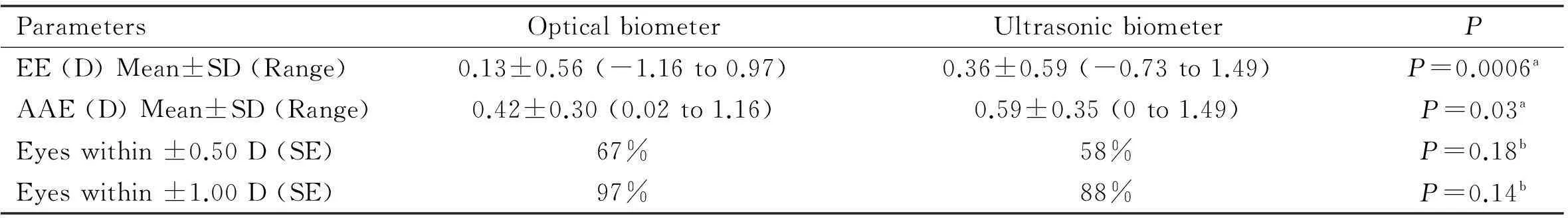
ParametersOpticalbiometerUltrasonicbiometerPEE(D)Mean±SD(Range)0.13±0.56(-1.16to0.97)0.36±0.59(-0.73to1.49)P=0.0006aAAE(D)Mean±SD(Range)0.42±0.30(0.02to1.16)0.59±0.35(0to1.49)P=0.03aEyeswithin±0.50D(SE)67%58%P=0.18bEyeswithin±1.00D(SE)97%88%P=0.14b
EE:Estimation error;AAE:Absolute estimation error;SE:Spherical equivalent;aStudentt-test;bWilcoxon test.
RESULTS
Forty eyes of 40 patients were included in the study. Three eyes were excluded due to the intra-operative complications such as posterior capsule rupture, vitreous loss and zonule dehiscence. Characteristics of patients are shown in Table 1.
Biometric parameters including K value, AL and ACD are shown on Table 2. The AL measured by Aladdin (23.4±0.7 mm) was significantly longer than AL by ultrasonic biometer (23.2±0.7mm) (P=0.01). The mean ACD measurement by Aladdin was also longer than the ACD by ultrasonic biometer, however the difference was not statistically significant (P=0.08). There was no statistically significant difference between two devices in measuring the mean K value (P=0.78).
The postoperative mean refractive error (spherical equivalent) was 0.13±0.66 D (range -1.50 to+1.25D). The results concerning the estimation error (EE), absolute estimation error (AEE), and percentages of eyes within target refraction for two devices are shown in Table 3. The mean EE obtained by using Aladdin was significantly smaller than EE by ultrasonic biometer (0.13±0.56 Dvs0.36±0.59 D) (P=0.0006). Also, the mean AEE obtained by using Aladdin was significantly smaller than AEE by ultrasonic biometer (0.42±0.30 Dvs0.59±0.35 D) (P=0.03). Aladdin optical biometer predicted more eyes with EE within ±0.50 and ±1.00 D of target refraction compared to ultrasonic biometer, however the differences were not statistically significant (P=0.18 andP=0.14).
DISCUSSION
Today’s cataract patients have greater expectations and request irreproachable results, especially about refractive subjects.IOL power calculation is essential for assessing the success of cataract surgery. Incorrect lens power calculation is the main cause of dissatisfaction and lens exchange in cataract surgery, especially in eyes with short or long axial lengths[8-11]. Ultrasound biometer is still used for IOL calculation;it is more favorable than optical biometer, especially in cases with dense cataract, media or corneal opacity and vitreous opacities. Optical biometer is a newer technology and has some advantages such as high accuracy, non contact and non invasive measurements, higher speed, higher patient comfort. As light has a very short wavelength compared to sound, the laser light has better resolution, because resolution improves as wavelength decreases. Therefore the accuracy of AL measured with ultrasound biometer is approximately 0.10-0.12 mm compared to 0.012 mm for measurement with optical biometer. Optical biometer is recommended to be used especially in cases those premium IOLs such as multifocal or toric IOLs will be used. But high cost of equipment, inability to measure dense cataracts, some corneal abnormalities and eyes with poor fixation are disadvantages of optical biometers[7].
Previous studies have compared two devices[3,12-21]. Fontesetal[3]found that both methods had high precision and reproducibility but there was a trend toward a subtle improvement in the prediction for postoperative refraction with optical biometer. Rajanetal[12]showed that optical biometer improved the predictive value for postoperative refraction in pseudophakic eyes. Kissetal[13], Packeretal[14], and Haigisetal[15]proved similar improvement in the prediction for postoperative refraction with optical biometer and US biometer.Bhattetal[16]showed similar results. Optical biometer was a beter predictor of postoperative refraction than US biometer, particularly within close ranges. In two study of Gantenbeinetal[17-18]and a study of Kutschan and Wiegand[19];the accuracy of postoperative refraction compared to the preoperative target was beter with US biometer compared to the optical biometer. In Gantenbeinetal’s[17-18]study optical biometer measured AL longer than US biometer and measured keratometry lower than Javal keratometer. Landers and Goggin[20]found that optical biometers predict more correct refractive outcomes compared to US biometer. In optical biometer, AL measurements were longer than US biometer, but they had similar results for ACD measurements. In another study, Nemethetal[21]showed that, AL and ACD was measured longer by optical biometer compared to US biometer.
Because of optical biometers’ high success in prediction of postoperative refraction, there is a widespread use of them for premium IOLs such as multifocal or toric IOLs. As we use commonly monofocal IOLs in our clinic, we wanted to evaluate and compared success of Aladdin and US biometer for monofocal IOLs.
In our study the Aladdin provided shorter ACD, but longer AL than US biometer. Difference in AL was statistically significant, but difference in ACD was not significant statistically. Most of previous studies also predicted longer ALs[17-18,20-21]. While US biometers use corneal epithelium and internal limiting membrane as reference points in measuring AL, optical biometers use the second principal plane of the cornea (0.05 mm deeper than the corneal apex) and photoreceptor layer (0.25 mm deeper than ILM). This may explain longer AL measurements with optical biometers. The power selection of implanted IOLs was determined based on the SRK/T formula, because it was one of the most common used formulas and it was found as one of the most successful formulas in determining postoperative refractive error[22]. Although there was no statistically significant difference, the Aladdin optical biometer obtained better refractive outcomes correlated with some previous studies[3,16,20]and predicted more eyes with EE within target diopters when compared US biometer. Also Aladdin optical biometer predicted smaller EE and AEE than ultrasonic biometer and the difference was statistically significant.
REFERENCES
1 Lee AC, QAzi MA, Pepose JS. Biometry and intraocular lens power calculation.CurrOpinOphthalmol2008;19(1):13-17
2 Joo J, Whang WJ, Oh TH, Kang KD, Kim HS, Moon JI. Accuracy of intraocular lens power calculation formulas in primary angle closure glaucoma.KoreanJOphthalmol2011;25(6):375-379
3 Fontes BM, Fontes BM, Castro E. Intraocular lens power calculation by measuring axial length with partial optical coherence and ultrasonic biometry.ArqBrasOftalmol2011;74(3):166-170
4 Wang JK, Chang SW. Optical biometry intraocular lens power calculation using different formulas in patients with different axial lengths.IntJOphthalmol2013;6(2):150-154
5 Rabsilber TM, Jepsen C, Auffarth GU, Holzer MP. Intraocular lens power calculation:clinical comparison of 2 optical biometry devices.JCataractRefractSurg2010;36(2):230-234
6 Chen YA, Hirnschall N, Findl O. Evaluation of 2 new optical biometry devices and comparison with the current gold standard biometer.JCataractRefractSurg2011;37(3):513-517
7 Mandal P, Berrow EJ, Naroo SA, Wolffsohn JS, Uthoff D, Holland D, Shah S. Validity and repeatability of the Aladdin ocular biometer.BrJOphthalmol2014;98(2):256-258
8 Jin GJ, Crandall AS, Jones JJ. Intraocular lens exchange due to incorrect lens power.Ophthalmology2007;114(3):417-424
9 Kiss B, Findl O, Menapace R, Wirtitsch M, Drexler W, Hitzenberger CK, Fercher AF. Biometry of cataractous eyes using partial coherence interferometry:clinical feasibility study of a commercial prototype I.JCataractRefractSurg2002;28(2):224-229
10 Wang L, Shirayama M, Ma XJ, Kohnen T, Koch DD. Optimizing intraocular lens power calculations in eyes with axial lengths above 25.0 mm.JCataractRefractSurg2011;37(11):2018-2027
11 Moschos MM, Chatziralli IP, Koutsandrea C. Intraocular lens power calculation in eyes with short axial length.IndianJOphthalmol2014;62(6):692-694
12 Rajan MS, Keilhorn I and Bell JA. Partial coherence laser interferometry vs conventional ultrasound biometry in intraocular lens power calculations.Eye(Lond) 2002;16(5):552-556
13 Kiss B, Findl O, Menapace R, Wirtitsch M, Petternel V, Drexler W, Rainer G, Georgopoulos M, Hitzenberger CK, Fercher AF. Refractive outcome of cataract surgery using partial coherence interferometry and ultrasound biometry:clinical feasibility study of a commercial prototype II.JCataractRefractSurg2002;28(2):230-234
14 Packer M, Fine IH, Hoffman RS, Coffman PG, Brown LK. Immersion A-scan compared with partial coherence interferometry:outcomes analysis.JCataractRefractSurg2002;28(2):239-242
15 Haigis W, Lege B, Miller N, Schneider B. Comparison of immersion ultrasound biometry and partial coherence interferometry for intraocular lens calculation according to Haigis.GraefesArchClinExpOphthalmol2000;238(9):765-773
16 Bhatt AB, Schefler AC, Feuer WJ, Yoo SH, Murray TG. Comparison of predictions made by the intraocular lens master and ultrasound biometry.ArchOphthalmol2008;126(7):929-933
17 Gantenbein CP, Ruprecht KW. Comparison between optical and acoustical biometry.JFrOphtalmol2004;27(10):1121-1127
18 Gantenbein C, Lang HM, Ruprecht KW, Georg T. First steps with the Zeiss IOLMaster:A comparison between acoustic contact biometry and non-contact optical biometry.KlinMonblAugenheilkd2003;220(5):309-314
19 Kutschan A, Wiegand W. Individual postoperative refraction after cataract surgery-a comparison of optical and acoustical biometry.KlinMonblAugenheilkd2004;221(9):743-748
20 Landers J, Goggin M. Comparison of refractive outcomes using immersion ultrasound biometry and IOLMaster biometry.ClinExperimentOphthalmol2009;37(6):566-569
21 Németh J, Fekete O, Pesztenlehrer N. Optical and ultrasound measurement of axial length and anterior chamber depth for intraocular lens power calculation.JCataractRefractSurg2003;29(1):85-88
22 Kaya F, Kocak I, Aydin A, Baybora H, Karabela Y. Comparison of different formulas for intraocular lens power calculation using a new optical biometer.JFrOphthalmol2015;38(8):717-722
摘要
关键词:Aladdin光学生物测量仪;超声波测量仪;眼轴长;人工晶状体屈光度数;术后屈光不正
DOI:10.3980/j.issn.1672-5123.2016.5.04
通讯作者:Ibrahim Koçak. ibrahimkocak@msn.com
目的:比较超乳手术时应用新型光学生物测量仪和标准超声波测量仪计算人工晶状体的屈光度及屈光结果。
方法:前瞻性研究。研究包含37例37眼白内障患者接受白内障超声乳化联合人工晶状体植入术。同一测量人员分别使用新型光学生物测量仪(Aladdin)与标准超声波测量仪(Sonomed AB 5500)对白内障患者进行检测。通过这两种设备记录生物测定参数,包括眼轴长度、角膜曲率、前房深度及人工晶状体屈光度数。分析术后实际屈光不正与两台设备根据SRK/T公式计算的误差,比较两台设备检查结果的平均估计误差(EE)、平均绝对估计误差(AEE)及生物测定参数。
结果:Aladdin测量仪(23.45±0.73 mm)较超声波测量仪(23.2±0.75 mm)检测出的眼轴显著较长(P=0.01)。Aladdin测量仪的EE与AEE均明显小于超声波测量仪(P=0.0006与0.03)。应用Aladdin测量的大多数眼与目标屈光度相差在±0.5(67%)及±1.00(97%)以内。
结论:Aladdin光学生物测量仪较超声波测量仪更精确,屈光结果更准确。
引用:Kaya F, Koçak I, Aydin A, Baybora H, Karadayi K. 比较标准超声波测量仪和新型光学生物测量仪计算人工晶状体屈光度的准确性.国际眼科杂志2016;16(5):807-810
·Original article·
猜你喜欢
中国眼镜科技杂志(2022年9期)2022-09-07
建材发展导向(2021年13期)2021-07-28
广东医科大学学报(2020年6期)2020-02-06
电子制作(2019年9期)2019-05-30
中医眼耳鼻喉杂志(2019年3期)2019-04-13
中学生数理化·中考版(2018年11期)2019-01-31
中国眼镜科技杂志(2017年13期)2017-08-16
中国眼镜科技杂志(2017年13期)2017-08-16
中国眼镜科技杂志(2017年13期)2017-08-16
电子制作(2017年7期)2017-06-05