Coding ultrasound based multi-depth blood flow measurement
2017-10-26 12:49LIShaoxingZHANGTianjiongCHENXinQINZhengdi
声学技术 2017年1期
LI Shao-xing, ZHANG Tian-jiong, CHEN Xin, QIN Zheng-di
Coding ultrasound based multi-depth blood flow measurement
LI Shao-xing1,2,3, ZHANG Tian-jiong1,2,3, CHEN Xin1,2,3, QIN Zheng-di1,2,3
(1. National-Regional Key Technology Engineering Laboratory for Medical Ultrasound, Shenzhen 518060, Guangdong, China;2. Guangdong Key Laboratory for Biomedical Measurements and Ultrasound Imaging, Shenzhen 518060, Guangdong, China;3. Department of Biomedical Engineering, Health Science Center, Shenzhen University, Shenzhen 518060, Guangdong, China)
Blood flow measurement and imaging are important contents in ultrasound diagnostics, which have been widely used in clinical practice because of their unique advantages. However, conventional Transcranial Doppler (TCD) ultrasound system in domestic markets is mainly an analog circuit system with complicated structure. Such instruments are susceptible to external changes and cannot offer a multi-depth blood flow examination. In this study, a multi-depth digital design of TCD ultrasound system is introduced, which can make up for the disadvantages of the traditional analog systemsThis system can achieve a multi-depth inspection and improve the detection sensitivity of the system, the average transmitted energy and system resolution. Furthermore, the feasibility of the system is proved by the phantom test, pump and clinical experiments. All of the results show that the system can display the blood flow information on the path of the ultrasound wave, detect and identify the vessel position. Meanwhile, it can also greatly improve the measurement sensitivity.
coded excitation; transcranial Doppler; multi-depth examination
0 INTRODUCTION
Transcranial Doppler (TCD) ultrasound system has been used successfully in diagnostics for decades. The advantages of the system, such as real-time detection and noninvasive clinical diagnosis, are appreciated by users. Compared with other imaging models, TCD plays an important role in the diagnosis of cardiovascular disease[1-2]. Currently, the techniques of I/Q demodulator, depth-sampling, and wall signal depressing are realized in analog circuits, so most TCD systems are still analog systems. In addition, digital processing portion has been used for spectrum analysis and result display, however strictly, this is just an analog plus digital system[3]. Due to the instability and inconsistency of the components in analog circuits, signal distortion could happen in the I/Q demodulator channel, which would cause the confusion in the direction of blood flow signal. Furthermore, use of analog demodulation can merely detect blood flow information at less depth points. To carry out multi-depth detection and increase sampling points, the number of devices and the volume of the TCD system will be greatly increased. As a result, using conventional TCD ultrasound system becomes impractical and impossible to achieve multi-depth detection and coded excitation imaging[4].
In this paper, a full digital design of TCD system using coded excitation is introduced. Ultrasonic echo signals input into an analog to-digital (A/D) converter, and store in PC's hard disk. Techniques of digital demodulation, decoding, and spectral estimation processing perform on the computer. Since most of data processing has been categorized by digital micro-processing chip, the system can greatly reduce the size of hardware circuits, system complexity and power consumption. When implemented with digital design this system could easily display the blood flow information on the path of the ultrasound wave. Meantime, the code excitation technology can be entered. In this design, the coded transmitting signal is an over-sampled 7-chip Barker code[5-7] modulated by 4 wavelengths basic pulse sequence. Finally, some experiments are performed to test the system.
1 METHODS
1.1 Data processing
After analog to digital converter, ultrasonic echo data save in a variable as a vector in MATLAB (v. 2009a: MathWorks, Natick, MA, USA). After band-pass filtering, the echo data processing techniques, such as digital I/Q demodulation, decoding and down-sampling, are used to generate a row vector. Then the vector is reshaped as a*dimensional matrix.is the sample data at a certain depth, and M is the number at a specified time. To reduce the Gibbs effect, a window of Hanning in the time domain is used[8].

Whererepresents the data after putting the window function of Hanning on, andrepresents a time domain window function. The data analysis ofbased on Fourier transform and suitable windowing operation in frequency domain is as follows[8]:

whereis the data after Fourier transform, andis a window function in frequency domain. Finally, the blood flow information on the path of the ultrasound wave is obtained from matrix.
1.2 Digital I/Q demodulation
The data processing of digital I/Q demodulation technique plays an important role in TCD system. Generally, when implemented with an analog demodulation method, it will bring a series of inevitable shortcomings appearing in gain balance, quadrature phase balance, DC offset, impedance matching and carrier leakage component drift. However digital I/Q demodulation can solve the problem that analog components encountered. The program of digital I/Q demodulation[9] is shown in Fig.1.
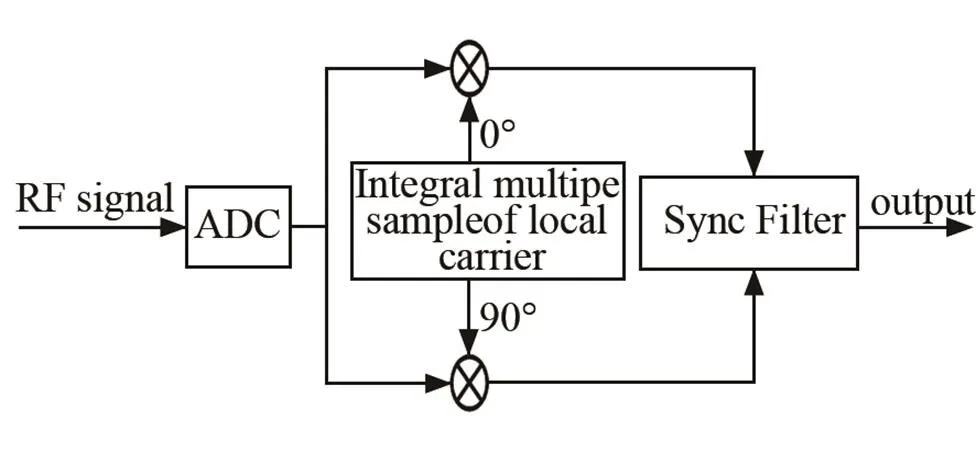
Fig.1 Block diagram of digital I/Q demodulator
To reduce data processing in the TCD system, the sampling frequency of the transmitting signal should be an integer multiple of a carrier frequency, and be in the same clock source. In the experimental system, the transmitting carrier frequency of the probe is 2 MHz, and the sampling frequency for the digital signal is four times. After digital I/Q demodulation, the complex signal also should be filtered to eliminate carrier leakage.
1.3 Coded excitation
Coded excitation can improve the system signal-to-noise ratio to the range of 15- 20 dB[10]without increasing the transmitting pulse peak power and affecting the resolution. In our experiments, the coded transmitting signal is an 7-chip Barker code (={1, 1, 1, -1, -1, 1, -1})[11]. Actually, coded excitation could be viewed as a signal modulation, so the process of coded excitation could be viewed as a secondary modulation. The coded sequence is over-sampled before it is loaded to wide emission threshold. Then, it is demodulated by a 2 MHz carrier signal. The process of an over-sampled 7-chip Barker code demodulated by 4 wavelengths base pulse sequence is shown in Fig.2.
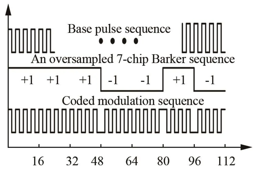
Fig.2 Schematic diagram of coded excitation waveform
2 RESULTS
2.1 Doppler phantom experiment
To verify the effectiveness of the system, a Doppler phantom experimental system is set up. As is shown in Fig.3, a Pulsating pump and a Doppler ultrasound phantom are the main instruments required for the experiment.
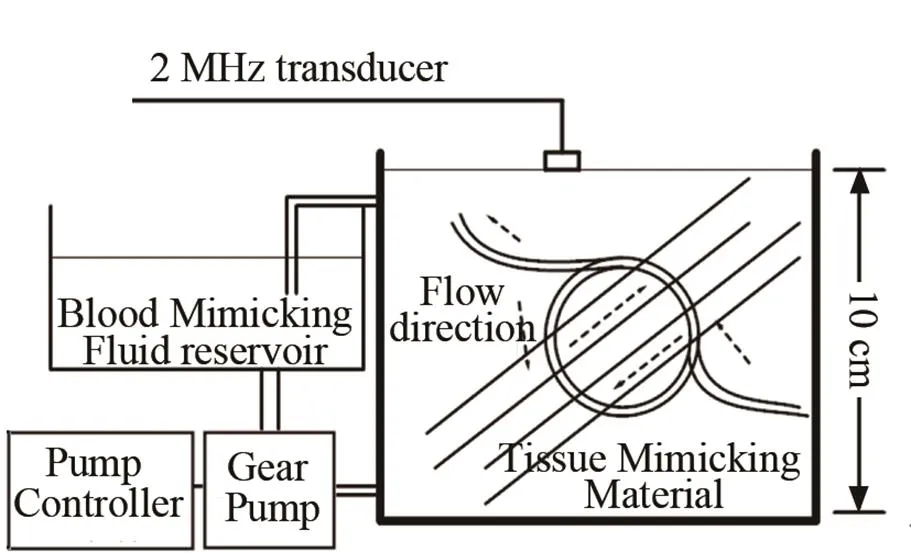
Fig.3 Schematic diagram of simulated blood circulation device
In this study, there are two kinds of excitation methods. One is single-pulse excitation, and the other is 7-chip Barker code excitation. Both of the excitation methods have the same excitation pulse width and RF energy. As illustrated in Fig. 3, there are two imitation blood vessels which are located just below the ultrasound transducer about 2.5 cm and 3.5cm. The purpose of the experiment is to achieve a multi-depth inspection, which could verify the effectiveness and practicality of this system, and probe whether code excitation could improve the axial resolution. According to the curves in Fig.4, Pnon-codedis the result from the single-pulse excitation system and Pcodedis the result from of the system using coded excitation.
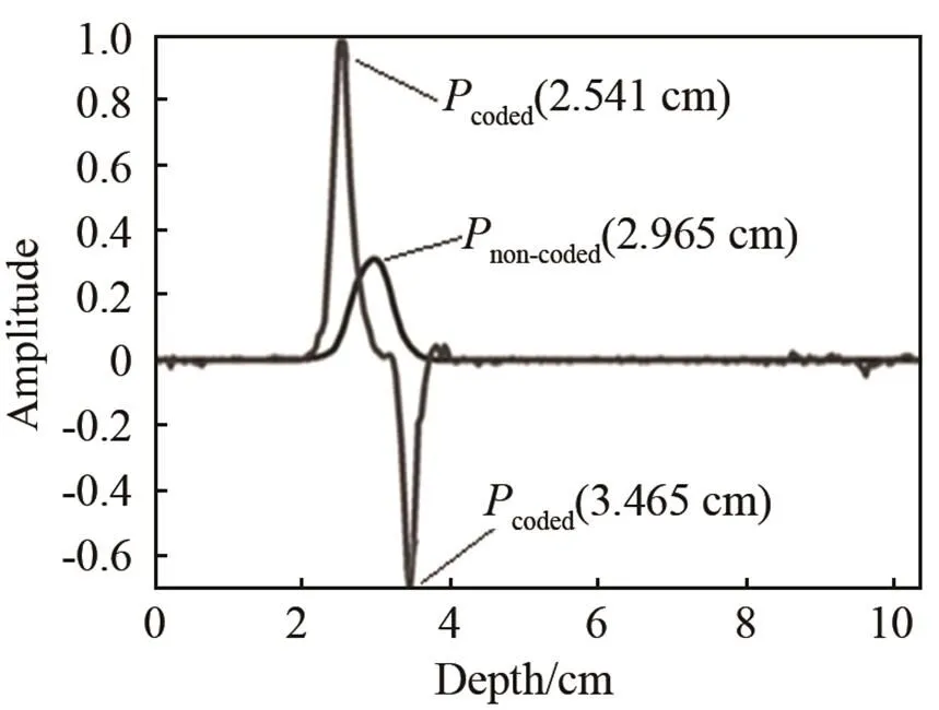
Fig.4 Results of pulse excitation and coded excitation
It could be easily observed that there is only one blood flow message in the single-pulse excitation system. Actually, blood flow signals aliasing is the direct reason, which results in confusion with imitation blood vessels. However, experimental results show that peak amplitude value is greatly improved by coded excitation, and the two opposing blood flow messages are clearly presented in Fig.4. It could be seen that the forward and reverse blood depth values of the coded system are about 2.541 cm and 3.465 cm, respectively, which are consistent with the real depths of two simulated blood vessels. Compared with single-pulse excitation, it is clear that the coded excitation system could significantly improve the vertical resolution. What’s more, as the transmission power is reduced, the axial resolution as well as the SNR is still better than the normal pulse system.
2.2 Analysis of human experiment and result
When implemented with full digital design of TCD, it could visually observe the blood flow information on the path of the ultrasound wave. Doctors could easily find the location of the blood vessel from the blood flow information. In order to find the differences between the single-pulse excitation and the code excitation, in our experiment, both of the excitation methods are designed to compare the sensitivity and reliability of detection. Fig.5 shows the blood flow information image from right temporal window detection of artery (MCA)[12] of the human body.
As is shown in Fig.5,non-codedis the result of single-pulse excitation, andcodedrepresents the result of coded excitation. The curves clearly show the differ- ence between the results of the two different excitation systems. It is obvious that the peak amplitude of thecoded excitation is higher than that of the single-pulse excitation. Under the same normalization condition, the sensitivity and axis resolution of the codes excitation is higher than those of the single- pulse excitation.
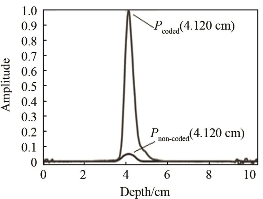
Fig.5 Results of single-pulse excitation and coded excitation through temporal window
3 DISCUSSIONS
In the experiment of the codes excitation system, the coded signal is generated by a 7-chip Barker code modulated by 4 wavelengths of base pulse sequence. According to the study of the coded sequence and the decoding filters, such as matched filter, inverse filter, and spike filter, it is found that the number of base pulse sequences is related to the SNR gain and the peak of sidelobe level after pulse compression. When implemented with a shorter coded sequence, it could get a better SNR gain. However, with the increasing of base sequence, the side lobes and random noise are no longer at a low level.
The base pulse sequence length is related to the SNR gain. It could be found that when the base pulse sequence is extended into a larger sequence, the peak of sidelobe level will be higher after pulse compression, which would result in an increase of accumulated time domain. Therefore, it should take a shorter base pulse sequence to improve performance of pulse compression. Due to limitations of the system bandwidth of the probe and other conditions, it is suitable to use the 4 wavelengths base pulse sequence[13].
In this paper, a full digital design of TCD ultrasound system using coded excitation is presented. Meanwhile, the effectiveness and the applicability of the system in theory and the experiment are verified. For this design, a lot of experiments are performed, such as Doppler phantom experiments, pulsating pump experiments, and clinical trials. To get the blood flow information, a large amount of echo data are collected and analyzed. Finally, the power spectrum of the blood flow information is drawn out by using the modern digital signal processing technology. The idea of coded excitation in the TCD system for the blood velocity detection could easily detect the depth of the blood vessel from the blood flow information and improve the axis resolution. In our future study, many new signal processing techniques will be also conducted with the full digital design.
[1] Aaslid R, Markwalder TM, Nornes H. Noninvasive transcranial Doppler ultrasound recording of flow velocity in basal cerebral arteries[J]. Journal ofNeurosurgery, 1982, 57(6): 769-774.
[2] GAO Shangkai. Medical imaging systems [M]. Beijing: Tsinghua University Press, 2000: 88-14.
[3] Li J, Diao X, Zhan K, Qin Z. A Full Digital Design of TCD ultrasound system using normal pulse and coded excitation[C]. Springer International Publishing, 2015, 47: 136-139.
[4] Misaridis T X, Gammelmark K, Jørgensen C H, Lindberg N,Thomsen A H, Pedersen M H, Jensen J A. Potential of coded excitation in medical ultrasound imaging [J]. Ultrasonics, 2000, 38(1-8): 183-189
[5] Barker R H. Group synchronization of binary digital systems [M]//Jackson W, eds. Communication Theory. London: Butterworths, 1953: 273-287.
[6] Welch L R, Fox M D. Practical spread spectrum pulse compression for ultrasound tissue imaging[J]. IEEE Transactions on Ultrasonics Ferroelectrics and Frequency Control, 1998, 45: 349-355.
[7] Qin Z D, Chen S P, Chen X. Coded transmission for ultrasound Doppler detection using truncated long code, Biomedical Engineering and Informatics (BMEI)[C]// 2010 3rd International Conference on, vol. 1, pp. 159-161, 16-18 Oct. 2010.
[8] WAN Mingxi. Biomedical ultrasonics[M]. Beijing:Science Press, 2010: 339-369.
[9] QI M C, GAO S K. Study on a Digital Doppler Ultrasonic Blood Flow Measurement System[J]. Chinese Journal of Biomedical Engineering, 2012, 31(3):349-353.
[10] O ' Donnell M. Coded excitation system for improving the penetration of real - time phased - array imaging systems[J]. IEEE transactions on ultrasonics , ferroelectrics , and frequency control, 1992, 39 (3): 341-351
[11] Peng Q Y, Gao S K. Coded excitation and its applications in medical ultrasound imaging[J]. Journal of Biomedical Engineering, 2005, 22(1): 175-180.
[12] Moehring M A, Spencer M P. Power. M-mode Doppler (PMD) for observing cerebral blood flow and tracking emboli[J]. Ultrasound in medicine & Biology, 2002, 28(1): 49-57.
[13] Xiang L, Gao S K. Coded excitation in pulsed wave Doppler ultrasound blood flow measurements[J]. Journal of Tsinghua University (Natural Science), 2008, 48(6): 1032-1035.
编码超声血流多深度检测
李绍兴1,2,3,张天炯1,2,3,陈昕1,2,3,覃正笛1,2,3
(1. 医学超声关键技术国家地方联合工程实验室,广东深圳518060; 2. 广东省生物医学信息检测与超声成像重点实验室,广东深圳518060; 3. 深圳大学医学部生物医学工程学院,广东深圳518060)
血流信息检测及其成像因其独特的优势,在临床上得到广泛应用。但常规经颅多普勒超声系统仍采用模拟和数字电路结合的传统技术,这类系统容易受到外界干扰且不能进行多深度检测。文章设计出一种全数字多普勒超声血流检测系统方案,弥补了传统模拟系统存在的问题。多普勒仿体和人体实验结果表明,该系统能够进行多深度检测,确定血管的深度;同时,还提高了检测灵敏度、超声穿透力和系统成像分辨率。
编码激励;经颅多普勒超声;多深度检测
R312
A
1000-3630(2017)-01-0038-04
10.16300/j.cnki.1000-3630.2017.01.008
Jun. 14, 2016; Revised: Aug. 25, 2016
*This paper is published in Chinese language in Technical Acoustics, Dec 2016, Vol.35, No.6, 527-530.
猜你喜欢
科学与社会(2022年4期)2023-01-17
体育科技文献通报(2022年3期)2022-05-23
科学与社会(2021年4期)2022-01-19
中国生物医学工程学报(2019年5期)2019-07-16
图书馆建设(2018年5期)2018-07-10
中国生物医学工程学报(2017年6期)2017-02-10
中外医疗(2016年15期)2016-12-01
中外医疗(2016年15期)2016-12-01
照明工程学报(2016年3期)2016-06-01
电子器件(2015年5期)2015-12-29