
Diagnostic and prognostic value of lncRNA cancer susceptibility candidate 9 in hepatocellular carcinoma
2019-02-12 12:58YongLianZengZhenYaGuoHuiZhaoSuFuDiZhongKeQingJiangGuanDouYuan
World Journal of Gastroenterology 2019年48期
Yong-Lian Zeng, Zhen-Ya Guo, Hui-Zhao Su, Fu-Di Zhong, Ke-Qing Jiang, Guan-Dou Yuan
Abstract
Key words: LncRNA cancer susceptibility candidate 9; Hepatocellular carcinoma;Prognosis; Kyoto Encyclopedia of Genes and Genomes; Gene Ontology; Competing endogenous RNA
INTRODUCTION
Liver cancer is a common digestive tract cancer[1]. An epidemiological investigation reported 800 thousand new and dead liver cancer patients in 2018, and liver cancer was ranked the sixth most common cancer worldwide and the second major cause of cancer death in humans[2]. Hepatocellular carcinoma (HCC), the most common liver cancer, is accompanied by insidious clinical manifestations in the early stage; thus,most HCC patients are already in middle or advanced stages when admitted to hospital, and are unsuitable for surgical treatment, which seriously affects patients'quality of life[3]. At present, there are no good diagnostic markers for early HCC, and alpha fetoprotein (AFP) is the most widely used serological marker for HCC diagnosis; however, 30% of patients with HCC do not show increased AFP, and can even be negative, which increases the diagnosis difficulty of HCC[4]. Therefore, it is particularly important to identify a biomarker of HCC with high specificity.
Due to its prominent use in various disciplines, lncRNA has been a hot research topic in recent years[5]. lncRNAs are non-coding RNAs with lengths exceeding 200 nucleotides. Initially, scholars considered that lncRNAs lacked prominent protein coding ability, but recent studies found that lncRNAs have regulatory functions in a variety of mechanisms, including epigenetic modification, transcriptional regulation and post-transcriptional modification[6-8]. A previous study demonstrated that lncRNA ZEB1-AS1 is a potential prognostic indicator of HCC due to its differential expression in this tumor[9]. Another study found that lncRNA MALAT1 promotes the proliferation, migration and invasion of HCC by antagonizing miR-142-3p[10].
Cancer susceptibility candidate 9 (CASC9), located on human chromosome 8q21.13,is a member of the lncRNA family[11]. Previous studies revealed that CASC9 was differentially expressed in esophageal cancer and lung adenocarcinoma, and was closely related to biological functions such as proliferation, invasion and metastasis[12,13]. However, there are few studies on whether CASC9 is expressed in HCC and whether it has clinical diagnostic and prognostic value. Therefore, this study analyzed CASC9 expression in HCC based on the Cancer Genome Atlas(TCGA) database and clinical verification, in order to provide a potential index for clinical diagnosis.
MATERIALS AND METHODS
Acquisition and analysis of TCGA data
Data on gene expression in HCC were downloaded for analysis by logging into TCGA, selecting Access TCGA Data and entering the database. The data were acquired from the core sample database of TCGA, and sequencing and analysis were performed on the acquired data based on a standardized processing scheme(http://can-cergenome.nih.gov/cancergenomics/tissuesamples). The data included a total of 424 patients, involving 374 cancer samples and 50 control samples. Clinical data on HCC patients were acquired from http://gdac.broadinstitute.org for analysis,and excluded patients without detailed data and a survival time less than 30 d.Original data on CASC9 were processed through log (X+1, 2) to analyze the differences between control samples and carcinoma tissues, and a survival curve for high and low expression groups was drawn according to the median value of CASC9.
Bioinformatic analysis of lncRNA
Starbase 3.0 was adopted to predict lncRNA CASC9 targeted microRNA (miR), and miRDB, miRTarBase, and TargetScan were adopted to predict target genes of potential miR on-line. Cytoscape software was adopted to draw the competing endogenous (ce)RNA network, and David software to analyze target genes based on the Kyoto Encyclopedia of Genes and Genomes and Gene Ontology (GO) enrichment analysis.
Collection of patient samples
A total of 80 HCC patients (50 males and 30 females with an average age of 54.6 ± 5.0 years) treated in The First Affiliated Hospital of Guangxi Medical University from May 2012 to January 2014 were enrolled in the patient group, and 50 healthy subjects(29 males and 21 females with an average age of 53.7 ± 4.1 years) were enrolled in the control group. This study was approved by the First Affiliated Hospital of Guangxi Medical University Ethics Committee. The patients meeting the following criteria were included: Patients diagnosed with HCC based on imaging and pathologic biopsy; patients meeting the TNM staging criteria for HCC by the American Joint Committee on Cancer in 2009[14]; patients who had not taken part in any previous targeted tumor research (surgery, radiotherapy and chemotherapy,etc.), understood the study and signed an informed consent form (including their families) and patients whose expected survival time was more than 3 mo. The following patients were excluded: Patients with other combined tumors, renal function diseases, infection before admission, and those unwilling to cooperate.
Sample collection and detection
Peripheral venous blood (5 mL) was obtained from the patients, centrifuged at 3000 rpm for 10 min after 30 min and the supernatant collected. Total RNA in the collected supernatant was extracted with TRIzol reagent (Carlsbad Invitrogen Company,California, United States), and the purity, concentration and integrity of the total RNA were determined using ultraviolet spectrophotometry and agarose gel electrophoresis. Reverse transcription was performed using TransScript®miRNA RT Enzyme Mix and 2×TS miRNA Reaction Mix in the TransScript Green Two-Step quantitative real-time polymerase chain reaction SuperMix kit (Beijing TransGen Biotech Co., Ltd., China) in strict accordance with the original kit instructions. The amplification system of CASC9 consisted of 1 μL of cDNA, 0.4 μL of upstream and downstream primers, respectively, 10 μL of 2X TransScript®Tip Green qPCR SuperMix, Passive Reference Dye (50X), and Nuclease-free water (added to 20 μL in total). The amplification conditions were as follows: Pre-denaturation at 94°C for 30 s,denaturation at 94°C for 5 s and annealing extension at 60°C for 30 s, with 40 cycles in total. Triplicate wells were prepared for each sample, and the experiment was repeated three times. Glyceraldehyde-3-phosphate dehydrogenase was used as an internal reference, and 2-Δctwas used to analyze the data. Experiments were carried out using a 7500 PCR instrument (ABI, United States).
Follow-up of patients
The patients were followed-up for 5 years by telephone interview and clinic reexamination. During the 1styear, the patients were followed-up at the 3rd, 6th, 9thand 12thmonth, respectively, and during the remaining 4 years, they were followed-up once every 4 mo.
Statistical analysis
In this study, the acquired data were statistically analyzed using the SPSS20.0 software package, and figures were drawn using the GraphPad 7 software package.The distribution of measurement data was analyzed using theK-Stest, and the data in normal distribution were expressed as mean ± SD. Comparisons between groups were performed using independent-samplesTtest. Comparisons within groups were analyzed by pairedttest, and expressed ast. Ranked data were analyzed using the rank sum test, and expressed asZ. Enumeration data were analyzed using the chisquare test. ROC curves of CASC9 to evaluate its diagnostic value in HCC were drawn, and a K-M curve for 5-year survival of patients was also drawn. The log-rank test was adopted for analysis. Multivariate Cox regression analysis was adopted to analyze independent risk factors for patients.P< 0.05 indicated a significant difference.
RESULTS
CASC9 expression in TCGA
Analysis of data from TCGA database on CASC9 expression in HCC patients showed that control samples had significantly lower CASC9 expression than carcinoma tissue samples (P< 0.001), and the grouping of patients according to median CASC9 expression revealed that the low CASC9 expression group had a higher survival rate than the high CASC9 expression group (P= 0.011, Figure 1).
Comparison of baseline data
Comparisons between the patient group and control group showed that there were no significant differences between the groups in terms of gender, age, body mass index(BMI), past medical history, smoking history, history of alcoholism and place of residence, while a difference in AFP expression was observed between the two groups(P< 0.001, Table 1).
CASC9 expression in patients and its clinical value
The expression of serum CASC9 in the patient group was significantly higher than that in the control group, and the ROC curve showed that the AUC was 0.933. Further analysis of the relationship between CASC9 and clinical pathological data showed that tumor size, combined hepatitis, TNM staging, lymph node metastasis,differentiation and AFP were closely related to CASC9 expression. In addition, the analysis of ROC curves showed that CASC9 was associated with tumor size, TNM staging, lymph node metastasis, differentiation and AFP (Figure 2 and Tables 2 and 3).
Relationship between CASC9 and patients' survival
All the patients were successfully followed-up for 5 years in terms of survival. The patient group showed a 5-year survival rate of 17.50% with 14 patients surviving. The patients were grouped into the high CASC9 expression group and the low CASC9 expression group according to the median CASC9 expression (3.305), and survival was compared. The high CASC9 expression group showed significantly lower 1-year,3-year and 5-year survival rates than the low CASC9 expression group (allP< 0.05,Figure 3).
Independent factors affecting prognosis of patients
Univariate Cox regression analysis of the pathological data showed that TNM staging,lymph node metastasis, differentiation, AFP and CASC9 were factors which affected the prognosis of patients, and multivariate Cox regression analysis of these factors showed that TNM staging, lymph node metastasis, differentiation, AFP, and CASC9 were independent factors affecting the prognosis of patients (Table 4). In addition, the survival curves in terms of TNM staging, lymph node metastasis, differentiation, AFP and patients' 5-year survival indicated that stage I+II patients with lymph node metastasis, low differentiation, and AFP > 200 ng/mL showed poor 5-year survival(Figure 4).
Bioinformatic analysis
Seventeen CASC9 potential targeted miRs were identified by Starbase 3.0, and 89 mRNAs were found by predicting the downstream mRNA of the 17 targeted miRs with miRDB, miRTarBase, and TargetScan. We used Cytoscape software to construct the interaction map between 1ncRNA-miRNA-mRNAs, and GO enrichment and Kyoto Encyclopedia of Genes and Genomes pathway enrichment analysis were performed on the 89 mRNAs in the ceRNA network using DAVID and KOBAS, and 18 GO functions withP< 0.05 and 8 signal transduction pathways withP< 0.05 were found (Tables 5 and 6).
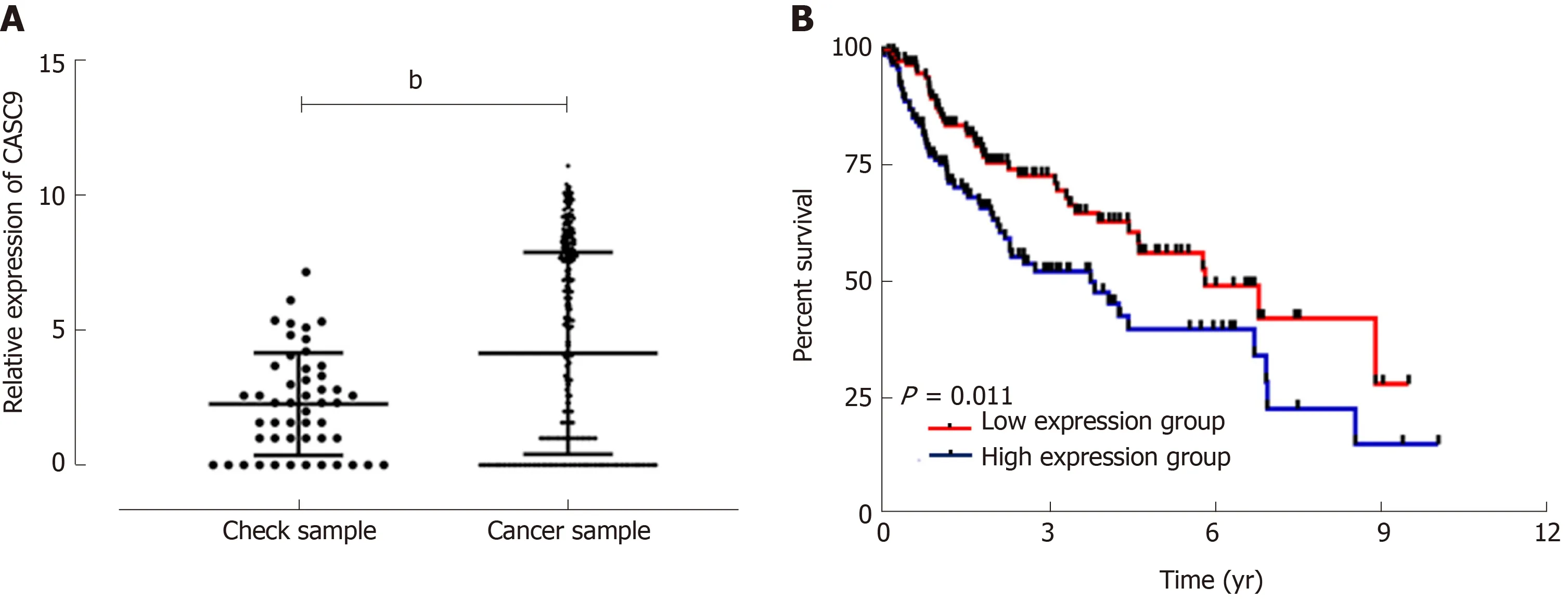
Figure 1 Cancer susceptibility candidate 9 expression and survival from TCGA database. A: Cancer susceptibility candidate 9 (CASC9) was highly expressed in cancer patients; B: The survival rate of the high CASC9 expression group was lower than that of the low CASC9 expression group (P = 0.011). bP < 0.01. CASC9:Cancer susceptibility candidate 9.
DISCUSSION
HCC, a digestive system neoplasm with high incidence and mortality, is a common disease in male patients, and morbidity in men compared to women is 2.81:1[15]. A Chinese tumor epidemiology survey reported that more than 400 thousand new and dead patients with HCC were observed in 2015; however, there is no effective diagnosis and treatment plan for this disease[16]. At present, AFP is the main clinical serodiagnostic index for HCC, but relevant studies have found that AFP expression increases in liver diseases such as hepatitis; thus, its specificity is low[17]. Therefore, the key to resolving the problem is to identify biological indices with high sensitivity and specificity.
LncRNA is a long-chain non-coding RNA. A previous study demonstrated that lncRNAs participate in the occurrence and development of various cancers[18]. A study by Maet al[19]found that the biological function of HCC was inhibited by regulating the expression of miR-122-5p in HCC based on lncRNA ANRIL knockout. CASC9 is a newly discovered tumor susceptibility gene. A study by Luoet al[20]found that CASC9 and CPSF3 could cooperatively regulate TGF-β signaling pathway conduction in colorectal cancer, and a study by Lianget al[21]reported that CASC9 promoted metastasis of esophageal squamous cell carcinoma by up regulating LAMC2 expression through an interaction with CREB binding protein. However, there are few reports on CASC9 in HCC at present. A study by Nohet al[22]showed that CASC9-mediated AKT ions promoted the survival of HCC cells, and there are no other relevant studies on whether CASC9 can be used as a diagnostic and prognostic indicator of HCC. Therefore, this study confirmed the expression and prognostic value of CASC9 in HCC based on the TCGA database, in order to provide a new potential clinical index.
TCGA database, one of the largest Cancer Genome Projects, contains a variety of tumor gene data[23]. In this study, we first extracted data on CASC9 expression in the tissues of HCC patients from TCGA database, analyzed these tissues, and found that CASC9 expression in carcinoma tissue samples was significantly higher than that in adjacent control tissue samples. The patients were divided into the high CASC9 expression group and the low CASC9 expression group according to the median CASC9 expression to determine survival of the patients. It was found that the low CASC9 expression group had a higher survival rate than the high CASC9 expression group. The above results indicated that CASC9 may be a potential diagnostic and prognostic indicator of HCC. Further clinical research demonstrated that the expression of serum CASC9 in HCC patients was consistent with the data from TCGA database. In addition, we found that the AUC of CASC9 expression (> 0.9) had very high diagnostic value based on ROC curves. We further analyzed the relationship between CASC9 and 1-year, 3-year and 5-year survival rates in HCC patients, anddifferences between the low CASC9 expression group and high CASC9 expression group in 1-year, 3-year and 5-year survival rates were found, which indicated that CASC9 could be adopted as an index for determining the short-term and long-term survival rates of HCC patients. We also analyzed the relationship between CASC9 and pathological data, and found that CASC9 was closely related to tumor size, TNM staging, lymph node metastasis, differentiation, and AFP, which indicated that CASC9 was closely related to the occurrence and development of HCC. In addition,CASC9 had certain diagnostic value in determining tumor size, TNM staging, lymph node metastasis, differentiation and AFP. A study by Gaoet al[24]revealed that CASC9 was also highly expressed in esophageal carcinoma, and had relatively high diagnostic value (AUC: 0.814). However, the AUC of CASC9 in our study group was 0.933. This indicated that CASC9 may have higher diagnostic value for HCC than for esophageal carcinoma, but whether it is a diagnostic index still requires further investigation.
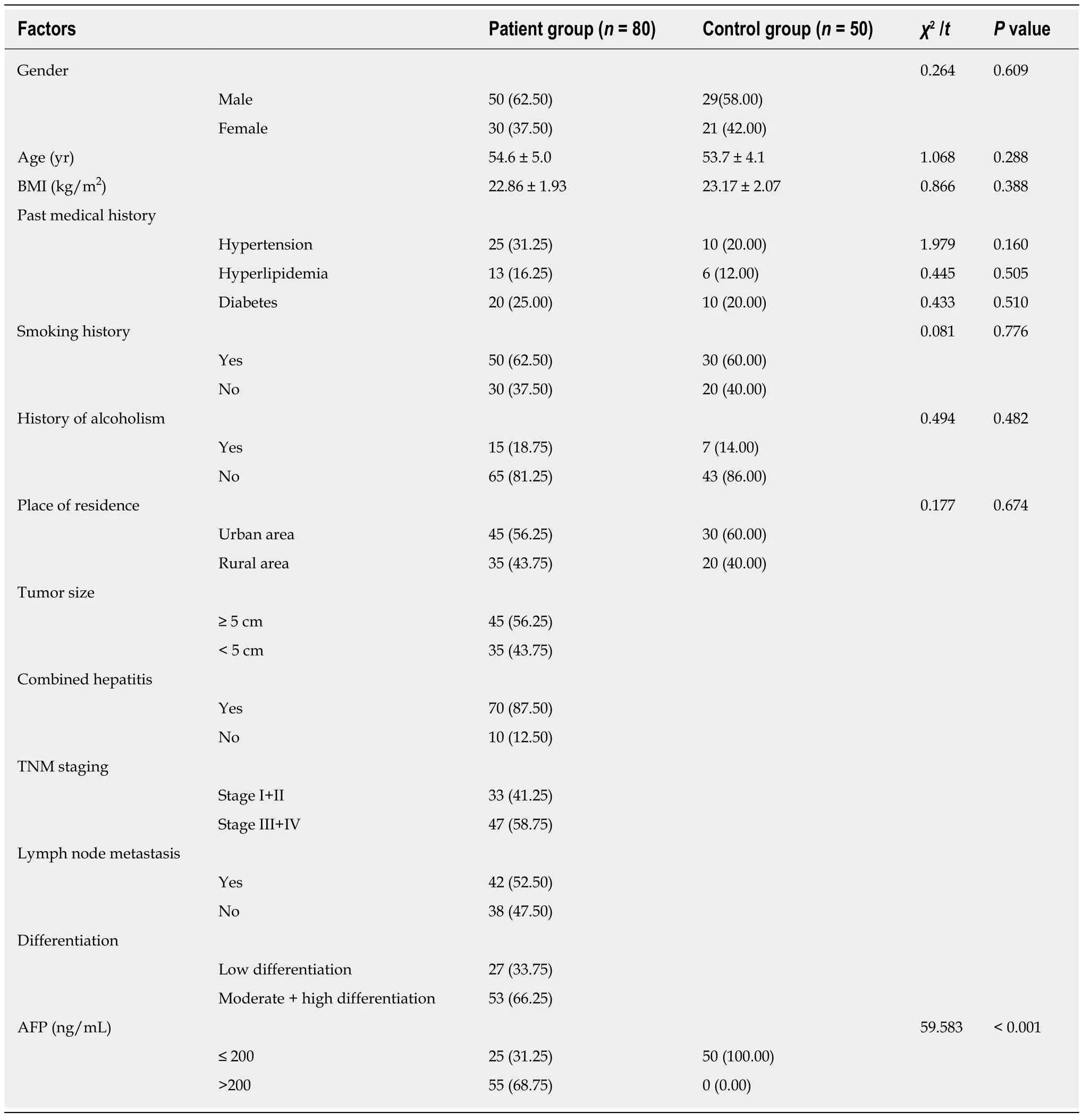
Table 1 Baseline analysis, n (%)
Pathological data on HCC patients were obtained to further analyze independent factors affecting the prognosis of patients, and TNM staging, lymph node metastasis,differentiation, AFP and CASC9 were found to be independent prognostic factors for HCC patients based on multivariate analysis. A study by Tan and Huang confirmed that TNM staging, lymph node metastasis, differentiation, and AFP were closely related to the prognosis of patients, which was consistent with the findings in our study[25,26]. However, we found that tumor size was not related to prognosis in this study, which was inconsistent with the previous study findings. We speculated that this may be related to the collected samples. The tumor size data in this study were relatively intensively distributed with an average size of 4.8 ± 0.8 cm, which may be the reason for this difference. Our study confirmed, for the first time, that CASC9 was an independent prognostic factor for HCC patients, indicating that CASC9 may be a prognostic and diagnostic index for HCC.
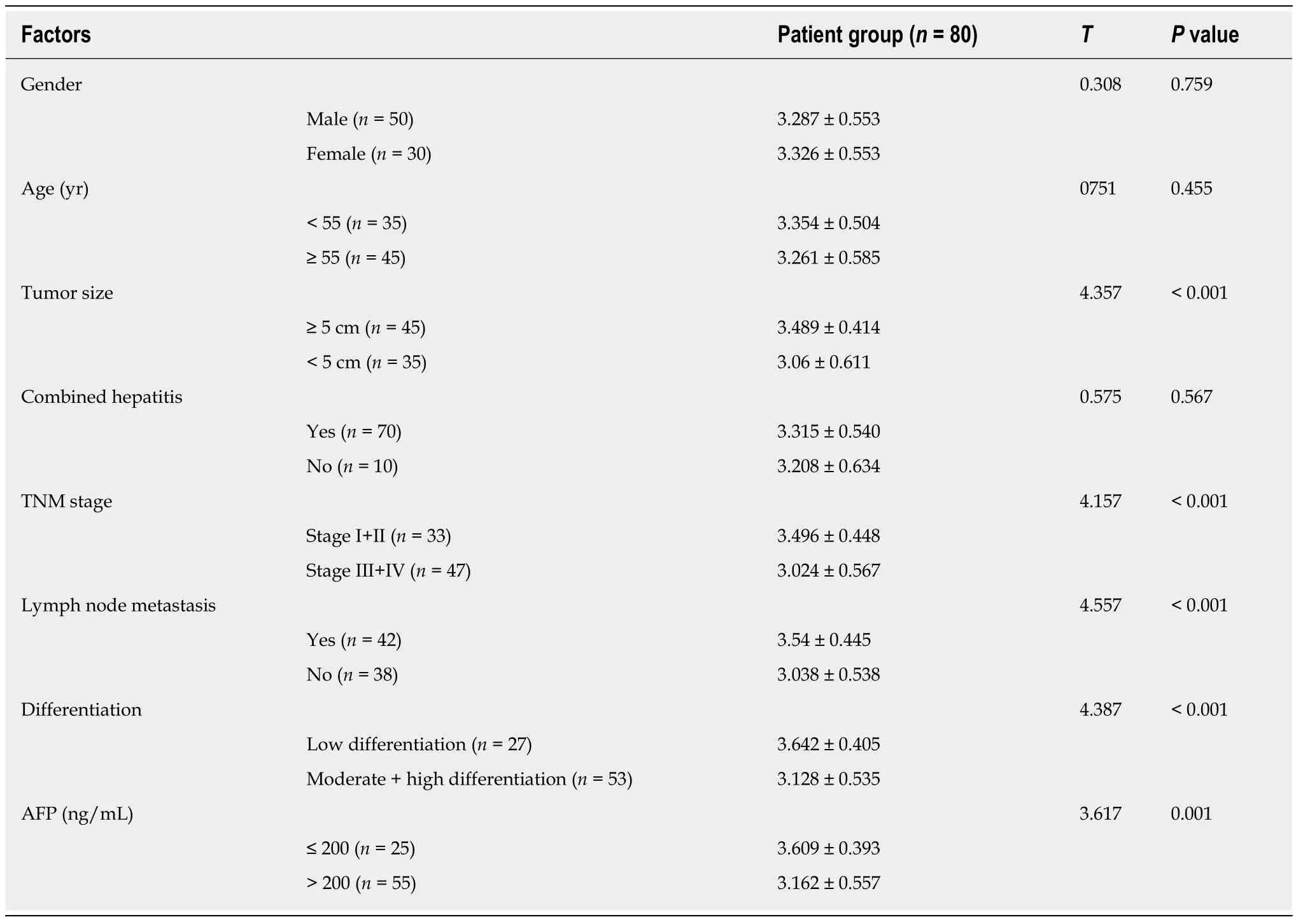
Table 2 Relationship between cancer susceptibility candidate 9 and pathological data
A co-expression network of lncRNA-miR-mRNA was constructed and 17 CASC9 targeted miRs, and 89 targeted mRNAs were observed. The top 10 functions in GO enrichment analysis were negative regulation of transcription from the RNA polymerase II promoter, positive regulation of transcription regulatory region DNA binding, cytoplasm, positive regulation of transcription, DNA-templated, osteoblast differentiation, neuron projection morphogenesis, transcriptional activator activity,RNA polymerase II core promoter proximal region sequence-specific, negative regulation of sequence-specific DNA binding transcription factor activity, and neuron projection, respectively, and the 8 different signal transduction pathways in the Kyoto Encyclopedia of Genes and Genomes analysis were microRNAs in cancer, pathways in cancer, p53 signaling pathway, Chagas disease (American trypanosomiasis),proteoglycans in cancer, cell cycle, PI3K-Akt signaling pathway, and Hippo signaling pathway, respectively. According to the above research, CASC9 can participate in various biological pathways by regulating the miR/mRNA axis, and it is noteworthy that signal pathways such as microRNAs in cancer, pathways in cancer, p53 signaling pathway, PI3K-Akt signaling pathway, and Hippo signaling pathway are important cancer-related signal transduction pathways[27-29]. CASC9 can also participate in the occurrence of these pathways by regulating the miR/mRNA axis, which provides an important cornerstone for our subsequent research.
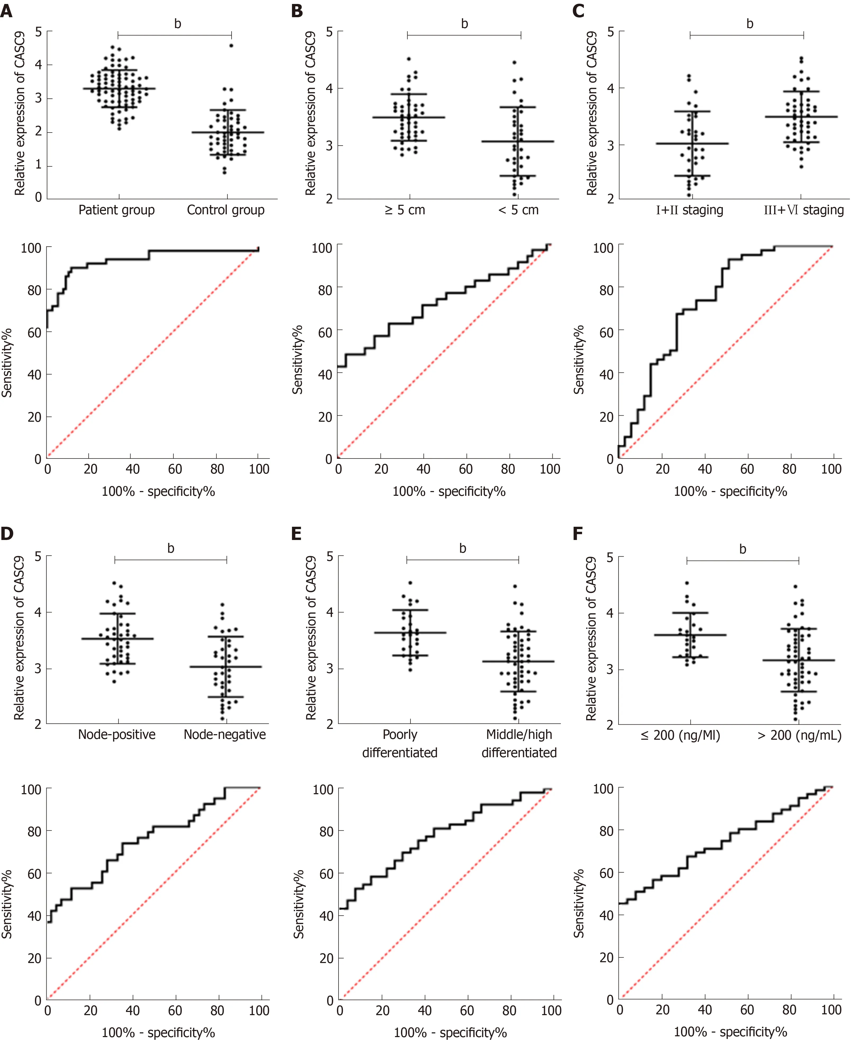
Figure 2 Cancer susceptibility candidate 9 expression in patients, its relationship with tumor size, Tumor, Node, Metastasis staging, lymph node metastasis, differentiation, and alpha fetoprotein expression, and its diagnostic value. A: Cancer susceptibility candidate 9 (CASC9) was highly expressed in the patient group, and the AUC was 0.933; B: Relationship between CASC9 expression and tumor size. The AUC was 0.726; C: Relationship between CASC9 expression and TNM staging. The AUC was 0.743; D: Relationship between CASC9 expression and lymph node metastasis. The AUC was 0.752; E: Relationship between CASC9 expression and differentiation. The AUC was 0.777; F: Relationship between CASC9 expression and AFP. The AUC was 0.738. bP < 0.01. CASC9:Cancer susceptibility candidate 9; AUC: area under the curve.
Although this study confirmed the value of CASC9 in HCC, it still has some limitations. Firstly, we only analyzed patients with HCC and healthy subjects, but did not determine CASC9 expression in patients with hepatitis; thus, whether CASC9 can distinguish HCC from hepatitis still requires further research in healthy subjects.Secondly, how CASC9 participates in the occurrence and development of HCC is unclear. Lastly, this study only focused on Chinese patients, and whether CASC9expression is increased in HCC patients of different races is unclear. Therefore, we hope to carry out bioinformatics and a basic study in the future to further analyze the way that CASC9 affects the occurrence and development of HCC, and obtain different pathological specimens for CASC9 detection in difference races, in order to address the shortcomings in this study. In conclusion, high CASC9 expression is beneficial for the prognosis of HCC patients, and CASC9 is expected to be a potential diagnostic and prognostic indicator of HCC.
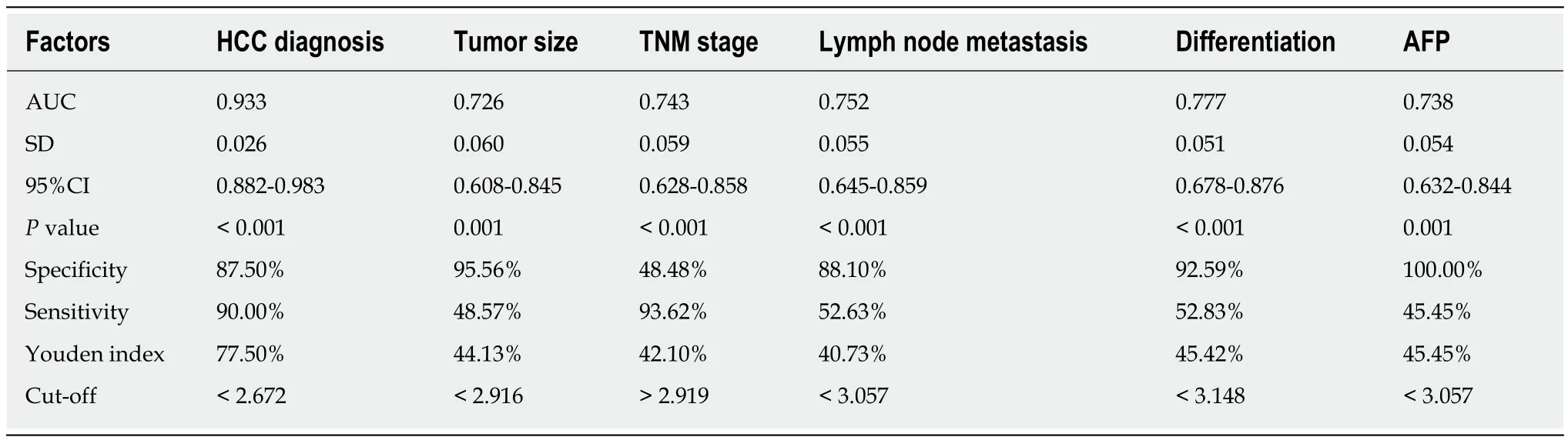
Table 3 Receiver operating characteristic parameters
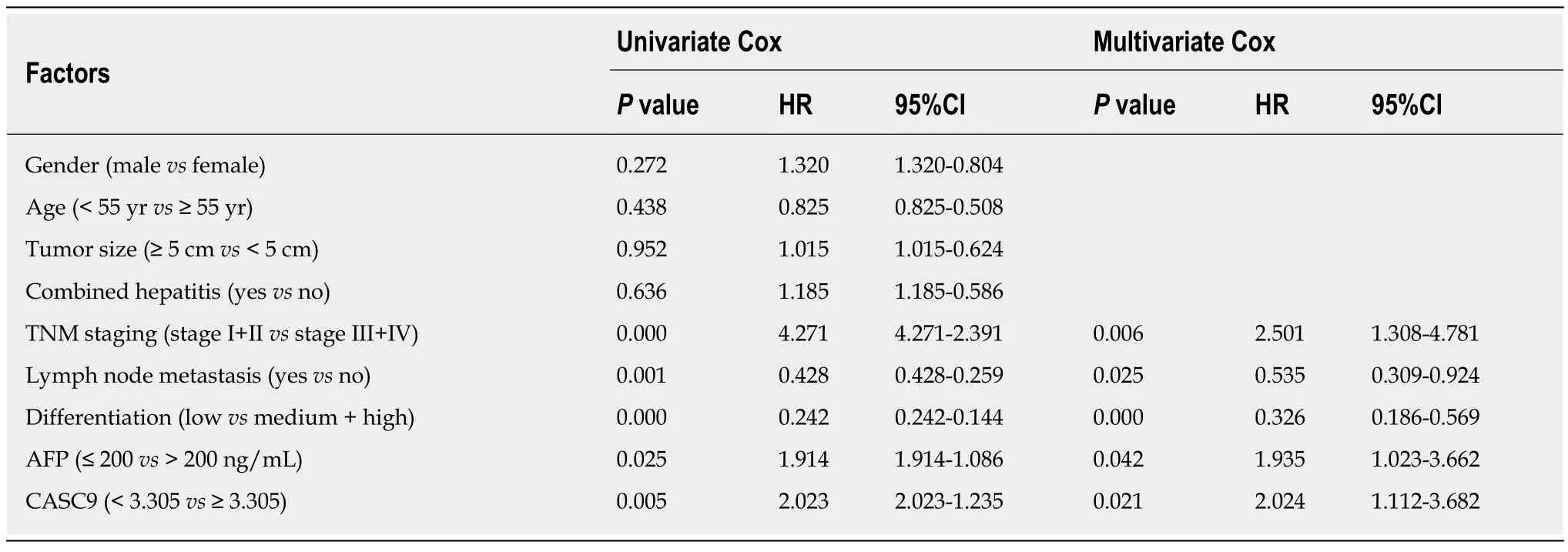
Table 4 Multivariate Cox regression analysis
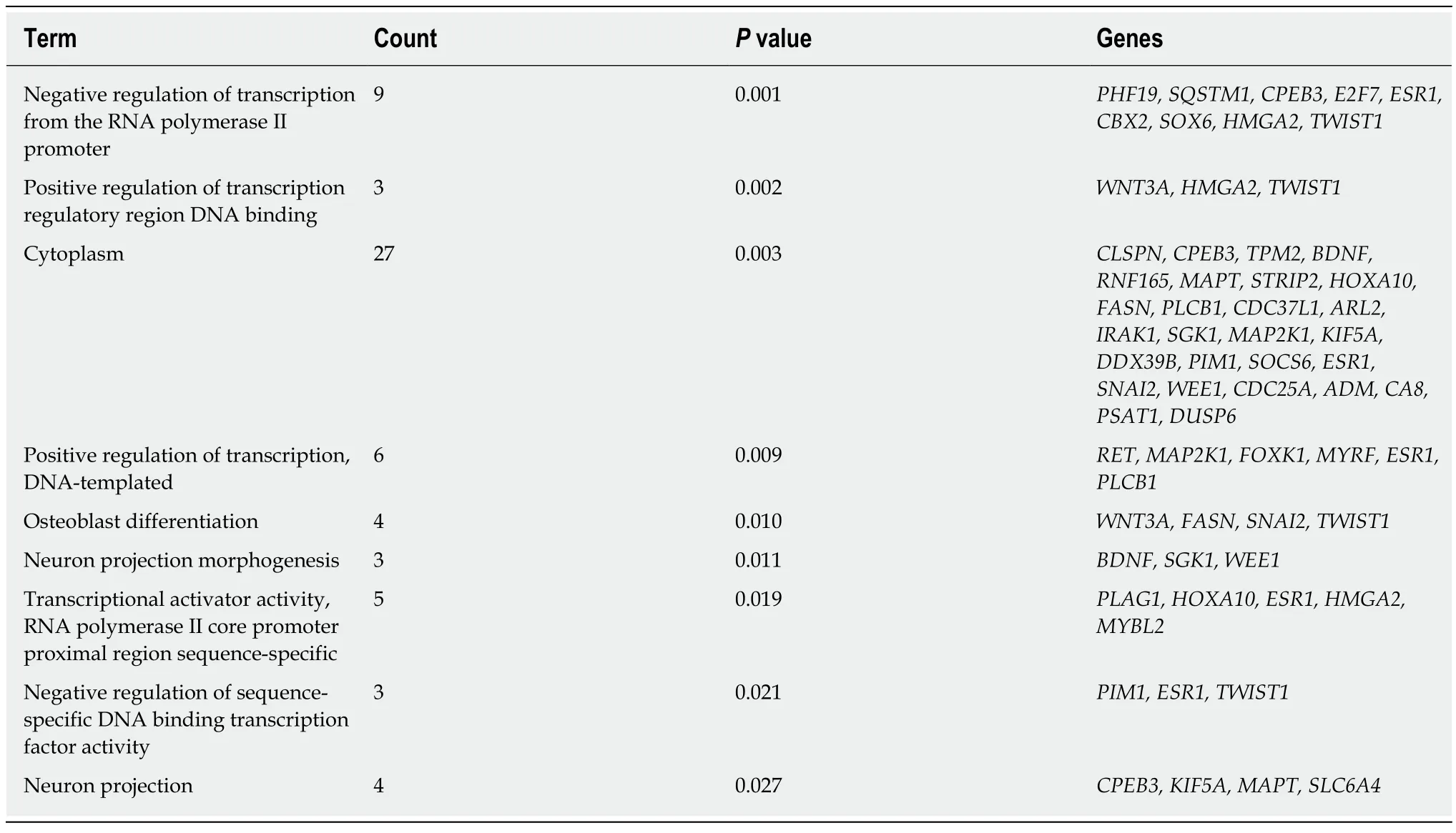
Table 5 The top 10 Gene Ontology enrichment functions
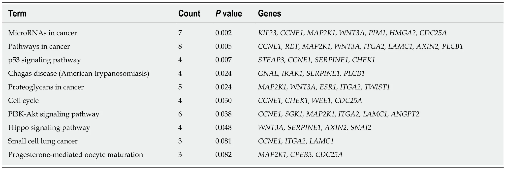
Table 6 The top10 Kyoto Encyclopedia of Genes and Genomes signal pathways
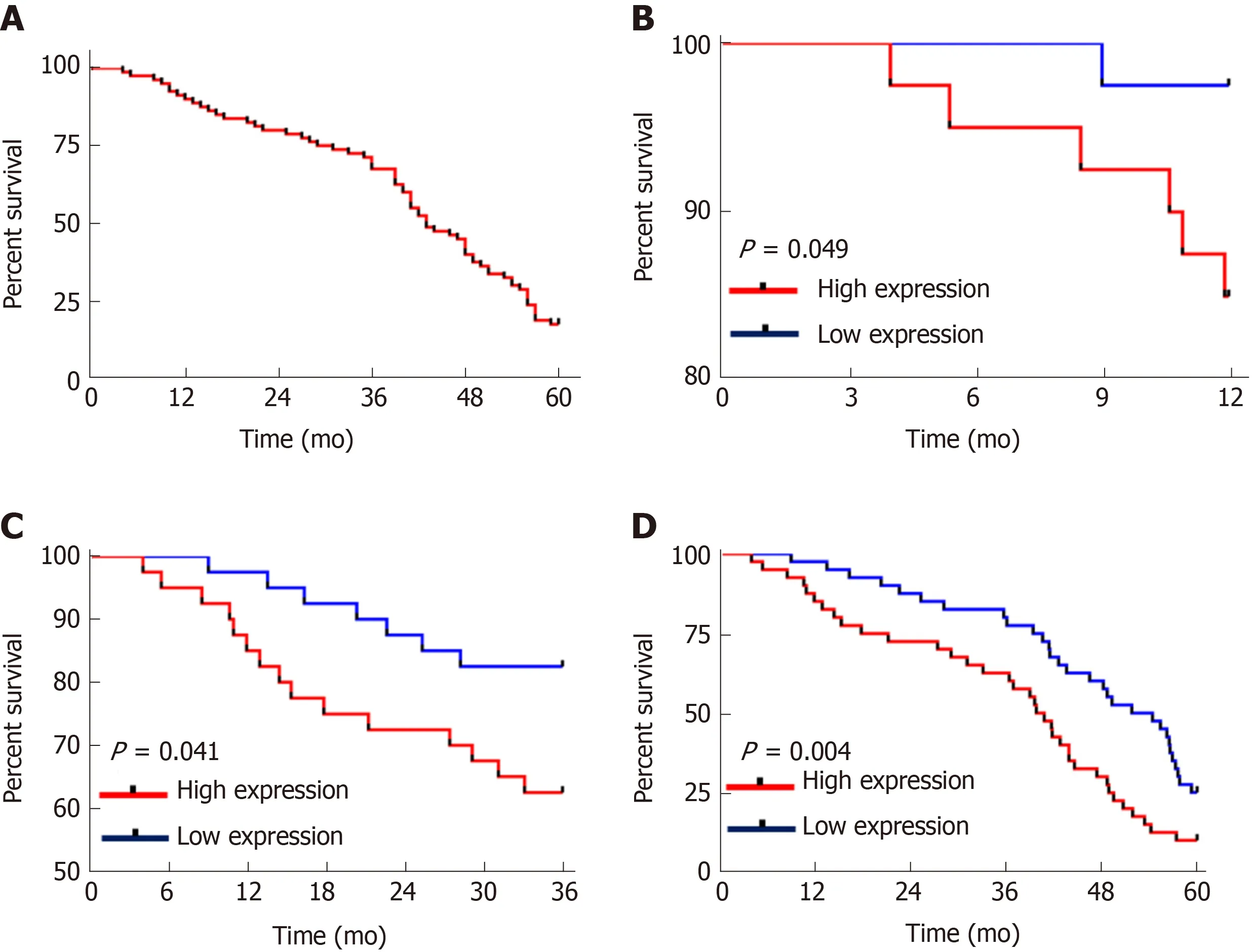
Figure 3 Relationship between cancer susceptibility candidate 9 and patients’ survival. A: The overall survival of patients; B: The 1-year survival of patients in the high and low Cancer susceptibility candidate 9 (CASC9) expression groups (P = 0.049); C: The 3-year survival of patients in the high and low CASC9 expression groups (P = 0.041); D: The 5-year survival of patients in the high and low CASC9 expression groups (P = 0.004). CASC9: Cancer susceptibility candidate 9.
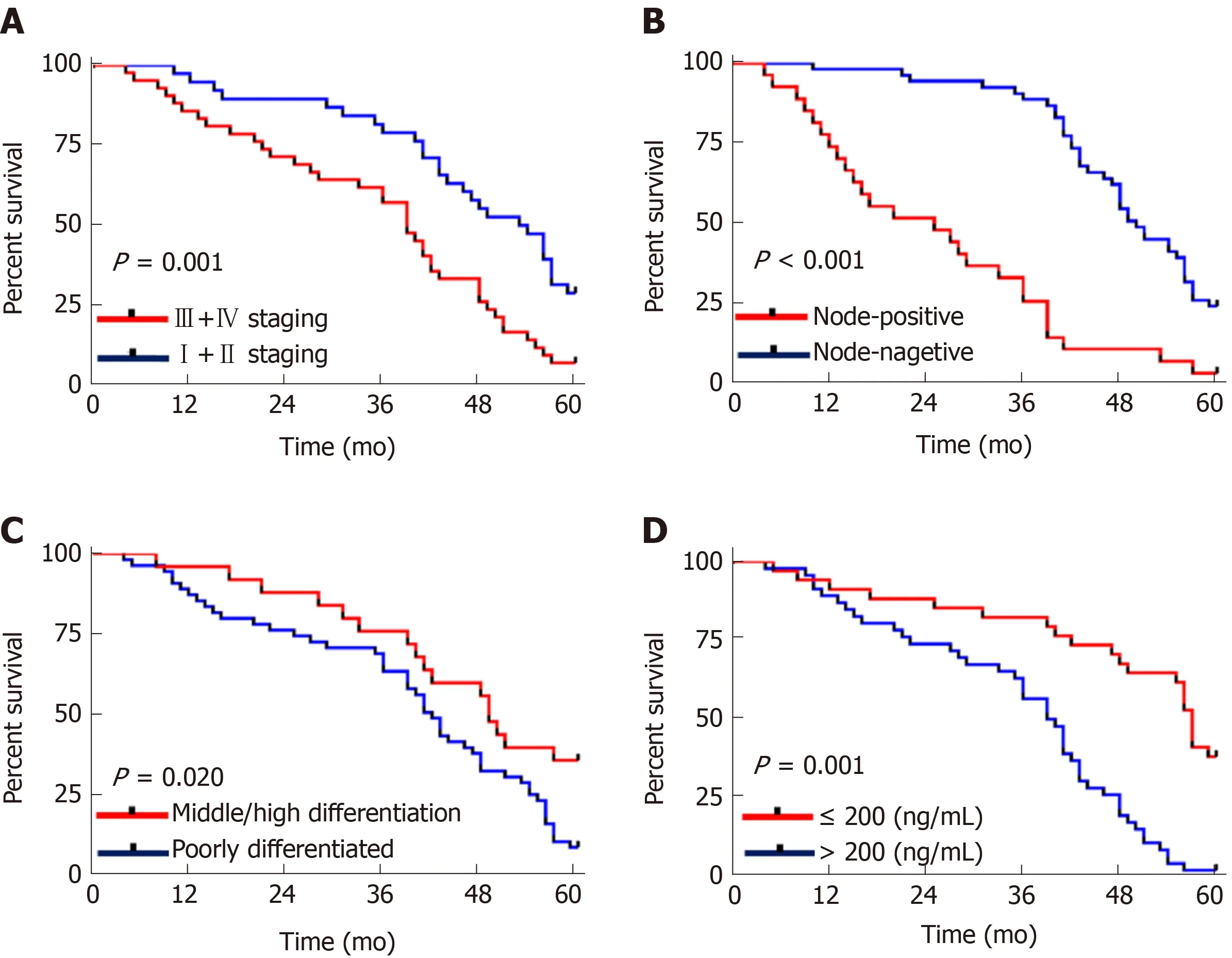
Figure 4 Relationship between tumor, node, metastasis staging, lymph node metastasis, differentiation, alpha fetoprotein and 5-year survival of patients.A: The 5-year survival rate of stage I+II patients was higher than that of stage III+IV patients (P = 0.001); B: The 5-year survival rate of patients without lymph node metastasis was higher than that of patients with lymph node metastasis (cP < 0.001); C: The 5-year survival rate of patients with moderate/high differentiation was higher than that of patients with low differentiation (P = 0.020); D: The 5-year survival rate of patients with AFP ≤ 200 was higher than that of patients with AFP > 200(P = 0.001).
ARTICLE HIGHLIGHTS
showed that lncRNA CASC9 could be used as an independent prognostic indicator in HCC patients. The bioinformatics analysis revealed that lncRNA CASC9 potentially targeted 17 miRs.A total of 89 mRNAs were found in mRNA prediction. GO enrichment and Kyoto Encyclopedia of Genes and Genomes pathway enrichment analysis revealed that lncRNA CASC9 participated in 18 GO functions and 8 signal transduction pathways.
Research conclusion
LncRNA CASC9 is highly expressed in HCC patients, and may be a potential diagnostic and prognostic indicator of HCC.
Research perspectives
It is necessary to further explore the value of lncRNA CASC9 in HCC, and prospective experiments and multi-center clinical studies are required to obtain more robust conclusions. In addition, we hope to further verify the relevant mechanisms of lncRNA CASC9 in HCC by carrying out relevant basic experiments, in order to address the deficiencies in this study.
 World Journal of Gastroenterology2019年48期
- World Journal of Gastroenterology的其它文章
- Gastric electrical stimulation: An emerging therapy for children with intractable gastroparesis
- Comprehensive multi-omics analysis identified core molecular processes in esophageal cancer and revealed GNGT2 as a potential prognostic marker
- Operative complications and economic outcomes of cholecystectomy for acute cholecystitis
- Hepatitis C virus eradication with directly acting antivirals improves health-related quality of life and psychological symptoms
- Significance of postoperative follow-up of patients with metastatic colorectal cancer using circulating tumor DNA
- Pulmonary tumor thrombotic microangiopathy of hepatocellular carcinoma: A case report and review of literature