
Successful management of tubular colonic duplication using a laparoscopic approach:A case report and review of the literature
2020-09-10 02:57GanBinLiJiaGangHanZhenJunWangZhiWeiZhaiYuTao
World Journal of Clinical Cases 2020年15期
Gan-Bin Li, Jia-Gang Han, Zhen-Jun Wang, Zhi-Wei Zhai, Yu Tao
Gan-Bin Li, Jia-Gang Han, Zhen-Jun Wang, Zhi-Wei Zhai, Yu Tao, Department of General Surgery,Beijing Chaoyang Hospital, Capital Medical University, Beijing 100020, China
Abstract
Key words:Colonic duplication;Diagnosis;Laparoscopy;Case report
INTRODUCTION
Duplications of the gastrointestinal tract can occur anywhere from the mouth to the anus[1-3];however, the ileum is the most common site and accounts for approximately 80% of all abnormalities[4].Several clinical studies have demonstrated that colonic duplication is rare, accounting for 6%-7% of cases[5].The manifestations vary greatly depending on the types and locations of the duplication[6]and include abdominal mass, constipation, chronic abdominal pain, and its associated complications, such as obstruction, perforation, intussusception, volvulus, or even malignancy[7,8].Surgery should be considered when the diagnosis is made.Herein, we report the case of a 17-year-old female who was later diagnosed with a tubular colonic duplication.
CASE PRESENTATION
Chief complaints
A 17-year-old female patient complaining of constipation and chronic abdominal pain visited our hospital.
History of present illness
The girl presented the above-mentioned symptoms since she was a child, and her constipation gradually developed to a degree that she had to take medicines to facilitate defecation.The girl had been disturbed by chronic intermittent abdominal pain without radiation for years.As conservative treatments failed to improve her symptoms, she sought definitive surgical intervention in our hospital.
Physical and laboratory examination
The physical examinations revealed left lower abdominal tenderness with a normal bowel movement, and the laboratory results showed no abnormalities.
Imaging examinations
The x-ray examination after oral intake of barium (Figure 1A) suggested two enlarged loops with accumulated barium in the left lower quadrant.An abdominal computed tomography (CT) (Figure 1B) revealed two dilated lumen with a massive amount of stored feces in the left abdominal region.Considering clinical manifestations and imaging results, we suspected a diagnosis of colonic duplication.
FINAL DIAGNOSIS
Tubular colonic duplication.

Figure 1 Related figures demonstrating the clinical characteristics of tubular colonic duplication.
TREATMENT
A laparoscopic exploration and left hemi-colectomy were then performed.During surgery, an intestinal loop was separated from the transverse colon adjacent to the splenic flexure and extended to the left iliac fossa with a dead end (Figure 1C).After dissociating the mesentery from the duplicated colon, a side-to-side anastomosis was made.The histopathologic examination revealed normal alimentary structures with well-formed mucosa and a smooth muscular layer, which further confirmed the diagnosis of a tubular colonic duplication (Figure 1D).
OUTCOME AND FOLLOW-UP
The patient was discharged after an uneventful post-operative clinical course.At the 6-mo follow-up evaluation, the patient was doing well without nausea or constipation.
DISCUSSION
Cystic and tubular duplication are the two common types of colonic duplication[9,10].Cystic duplication is the most common type;only 20% of colonic duplications are tubular[11], which can be further divided into T- and Y-shaped duplications[12].Tubular colonic duplication usually shares a common wall or has a direct communication with the native tract, as in our patient, unlike a cystic duplication[2,3].
To discuss the diagnosis and treatment of colonic duplication, a search was conducted in the PubMed database using the terms “colonic duplication”, and we made a list about the information, shown in Table 1.The clinical characteristics of the included literature are shown in Table 2.A total of 99 case reports were included,and approximately 57.6% were female.The common site of duplication was reported to be the sigmoid (28.2%), ascending (21.2%), complete (21.2%), transverse (15.2%), and descending colon (11.2%).Approximately 30.3% of cases were diagnosed and treated at The manifestations of colonic duplication are non-specific, including abdominal mass, chronic constipation and abdominal pain, an acute abdomen, obstruction,perforation, and malignancy[4-6,13,14].Our patient was disturbed by chronic abdominal pain and constipation since she was a child.We speculated that her constipation was caused by excessive feces accumulated in the duplicated colon with a dead end that made it more difficult to defecate.Most of the cases are diagnosed and treated before 2 years of age;colonic duplication occurring in adults is extremely rare and many of the patients are asymptomatic[14-16].Due to the non-specific presentation and low incidence,it is a challenge to make an accurate diagnosis before surgery[17,18]. Malignancy arising from colonic duplication is rare;only 13 cases have been previously reported[18]and adenocarcinoma is the most common type[19].Kanget al[18]reported a 23-year-old female with complaint of a huge unfixed abdominal mass;CT scan revealed a cystic mass located lateral to the ascending colon.The mass was resected laparoscopically, and the pathologic diagnosis was a malignancy.A rare case of malignancy arising from colonic duplication that metastasized to the omentum was also reported[17].As a result, much attention should be paid to patients who present with an abdominal mass, and combined resection of the normal and duplicated colon is necessary in case of malignancy. Many tools for the diagnosis are available.A recent review concluded that the primary imaging method for the diagnosis of colonic duplication was ultrasonography[14].The typical presence of duplication under ultrasonography was usually a cyst adjacent to the tract with a double wall.Ultrasonography was also helpful for differentiating solid and cystic masses[20]. Abdominal x-ray is a primary tool for differential diagnosis.Jimenezet al[21]reported a patient with intestinal obstruction that was caused by colonic duplication, and an xray revealed extremely dilated loops full of stool-like substance.Similarly, the x-ray in our study also presented two large separated loops full of barium in the left lower abdomen.We speculated that this phenomenon was probably caused by excessive accumulation of barium in the duplicated colon, which had a direct communication with the native gut.Abdominal CT is another necessary examination for duplication,which might reveal a low-density cystic structure or dilated lumen running parallel to the native tract[22,23].Sobhaniet al[2]reported a patient in whom the abdominal CT showed an extremely dilated and air-filled loop of bowel adjacent to the sigmoid colon;colonic duplication was later diagnosed intra-operatively.In our patient, two enlarged intestinal loops running parallel from the splenic flexure to the sigmoid colon were demonstrated, and the diagnosis was confirmed during surgery.In addition,colonoscopy is an alternative, especially helpful for tubular duplication because it can be easily detected[7].An intraluminal transparent spherical lesion was found by colonoscopy[10].A case presented with non-specific abdominal pain was reported by Asouret al[7];the patient was accurately diagnosed with tubular colonic duplication according to colonoscopy.We suggest that the pre-operative diagnosis can only be made with prior awareness of the disease regardless of which imaging tool is used. Traditional treatment of alimentary duplication is surgical resection of both the duplicated and normal colon with an end-to-end anastomosis[24,25].Most surgeons advocate that symptomatic patients should undergo elective surgery following accurate diagnosis[3,4,20,26].It has been reported that symptomatic patients are treated successfully by surgery[8,16,19,26,27].Our patient also underwent surgery and was doing well post-operatively without constipation or abdominal pain;however, the management of asymptomatic patients remains controversial.Some surgeons advocate conservative treatments, while others suggest surgical resection when the diagnosis is made[28,29].We propose that surgery should be considered as first-line treatment when the duplication is diagnosed, even though some patients were asymptomatic. Surgery is the mainstay of treatment for colonic duplication.Greater than 90% of patients undergo laparotomy, only 10% undergo laparoscopic surgery.Our patienthad a laparoscopic exploration with an excellent post-operative recovery.Compared to open surgery, minimally invasive surgery has the advantages of smaller incision,quicker recovery, less pain, and reduced blood loss[30].Although sufficient evidence to demonstrate the superiority of laparoscopy for the treatment of colonic duplication is lacking, laparoscopic surgery should be considered for asymptomatic or stable patients. Table 1 Clinical characteristics of colonic duplication reported in the literature 1Diarrhea 2 (2.0%), fever 2 (2.0%), vomiting 4 (4.0%), anorectal malformation 3 (3.0%), volvulus 2 (2.0%), imperforate anus 3 (3.0%), intussusception 1(1.0%). Of the patients reported in the literature, 75.8% had an uneventful follow-up.A female patient diagnosed with cystic colonic duplication who underwent surgery was regularly followed and the symptom of constipation was significantly improved[20].Our patient was also doing well, and the constipation or abdominal pain did not recur during a 6-mo follow-up.Post-operative complications or recurrence of manifestations have also been reported.Kauret al[24]reported a case with recurrence of constipation and a rectovestibular fistula after surgical resection of the duplicated colon.Recurrence of symptoms has also been reported by other surgeons[13,16].The recurrence might be related to surgical technique, as reported by Prasilet al[31],who considered that local excision and closure of a recto-vaginal fistula caused by complete duplication might lead to a recurrence. Colonic duplication is a rare congenital disease in adults.It is a great challenge to make an accurate diagnosis before surgery due to the non-specific manifestations and low incidence.Surgery should be considered as first-line treatment to prevent complications and malignancy.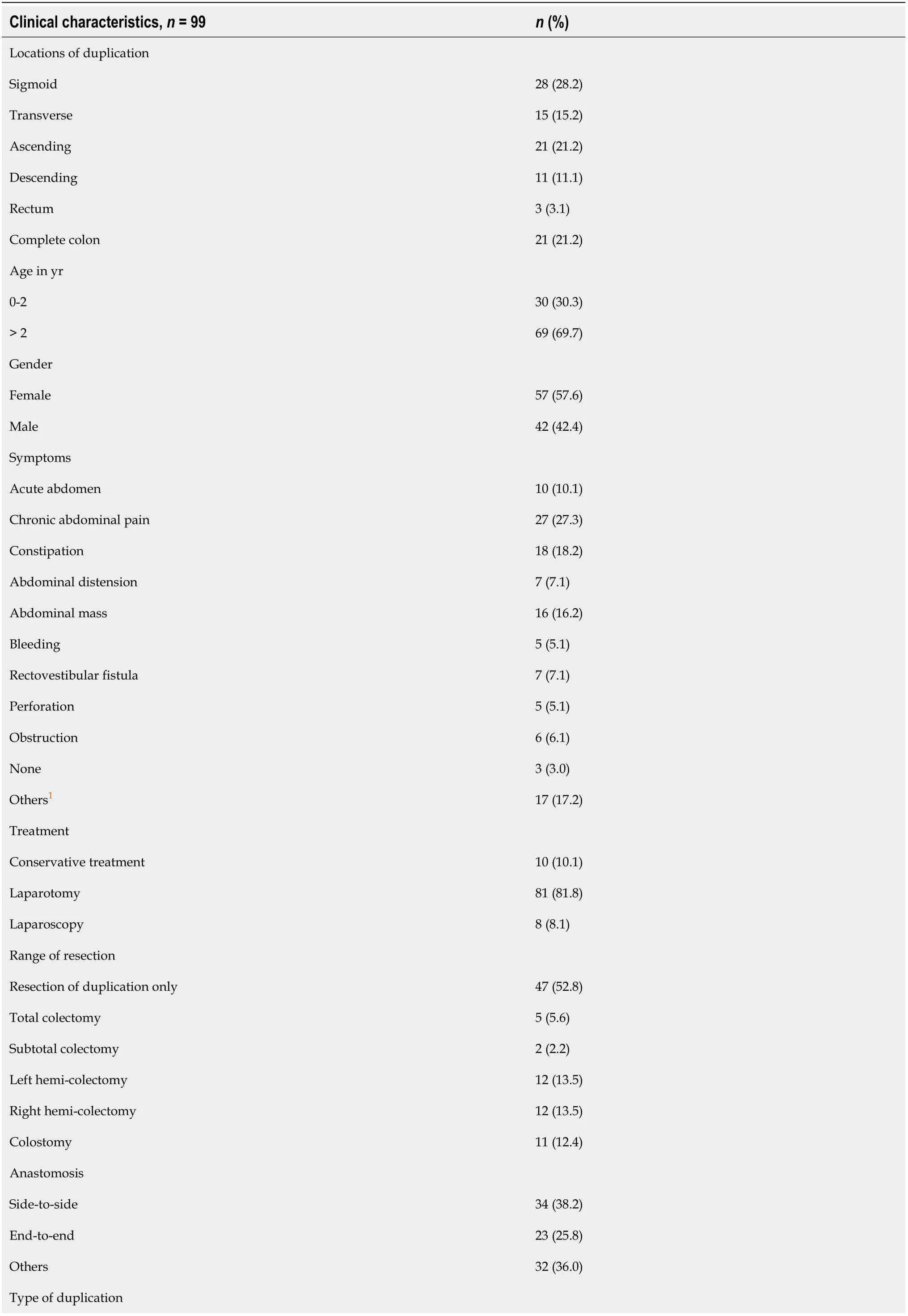

CONCLUSION
 World Journal of Clinical Cases2020年15期
- World Journal of Clinical Cases的其它文章
- Impacts and challenges of United States medical students during the COVID-19 pandemic
- Recent advances in the management of gastrointestinal stromal tumor
- Medical research during the COVID-19 pandemic
- Progress of intravoxel incoherent motion diffusion-weighted imaging in liver diseases
- Typical and atypical COVID-19 computed tomography findings
- Review of possible psychological impacts of COVID-19 on frontline medical staff and reduction strategies