
Pluripotent stem cell derived inhibitory interneurons - principles and applications in health and disease
2020-09-18 07:04FrancescaKeefe,MengLi
中国神经再生研究(英文版) 2020年2期
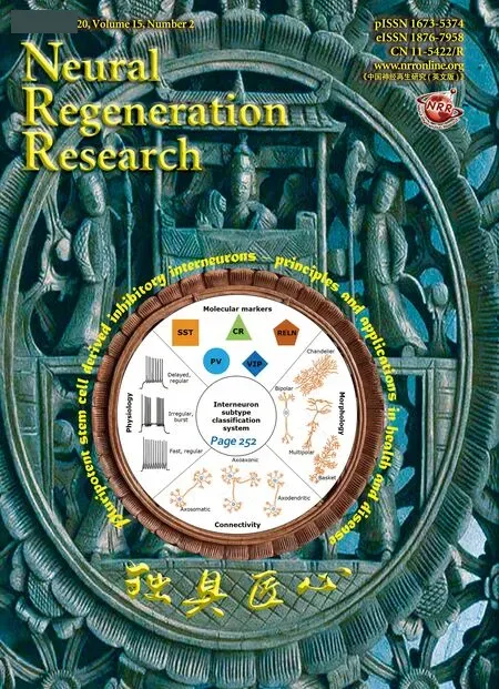
Inhibitory interneurons are gamma-aminobutyric acid-ergic (GABAergic)nerve cells that act to maintain the appropriate excitation-inhibition balance, and synchronise the output of principle cells to generate rhythmic patterns of firing (Kessaris et al., 2014). This critical role, along with their brain-wide distribution, has led to the implication of interneurons in many neuropathologies, including schizophrenia, autism, dystonia and epilepsies(Marín, 2012). This has in turn fuelled a growing interest into their investigation. The molecular and functional heterogeneities within this class of neurons have resulted in a complex multifactorial classification system to assign interneurons into multiple subtypes (Figure 1A). Our journey towards understanding the interneuron diversity behind the classification system has brought to light the following principles. First, the subtype reflects the birthplace of the interneuron. Within the developing brain, inhibitory interneurons are born within focal regions, including the caudal and medial ganglionic eminences (CGE and MGE, respectively). Fate-mapping experiments in rodents demonstrated that calretinin (CR), vasoactive intestinal peptide and reelin positive interneurons are predominantly derived from CGE progenitors. Whereas, somatostatin (SST) and parvalbumin (PV) subtypes are MGE-derived, with an apparent SST: PV ratio shift along the MGE dorsoventral gradient (Kessaris et al., 2014). Second, interneuron subtypes exhibit differential vulnerability in neurological diseases (Marín, 2012).Third, there are brain region-specific differences in interneuron subtype composition, which are also species-dependent (Wu and Parent, 2000). The significance of these findings is unclear, as the manner to which interneuron lineages are determined by intrinsic and extrinsic cues remains under investigation (Kessaris et al., 2014).
The importance of interneurons in neurological diseases, and the emerging evidence of species-specific subtype diversity and regional composition,has emphasized the need for a human interneuron model system for research and drug discovery. Over recent years several independent groups have been dedicated towards achieving robust, and efficient protocols to generate inhibitory interneurons from human pluripotent stem cell (hPSC)in vitro (Maroof et al., 2013; Nicholas et al., 2013; Noakes et al., 2019). In general, interneuron differentiation paradigms share a common principle of driving interneuron fate by mimicking the native MGE/CGE environment through inducing Sonic Hedgehog signalling. The interneuron-like progenitors generated give rise to a heterogeneous population of GABAergic interneurons following maturation in vitro and/or ex vivo after transplantation into the rodent brain (Maroof et al., 2013; Nicholas et al., 2013; Noakes et al., 2019).
With most studies focusing on cortical interneuron phenotypes, we transplanted hPSC-derived MGE/CGE progenitors into the neonatal rat striatum, and examined the morphological and molecular properties of the differentiated progeny in comparison to the sister cells maintained in vitro(Noakes et al., 2019). We found that in the host brain the hPSC-derivatives had adopted morphologies and molecular profiles akin to those found in the human striatum, with CR positive interneurons as the predominant phenotype present (Wu and Parent, 2000). Choline acetyltransferase interneurons, another subtype enriched in the human striatum, were also present within the graft, but absent when the culture was maintained in vitro. Moreover, no graft-derived SST and PV positive cells were detected. In contrast, the in vitro matured sister cells gave rise to a GABAergic population mainly comprised of SST positive interneuron-like cells, with a smaller proportion of CR and PV subtypes. So why is there a difference between the subtype composition of hPSC-derived interneurons matured in the host striatum and in vitro? One potential reason was a subtype-dependent survival bias. Post-transplantation into the host striatum, the graftcells must successfully integrate with the host circuits in order to survive.Under this principle, progenitors committed to the region-specific fate,such as CGE-like progenitors committed to CR fate, would have an innate advantage over PV and SST committed MGE-like progenitors. This would have led to CGE-like progenitor selected survival, integration and maturation within the host striatum, while MGE-like progenitors perished. On the other hand, if fate commitment of the progenitors was undetermined at the point of transplantation, an environment-driven redirection of interneuron fate could have led to a shift in the subtype composition to match the region(Figure 1B, model 2). The latter reasoning was used to explain a similar finding demonstrated with heterotopic transplantation of MGE tissues in rodents (Quattrocolo et al., 2017). The degree of fate commitment present at the progenitor stage could potentially be addressed by transplanting purified hPSC-derived CGE-like progenitors and MGE-like progenitors into the neonatal striatum (and cortex) using interneuron type-specific hPSC reporter lines. The relative survival and subtype composition achieved by these grafts (and between regions) would shed light on lineage potential of hPSC-derived progenitors, and relative influence of intrinsic and extrinsic cues have on interneuron fate.
An alternative, and complementary means, to battle the question of how and when interneuron lineage commitment, and divergence into lineage dictated subpopulations, occurs has been to perform longitudinal transcriptome analysis (Close et al., 2017; Mi et al., 2018). To date our understanding of the molecular steps driving interneuron lineage divergence, to identify the critical lineage determining transcription factors, remains incomplete.Consequently, opposing theories over intrinsic versus external influences on interneuron fate determination ensues (Figure 1B). To fully understand how interneurons diverged out into different subtypes requires us to tease apart the molecular heterogeneity within the population. This requires transcriptome analysis at single cell resolution. The first comprehensive example of single cell transcriptome analysis of hPSC-derived interneurons provided novel insight into the heterogeneity within the interneuron-like population, both at the progenitor and post-mitotic stages of interneuron differentiation in vitro (Close et al., 2017). A series of studies have also been carried out using primary mouse MGE/CGE tissue to give insight into the authentic interneuron population heterogeneity across mouse development (Mi et al., 2018). Initial findings from single cell RNA sequencing analysis in mouse identified, for the first time, molecular heterogeneity driven by origin-of-birth specific transcriptomic molecular signatures. The origin-of-birth reflected gene profiles were retained through development,remaining as an integral marker of each MGE/CGE-derived interneuron subtype when characterized within the adult mouse brain (Mi et al., 2019).This led to the conclusion that the denominating factor in interneuron lineage determination was an intrinsically instructed expression pattern, rather than one moulded by the final environment (Figure 1B, model 1). Currently, efficient generation of certain interneuron subtypes (typically PV) has proved challenging in vitro. Knowledge of lineage determining transcription factors would enable the generation of more authentic and defined interneuron subtypes in vitro, thereby enhancing the validity and scope of the model. This has spurred on the search for lineage determining transcription factors, which could subsequently help to refine hPSC differentiation protocols. However, the relative influence that intrinsic and extrinsic factors play in directing interneuron fate remains open to discussion, with supporting evidence for both theories (Kessaris et al., 2014). Moreover, whether these findings in mouse are replicated in human is still eagerly awaited.
Along with providing an in vitro model of human neural development,hPSC-derived neurons have many additional applications. Our ex vivo work demonstrated the successful functional maturation of hPSC-derived interneuron grafts in the rodent brain (Noakes et al., 2019). This adds to many other proof-of-principle studies that would encourage the use of hPSC as a valid alternative source to foetal tissue in cell replacement therapies (Tornero et al., 2017). However, there are additional questions to address, beyond graft survival and maturation, in order to validate the use of hPSC-derived neurons in regenerative medicine. The significance of successful and appropriate graft-host integration has become apparent in the literature through reports of survival alone being an insufficient predictor of behavioural improvements in rodent disease models. This has encouraged the use of novel techniques to establish functional connectivity between graft and host. One increasingly popular approach is the application of viral tracers, combined with optogenetic reporter, to allow both graft-host connectivity to be traced visually (to identify connecting partners), and the traced circuits to be activated or suppressed on demand (Tornero et al., 2017). This approach has been successfully described for multiple hPSC-derived neuronal classes, but the viral tracer system has not yet been reported to evaluate hPSC-derived interneuronal graft-host integration, or to determine circuit output at the behavioural level.

Figure 1 Inhibitory interneuron diversity - classification system and lineage commitment models.
Moreover, hPSC-derived neurons represent a useful and relatively inexpensive platform for in vitro disease modelling and drug screening. Induced pluripotent stem cells (iPSC) are a recent addition to hPSC research platform (Yu et al., 2007). Advancements in cell biology and technology paved our way towards being able to revert somatic cells (most typically skin fibroblasts) back to an unspecialised, pluripotent state (Yu et al.,2007). Pioneering research using neurons derived from iPSC of patients with neurodevelopmental disorders has greatly aided our understanding of these complex pathologies. With schizophrenia being a prime example of where patient derived iPSC work has provided an otherwise impossible insight into the neurodevelopmental potential of patient derived neural progenitors, and the functional deficits in the neurons generated (Shao et al., 2019). Whilst most of the iPSC modelling studies on neuropsychiatric disorders focused on cortical projection neurons, a recent report by Shao et al characterised disease-relevant phenotypes in patient iPSC derived interneuron-like cells (Shao et al., 2019). Genome-wide RNA sequencing in turn enabled the molecular pathways underpinning the phenotypic differences between patient and control interneuron-like cells to be identified (Shao et al., 2019). Pharmacological attempts to compensate for the dysregulated pathway, and rescue the phenotypic deficits in interneuron-like cells, by the authors highlights the use of in vitro models as one of many stepping-stones in the development of more efficacious therapies for neuropathologies.
In summary, hPSC-derived interneurons are useful tools towards understanding human interneuron development in health and disease. With the growing application of in vitro models, together with novel molecular techniques, many of the mysteries surrounding interneurons are finally being decoded.
This work was supported by grant from the UK Medical Research Council and EU Framework Programme 7 Repair-HD, to ML; FK is a recipient of a Wellcome Trust PhD studentship.
Francesca Keefe*, Meng Li*Neuroscience and Mental Health Research Institute, School of Medicine and School of Bioscience, Cardiff University, Cardiff, UK
*Correspondence to:Francesca Keefe, KeefeF@cardiff.ac.uk;Meng Li, PhD, lim26@cardiff.ac.uk.
orcid:0000-0001-7262-5307 (Francesca Keefe)
Received:June 19, 2019
Accepted:July 1, 2019
doi:10.4103/1673-5374.265547
Copyright license agreement:The Copyright License Agreement has been signed by both authors before publication.
Plagiarism check:Checked twice by iThenticate.
Peer review:Externally peer reviewed.
Open access statement:This is an open access journal, and articles are distributed under the terms of the Creative Commons Attribution-NonCommercial-ShareAlike 4.0 License, which allows others to remix, tweak, and build upon the work non-commercially, as long as appropriate credit is given and the new creations are licensed under the identical terms.
Open peer reviewer:Chong Gao, The University of Hong Kong, China.
- 中国神经再生研究(英文版)的其它文章
- Ethanol extract from Gynostemma pentaphyllum ameliorates dopaminergic neuronal cell death in transgenic mice expressing mutant A53T human alpha-synuclein
- Peripheral nerve injury induced changes in the spinal cord and strategies to counteract/enhance the changes to promote nerve regeneration
- Genetic targeting of astrocytes to combat neurodegenerative disease
- Pathological significance of tRNA-derived small RNAs in neurological disorders
- Applications of advanced signal processing and machine learning in the neonatal hypoxic-ischemic electroencephalography
- Protective effect of hydrogen sulfide on oxidative stress-induced neurodegenerative diseases