
Particulate matter exposure exacerbates susceptibility to SARS-CoV-2 infection in humanized ACE2 mice
2021-06-17 12:40Teng-YuZhu,HuanQiu,Qi-QiCao等
Zoological Research 2021年3期
The global outbreak of coronavirus disease 2019 (COVID-19),which is caused by severe acute respiratory syndrome coronavirus 2 (SARS-CoV-2), as of 8 May 2021, has surpassed 150 700 000 infections and 3 279 000 deaths worldwide. Evidence indicates that SARS-CoV-2 RNA can be detected on particulate matter (PM), and COVID-19 cases are correlated with levels of air pollutants. However, the mechanisms of PM involvement in the spread of SARS-CoV-2 remain poorly understood. Here, we found that PM exposure increased the expression level of angiotensin-converting enzyme 2 (ACE2) and transmembrane serine protease 2(TMPRSS2) in several epithelial cells and increased the adsorption of the SARS-CoV-2 spike protein. Instillation of PM in a hACE2 mouse model significantly increased the expression ofACE2andTmprss2and viral replication in the lungs. Furthermore, PM exacerbated the pulmonary lesions caused by SARS-CoV-2 infection in the hACE2 mice. In conclusion, our study demonstrated that PM is an epidemiological factor of COVID-19, emphasizing the necessity of wearing anti-PM masks to cope with this global pandemic.
The global outbreak of coronavirus disease 2019 (COVID-19), which is caused by severe acute respiratory syndrome coronavirus 2 (SARS-CoV-2), continues to significantly impact the global economy, health, and politics. Current studies have indicated that daily COVID-19 incidence is positively correlated with the concentration of air pollutants, especially PM (Fronza et al., 2020; Zoran et al., 2020). However, the mechanisms underlying PM involvement in the spread of SARS-CoV-2 remain poorly understood.
COVID-19 is spread through droplets and mainly results in lung injury, inflammation, and subsequent respiratory distress(Zhu et al., 2020). Here, we first investigated the adsorption of the spike protein of SARS-CoV-2 after PM treatment in human pulmonary alveolar epithelial cells (HPAEpiC). Notably,immunofluorescence analysis showed that more spike protein was adsorbed in the HPAEpiC cells after PM treatment(Figure 1A). Angiotensin-converting enzyme 2 (ACE2) and transmembrane serine protease 2 (TMPRSS2) are important entry factors of SARS-CoV-2 that bind to and cleave the viral spike protein (Matsuyama et al., 2010). ACE2 and TMPRSS2 are also highly expressed in other organs like the intestines and kidneys (Hamming et al., 2004). We, therefore, compared the protein expression levels of ACE2 and TMPRSS2 in HPAEpiC cells with or without PM administration. As shown in Figure 1B, the expression levels ofACE2andTMPRSS2in the HPAEpiC cells were significantly up-regulated after PM administration. Moreover, immunoblotting also revealed that the levels of ACE2 and TMPRSS2 were consistently upregulated in HPAEpiC cells after PM treatment (Figure 1C).Increased expression levels ofACE2andTMPRSS2were also observed in human colon adenocarcinoma (Caco2),human ileocecal carcinoma (Hct-8), and African green monkey kidney cell lines (Vero) (Supplementary Figure S1). Thus, we speculated that PM-elevated ACE2 and TMPRSS2 levels may be beneficial for SARS-CoV-2 infection.
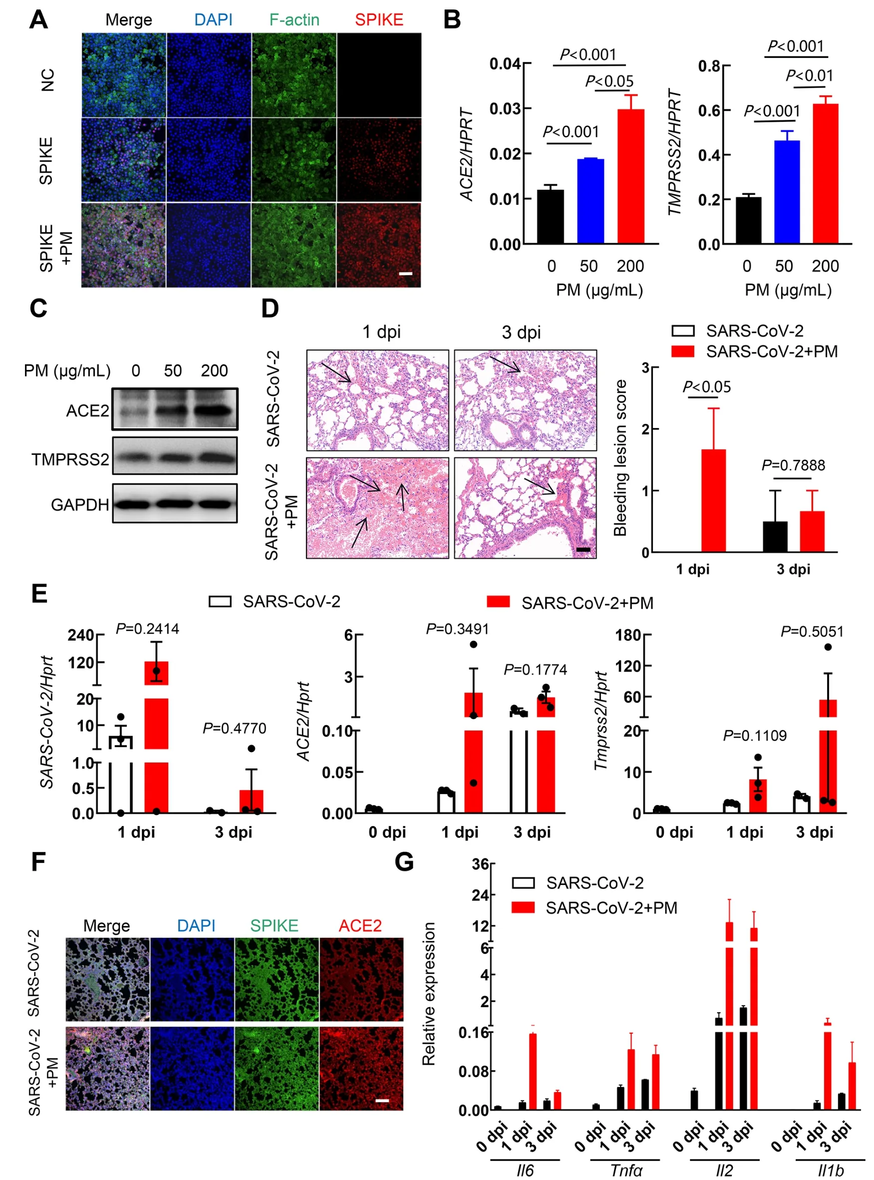
Figure 1 PM exposure induces ACE2 and TMPRSS2 expression and elevates susceptibility of hACE2 mice to SARS-CoV-2 infection
As SARS-CoV-2 has high affinity to human ACE2 but not mouse ACE2, we chose the recently established ACE2 humanized (hACE2) inbred mouse model to further examine whether PM plays a role in the pathogenesis of SARS-CoV-2 in vivo (Liu et al., 2021). Urban PM (1648a, NIST) was used in this study and was suspended in phosphate-buffered saline(PBS) to a final concentration of 10 mg/mL. The mice were intranasally inoculated with 400 μg PM (40 μL) or an equal volume of PBS (control mice) daily. After exposure to PM for 3 days, specific pathogen-free male hACE2 (C57BL/6NAce2em2(hACE2-WPRE,pgk-puro)/Gdl) mice were infected with SARSCoV-2 strain 107 (NMDCN0000HUI) at a dose of 2×10⁶TCID50intranasally. All animal procedures were reviewed and approved by the Institutional Committee for Animal Care and Biosafety from the Animal Care and Use Committee of the Kunming Institute of Zoology, Chinese Academy of Sciences,Yunnan, China (SMKX-20 200 402-09). No obvious gross pathological changes were observed in any mice at 1-day post-infection (dpi). Thickening of the alveolar septa with inflammatory cell infiltration in the alveolar cavities also showed little difference after PM exposure (Supplementary Figure S2A. However, SARS-CoV-2-infected hACE2 mice exposed to PM exhibited more severe congestion compared to unexposed mice at 1 dpi (Figure 1D). As shown in Figure 1E, viral loads andACE2andTmprss2expression at 1 and 3 dpi were higher in the lungs of SARS-CoV-2-infected hACE2 mice exposed to PM than in the control mice.Immunofluorescence analysis of the left lungs of SARS-CoV-2-infected hACE2 mice showed similar results (Figure 1F). In conclusion, after the SARS-CoV-2-infected hACE2 mice were exposed to PM, the expression ofACE2andTmprss2significantly increased in the lungs. Consistent with the previous observation, we found no significant differences in weight loss or body temperature between the two groups of mice (Supplementary Figure S2B, C). As shown in Figure 1G, the expression levels of COVID-19-related inflammatory factors, such asIl6,Tnfa,Il2, andIl1b, were significantly induced in the lungs of SARS-CoV-2-infected hACE2 mice exposed to PM compared with that in the control mice. Combined with other data, we found that instillation of PM exacerbated pulmonary lesions and viral loads and may have led to more severe pneumonia in hACE2 mice post SARS-CoV-2 infection. Recent research from Italy also demonstrated that COVID-19 cases are significantly associated with various air-borne pollutants (NO2, O3, PM2.5,PM10) (Fattorini & Regoli, 2020; Kim et al., 2020).Furthermore, cigarette smoke exposure is reported to increase the expression of ACE2 by a similar mechanism (Smith et al.,2020). Our novel results provide further insight into the strength of the correlation between PM and SARS-CoV-2 entry factors.
This study clarified the relationship between PM and SARSCoV-2 entry factors and provided new evidence regarding the underlying mechanism related to the increase in COVID-19 prevalence following PM exposure. However, under a stable atmosphere and high PM concentration, whether SARS-CoV-2 can cluster with PM particles and enhance the persistence of the virus in the atmosphere by reducing the diffusion coefficient has not yet been elucidated. In conclusion, our study demonstrated that PM may be an epidemiological factor of COVID-19, thus emphasizing the necessity of wearing anti-PM masks to help cope with this global pandemic.
SUPPLEMENTARY DATA
Supplementary data to this article can be found online.
COMPETING INTERESTS
The authors declare that they have no competing interests.
AUTHORS’ CONTRIBUTIONS
L.J., W.J.D. and R.L. designed the study. T.Y.Z., L.J. and H.Q.performed most of the experiments and analyzed the data.L.J. and Z.L.D. contributed to the animal experiments in Animal Biosafety Level 3 (ABSL-3). F.L.L., T.Z.S., G.M.W. and Y.T.Z. assisted in the animal experiments. Q.Q.C., Y.L. and Y.Q.F. assisted in RNA isolation and quantitative PCR detection. T.Y.Z. and L.J. wrote the manuscript. All authors read and approved the final version of the manuscript.
ACKNOWLEDGMENTS
The authors would like to thank Prof. Jie-Kai Chen(Guangzhou Institutes of Biomedicine and Health, Chinese Academy of Sciences) for the hACE2 mice and Dr. Peng Wang (Dalian Institute of Chemical Physics, Chinese Academy of Science) for the HPAEpiC cells.

- Zoological Research的其它文章
- Species bias and spillover effects in scientific research on Carnivora in China
- A review of the Cypriniform tribe Yunnanilini Prokofiev,2010 from China, with an emphasis on five genera based on morphologies and complete mitochondrial genomes of some species
- Potential aquatic environmental risks of trifloxystrobin:Enhancement of virus susceptibility in zebrafish through initiation of autophagy
- Northern pig-tailed macaques (Macaca leonina)infected with SARS-CoV-2 show rapid viral clearance and persistent immune response
- Cataract-causing allele in CRYAA (Y118D) proceeds through endoplasmic reticulum stress in mouse model
- Molecular mechanisms of intermuscular bone development in fish: a review