
根样釉珠1例
2016-07-26 03:39毛小泉蒙亚娇李海芳
国际口腔医学杂志 2016年4期
毛小泉 蒙亚娇 李海芳
中南大学湘雅医学院附属海口医院口腔中心•海南省口腔医学中心 海口 570208
·病例报告·
根样釉珠1例
毛小泉 蒙亚娇 李海芳
中南大学湘雅医学院附属海口医院口腔中心•海南省口腔医学中心 海口 570208
釉珠是出现于磨牙区的釉质突起,本文报道1例1.8 mm宽、8 mm长的扁平状釉珠。本病例是1例少见的、较长的牙根样的釉珠。
釉珠; 根尖片; 锥形束CT
釉珠是附着在牙骨质表面的釉质小块,是牙齿发育时小团错位的成釉细胞或者上皮根鞘某处异常分化,再度出现成釉功能而形成的釉质,常见为磨牙根分叉区或近颈部牙骨质上单个栗粒大小球形或小舌状的突起,牢固附着于牙根表面,为不透明结节。牙齿内部的釉珠呈圆形放射影像,范围从釉牙本质界到冠部牙本质。。
1 病例报告
患者,女,42岁,因牙痛就诊于中南大学湘雅医学院附属海口医院牙体牙髓病科。
临床检查为左侧下颌第一磨牙远中龋,根尖压痛和扣痛及颊侧深牙周袋,牙髓电活力检查阴性,近中根尖放射透明影。
X线检查根分叉区有不透明突起,扁形,1.8 mm宽、8 mm长(图1~3)。
患牙被诊断为根尖周炎,盐酸甲哌卡因/肾上腺素注射液(斯康杜尼)麻醉后,橡皮障隔湿,开髓,找到3个根管口。根尖片可见牙槽骨内有一

图 1 根分叉釉珠Fig 1 Enamel pearl in furcation area

图 2 CT水平面观Fig 2 CT horizontal section view

图 3 CT颊舌切面观Fig 3 CT buccolingual section view
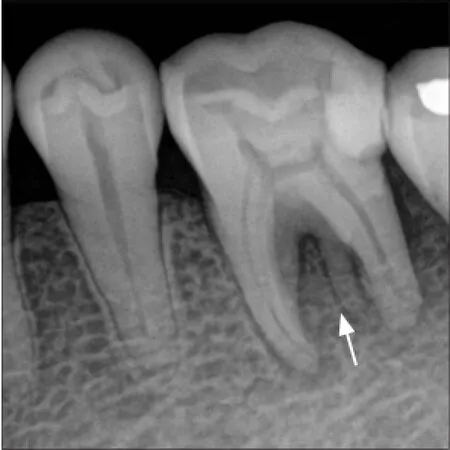
图 4 根样釉珠Fig 4 Root-like enamel pearl
所有根管使用不锈钢K锉和机用马达预备,质量分数为3%的过氧化氢冲洗,彻底干燥,根管内置樟脑酚,氧化锌暂封。1周后患者症状完全消失,拍片进行根管长度确定后,使用牙胶进行根管充填。随后进行烤瓷冠修复,3个月后无临床症状。
2 讨论
釉珠是沉积于恒牙根分叉区的异常釉质,尤其是上颌第二、第三磨牙[1],每颗牙通常仅有1颗,但也有观察到3~4颗者[2],发生率在1.1%~9.7%[3]。一般不发生于单根牙,也有报道发生于前磨牙、尖牙、切牙者[4-6],主要附着于牙根表面,也有发生于牙本质者[7],直径在0.3~4 mm[8]。而笔者报道1例根样釉珠,较少见:附着于分叉区,呈牙根样,1.8 mm宽、8 mm长。
临床上发现的釉珠大小不一,其内部结构也不尽相同。放大观察,较小的釉珠有时仅为釉质构成,而有时含有牙本质;体积较大时,往往可见纤细的牙髓组织进入釉珠内。锥形束CT(cone beam computed tomography,CBCT)有助于釉珠的诊断[9-10]。一般来说,釉珠会影响牙龈和牙体之间的良好附着关系,引起牙周疾病,且诊治时妨碍龈下刮治;釉珠下方有时可见多个小孔,因此有可能成为牙髓-牙周联合病变的感染途径[11-12]。
现已明确描述的釉珠有3种类型[13-14]:1)完全由釉质组成;2)复合釉珠或釉质牙本质型釉珠,包含管状牙本质;3)釉质牙本质型釉珠含有髓角,可能延伸到冠髓腔或根管。
釉珠是临床上一种较为少见的牙齿形态异常,局限性釉质增生的小球形团块。其发生的病因仍然不清楚,通常认为是上皮根鞘上皮剩余发育的结果,牙根发育过程中,这些细胞分化为有功能的成釉细胞,产生釉质沉积于牙根并牢固附着于牙根表面[15-16]。
3 结论
本例釉珠较为少见,像牙根。釉珠可以是完全性釉质或者含有牙本质或牙髓组织,下方有时可见多个小孔,成为牙髓和牙周组织的交通通道。由于釉珠可能形成牙本质小管/髓腔,因此治疗牙髓病时要仔细检查髓室底,沉积的牙本质必须仔细磨除以防止根管遗漏[1],并拍摄CBCT,评估釉珠内是否含有牙本质小管。对于引起牙周病的釉珠临床上可磨除,必要时断面局部备洞填充[17]。
[1] Shojaeian S, Ghoddusi J, Hajian S. A case report of maxillary second molar with two palatal root canals and a furcal enamel pearl[J]. Iran Endod J, 2013, 8 (1):37-39.
[2] 吴丽. 右上第三磨牙多发釉珠1例[J]. 实用口腔医学杂志, 2013, 29(4):510. Wu L. The multiple enamel pearls in right maxilla third molar[J]. J Pract Stomatol, 2013, 29(4):510.
[3] Chrcanovic BR, Abreu MH, Custódio AL. Prevalence of enamel pearls in teeth from a human teeth bank[J]. J Oral Sci, 2010, 52(2):257-260.
[4] Sehic A, Risnes S, Khan QE, et al. Gene expression and dental enamel structure in developing mouse incisor[J]. Eur J Oral Sci, 2010, 118(2):118-130.
[5] Sehic A, Peterkova R, Lesot H, et al. Distributionand structure of the initial dental enamel formed in incisors of young wild-type and Tabby mice[J]. Eur J Oral Sci, 2009, 117(6):644-654.
[6] Sehic A, Peterkova R, Lesot H, et al. P32-distribution and structure of the initial dental enamel formed in incisors of young wild-type and Tabby mice[J]. Bull Group Int Rech Sci Stomatol Odontol, 2010, 49 (3):104-106.
[7] Sehic A, Nirvani M, Risnes S. Incremental lines in mouse molar enamel[J]. Arch Oral Biol, 2013, 58 (10):1443-1449.
[8] Versiani MA, Cristescu RC, Saquy PC, et al. Enamel pearls in permanent dentition: case report and micro-CT evaluation[J]. Dentomaxillofac Radiol, 2013, 42 (6):20120332.
[9] Akgül N, Caglayan F, Durna N, et al. Evaluation of enamel pearls by cone-beam computed tomography (CBCT)[J]. Med Oral Patol Oral Cir Bucal, 2012, 17 (2):e218-e222.
[10] Kottoor J, Hemamalathi S, Sudha R, et al. Maxillary second molar with 5 roots and 5 canals evaluated using cone beam computerized tomography: a case report [J]. Oral Surg Oral Med Oral Pathol Oral Radiol Endod, 2010, 109(2):e162-e165.
[11] Aggarwal V, Singla M, Logani A, et al. Endodontic management of a maxillary first molar with two palatal canals with the aid of spiral computed tomography: a case report[J]. J Endod, 2009, 35(1):137-139.
[12] Romeo U, Palaia G, Botti R, et al. Enamel pearls as a predisposing factor to localized periodontitis[J]. Quintessence Int, 2011, 42(1):69-71.
[13] Cavanha AO. Enamel pearls[J]. Oral Surg Oral Med Oral Pathol, 1965, 19:373-382.
[14] Saini T, Ogunleye A, Levering N, et al. Multiple enamel pearls in two siblings detected by volumetric computed tomography[J]. Dentomaxillofac Radiol, 2008, 37(4):240-244.
[15] Khan QE, Sehic A, Khuu C, et al. Expression of Clu and Tgfb1 during murine tooth development: effects of in-vivo transfection with anti-miR-214[J]. Eur J Oral Sci, 2013, 121(4):303-312.
[16] Khan QE, Press CM, Sehic A, et al. Expression of prion gene and presence of prion protein during development of mouse molar tooth germ[J]. Eur J Oral Sci, 2010, 118(6):559-565.
[17] 张震康, 俞光岩. 实用口腔科学[M]. 北京: 人民卫生出版社, 2009:37-38. Zhang ZK, Yu GY. Practice of stomatology[M]. Beijing: People’s Medical Publishing House, 2009: 37-38.
(本文编辑 王姝)
A case report of root-like enamel pearl
Mao Xiaoquan, Meng Yajiao, Li Haifang. (Stomatology Center, Affiliated Haikou Hospital, Xiangya Medical College of Central South University, Oral Medicine Center in Hainan, Haikou 570208, China)
Enamel pearls are generally found in maxillary molars as a small globule of enamel. However, we report here in an enamel pearl exhibiting a prolate spheroid shape and is 1.8 mm wide and 8 mm long. This rare enamel pearl is very long and resembles a dental root.
enamel pearl; periapical radiographic image; cone beam computed tomography
R 781.34
B
10.7518/gjkq.2016.04.009
2015-05-19;
2015-10-30
毛小泉,副主任医师,硕士,Email:horse.m@163.com
毛小泉,副主任医师,硕士,Email:horse.m@163.com个根样影像(图4),故再次寻找根管口,但未找到。
猜你喜欢
口腔医学(2021年10期)2021-12-02
科技进步与对策(2021年12期)2021-06-23
安全(2021年4期)2021-05-19
湖南包装(2020年6期)2021-01-20
口腔疾病防治(2020年2期)2020-02-26
今古传奇·双月号(2019年2期)2019-06-11
文贝:比较文学与比较文化(2015年1期)2015-11-14
医学研究杂志(2015年9期)2015-07-01
