
Effect of acupuncture in intervening heroin-induced brain damage via regulating ubiquitin-proteasome pathway
2018-04-28 03:27ZhangLida张利达CaoJiangpeng曹江鹏CaiXinghui蔡兴慧WuShengbing吴生兵HouXiaorong侯晓蓉GaoYonglong高永龙ZhangRongjun张荣军SongXiaoge宋小鸽
Zhang Li-da (张利达), Cao Jiang-peng (曹江鹏), Cai Xing-hui (蔡兴慧), Wu Sheng-bing (吴生兵), Hou Xiao-rong (侯晓蓉),Gao Yong-long (高永龙), Zhang Rong-jun (张荣军), Song Xiao-ge (宋小鸽)
1 School of Graduate, Anhui University of Chinese Medicine, Hefei 230038, China
2 School of Acupuncture, Moxibustion and Tuina, Anhui University of Chinese Medicine, Hefei 230038, China
3 Key Laboratory of Xin’ an Medicine Constructed by Anhui Province and Chinese Ministry of Education, Anhui University of Chinese Medicine, Hefei 230038, China
4 School of Ⅰntegrated Traditional Chinese and Western Medicine, Anhui University of Chinese Medicine, Hefei 230038, China
5 Ⅰnstitute of Acupuncture and Meridian, Anhui University of Chinese Medicine, Hefei 230038, China
Research has shown that cell apoptosis is involved in the heroin-induced loss or damage of neuron function;major pathological manifestations of heroin-induced toxic brain damage include degeneration and necrosis of nerve cells at multiple sites and dissolved or disrupted microvascular basement membrane[1-4].Acupuncture, as a unique therapy used in preventing and treating drug addiction in our country, has shown its advantages[5]. It’s been proved by studies that acupuncture can mitigate the protracted withdrawal symptoms of heroin addicts and inhibit the nerve cell apoptosis in heroin-addicted rats[6-7].
Ubiquitin-proteasome pathway (UPP) is a highefficient mechanism for protein catabolism, composed of ubiquitin (Ub), ubiquitin activating enzyme (E1),ubiquitin conjugating enzyme (E2), ubiquitin protein ligase (E3), 26S proteasome and ubiquitin recycling enzyme[8]. In the form of polyubiquitin chain, Ub binds to and labels the to-be-degradated substrate protein for further degradation; E3 directly or indirectly promotes Ub to transfer to the target protein and form up polyubiquitin chain; 26S protein proteasome decomposes the ubiquitinized target protein into small peptides or amino acids. It’s reported that UPP has mediated cell apoptosis in multiple ways[9]. Taking this as the focus, this study was to observe the effect of acupuncture on UPP in heroin relapse rats and discuss the action mechanism of acupuncture in intervening heroin-induced brain damage.
1 Materials
1.1 Experimental animals
Thirty male Sprague-Dawley (SD) rats, 3-month-old and weighing (200±20) g, were provided by Anhui Experimental Animal Center [certificate of approval:SCXR (Wan) 2011-002; quality tested by: Anhui Laboratory Animal Quality and Inspection Center]. The rats were housed separately in a quiet environment,with the room temperature controlled at (22±1) ℃and relative humidity at (55±5)%, with natural light and free access to food and water. The plastic cages were paved with sterilized wood dust. The adaptive feeding lasted for 1 week before the beginning of the experiment. The whole process of experiment conformed to theGuiding Opinions on the Treatment of Experimental Animalsissued by the Ministry of Science and Technology, in order to avoid any unnecessary harm to the rats and reduce pain and sufferings.
1.2 Main reagents
Heroin (provided by Anhui Drug Prohibition Office,purity 85%); normal saline injection (Jiangsu Chiatai Tianqing Pharmaceutical Co., Ltd., China); primer synthesis (Invitrogen, USA); fluorescent quantitative reagents and consumable materials (Qiagen, Germany);Trizol (Life Technologies, USA); RNeasy® MinEluteTM cleanup kit (Qiagen, Germany); reverse transcriptase(Promega, USA); real-time polymerase chain reaction(RT-PCR) kit (Thermo, USA); in situ cell apoptosis detection kit (Roche, USA); bs-9347R primary antibody 26S (1:11), bs-1549R Ub (1:100) and bs-4895R E3 (1:100)(Beijing Bioss Biotechnology Co., Ltd., China); ZB-2301 HRP-labeled goat anti-rabbit IgG (H+L) (1:10 000, Beijing ZSGB-Bio, China).
1.3 Experiment instruments
DP-801 YB-7B paraffin embedding machine (Taiwei Electronics Co., Ltd., China); Leica RM2135 automatic microtome (Leica Incorporation, Germany); Olympus BX61 optical microscope (Olympus Corporation, Japan);DP-801 microscope image processing system (Jiangsu JEDA Science-technology Development Co., Ltd., China);quantitative fluorescence PCR instrument and PIKO Plate Illuminator (Thermo, USA); QuantiFast SYBR Green PCR kit (Qiagen, Germany).
2 Methods
2.1 Modeling
2.1.1 Heroin addicted rat model
The rats for heroin addicted model establishment received intramuscular heroin injection for successive 8 d at a progressively increased dose [daily heroin injection dose was 3, 3, 4, 4, 5, 8, 10 and 10 mg/(kg·bw)respectively from the 1st day to the 8th day]. During day 1-4, the daily injection was finished by 1 time;during day 5-8, the daily injection was finished by two injections, at 7:00 and 19:00 respectively.
2.1.2 Heroin relapse rat model
By following the steps of establishing heroin addicted rat model, the rats received 8-day intramuscular heroin injection at a progressively increased dose for addiction,followed by 5-day suspension of the injection for withdrawal. The addiction-withdrawal process was repeated 3 times, i.e. addiction → withdrawal→addiction→withdrawal→addiction→withdrawal, to establish a heroin relapse rat model[10-11].
2.2 Grouping and Intervention
During the experiment, the rats were divided into 3 groups by using the random number table: a control group, a model group and an acupuncture group, with 10 rats in each group.
2.2.1 Control group
Based on the cycles in the establishment of heroin relapse rat model, rats in the control group received intramuscular injection of normal saline at the addiction stage, 0.2 mL each time for each rat, once a day. The rats didn’t receive any interventions at the withdrawal stage.
2.2.2 Model group
Based on the cycles in the establishment of heroin relapse rat model, rats in the model group received 8-day intramuscular heroin injection at a progressively increased dose at the addiction stage, and didn’t receive any interventions at the withdrawal stage.
2.2.3 Acupuncture group
Based on the cycles in the establishment of heroin relapse rat model, rats in the acupuncture group received 8-day intramuscular heroin injection at a progressively increased dose at the addiction stage, and acupuncture treatment at the withdrawal stage.
Acupuncture method: Rat was placed gently on a specially-made experiment frame. When it’s sober and quiet, Baihui (GV 20) was punctured horizontally and Dazhui (GV 14) was punctured obliquely both by around 12 mm with filiform needles of 0.25 mm in diameter and 25 mm in length by referring to theAtlas of Acupoints for Rat[12]. The needles were retained for 30 min and manipulated every 10 min. The treatment was performed once a day, for successive 5 d, carried out at each withdrawal stage of the three cycles.
2.3 Observation items and methods
2.3.1 Sampling
The model obtained in this study is mature, with a high replication rate. There was no death of animal in this experiment.
The rats were sacrificed on the 39th day of the experiment. They were first anesthetized via intraperitoneal injection of 10% chloral hydrate at 3.6 mL/(kg·bw). Under successful anesthesia, rat’s thoracic cavity was opened by surgical scissors to expose the heart area. A perfusion needle was then inserted along the left ventricle into the heart nearby the apex to inject 0.9% NaCl 250 mL. When liver and lungs turned pale and the effluent from the right atrium became clear, 4% paraformaldehyde in phosphate buffer solution was used for perfusion until rat presented twitched limbs, stiff limbs and tail, and rigid body. Rat’s head was then chopped off and craniotomy was performed. The complete brain tissues were extracted and placed on an ice bag. The hippocampus and ventral tegmental area (VTA) were obtained according to theStereotaxic Atlas of the Rat Brain[13]and fixed in paraformaldehyde for 24 h prior to paraffin embedding.
2.3.2 Detection of cell apoptosis
Each group contributed paraffins from 3-5 rats. The paraffins of hippocampus and VTA were sliced at 5 µm and each rat had 4-6 slices for the detection of cell apoptosis in hippocampus and VTA by using terminal deoxynucleotidyl transferase-mediated nick and labeling (TUNEL). The number of apoptotic cells in rat VTA and hippocampus from each group was counted by using DP-801 microscope image processing system. Five discrete fields were selected from each slice for cell counting through microscope at a magnification of 400 times. Brown stained granules in cytoplasm were considered as positive staining. The numbers of positively stained cells were gathered for statistics.
2.3.3 Protein expressions of Ub, E3 and 26S in rat VTA and hippocampus
Each group contributed paraffins of the left brain of 3-5 rats. The paraffins of hippocampus and VTA were sliced at 5µm and each rat had 4-6 slices to determine the protein expressions of Ub, E3 and 26S by following the instruction of immunohistochemistry (IHC) kit. The Image-Pro plus 6.0 was adopted to detect the positive expressions of Ub, E3 and 26S in the IHC slices of rat VTA and hippocampus through microscope at a high magnification of 400 times. The mean optical density(MOD) was collected for statistics. Five discrete fields were randomly selected to calculate the mean value,which was taken as the positive expression of Ub, E3 and 26S proteins, respectively.
2.3.4 Expressions of Ub, E3 and 26S mRNAs in rat VTA and hippocampus
Ten rats from each group had their VTA and hippocampus of the right brain examined by quantitative real-time PCR (RT-qPCR) for the expressions of Ub, E3 and 26S mRNAs. The collected samples were put in cryovials and preserved at –80 ℃ for stand-by.The total RNA was extracted from the tissues ground in liquid nitrogen by Trizol. The mass of the total RNA was about 3 µg. After nucleic acid gel electrophoresis,OD260 and OD280 of RNA were detected by using ultraviolet (UV) spectrophotometer. The content and purity were calculated. A content above 1 µg/µL and purity between 1.8 and 2.0 indicated that the extracted RNA was not decomposed and had a good integrity.Reverse transcription was performed by using reverse transcription kits to obtain cDNA. QuantiFast SyBr Green PCR kit was adopted for PCR reaction. The sequence of primer is shown in Table 1. The reaction system was 10 µL. The condition for amplification was:40 cycles of denaturalization at 95 ℃ for 5 min, 95 ℃for 10 s and 60 ℃ for 30 s. The CT values of Ub, E3 and 26S mRNAs were obtained at the end of the reaction.The relative expressions of the target genes were calculated by using 2-ΔΔCT.
2.4 Statistical methods
Statistics were conducted by using SPSS 17.0 for windows. The data were expressed by mean ± standard deviation, and one-way ANOVA was used for the inter- group comparison of mean value; for multiple comparison of mean value, the least significant difference was adopted when the variance was homogeneous, otherwise, Tamhane’s T2 would be used.P<0.05 was indicative of a statistical significance.Statistical graphs were drawn by using GraphPad Prism 6.
3 Results
3.1 Nerve cell apoptosis in rat VTA and hippocampus
The apoptotic cells positively stained by TUNEL often distributed independently, with dense concentrated and brown-stained nucleus. The cells negatively labeled by TUNEL had normal nucleus stained light blue. The control group had less positively stained cells and most of the nuclei were in an oval or round shape and stained light blue. The model group had more positively stained cells and the nuclei were majorly stained brown(Figure 1 and Figure 2).
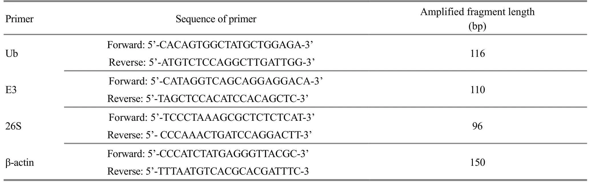
Table 1. Primer sequences in the RT-qPCR reaction
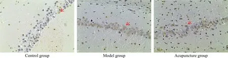
Figure 2. Apoptotic nerve cells in rat hippocampus (TUNEL, ×400)
Comparison of the positively stained cells in rat VTA and hippocampus: compared with the control group,the total number of the positively stained cells by TUNEL in rat VTA and hippocampus was significantly higher in the model group (P<0.01); compared with the model group, the total number of the positively stained cells by TUNEL in rat VTA and hippocampus significantly dropped in the acupuncture group (P<0.01), (Figure 3).
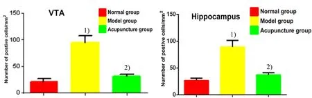
Figure 3. Comparison of the positive cell count in rat brain slices
3.2 Expressions of Ub, E3 and 26S proteins in rat VTA and hippocampus
Ub, E3 and 26S are resident in cytoplasm. Cells with its cytoplasm stained brown were positive ones, and those with its cytoplasm stained light blue were negative one (Figure 4-Figure 9).
The expressions of Ub, E3 and 26S proteins in rat VTA and hippocampus were significantly higher in the model group than in the control group (P<0.01); the expressions of Ub, E3 and 26S proteins in rat VTA and hippocampus were significantly lower in the acupuncture group compared with those in the model group (P<0.01). The results suggest that heroin re-addiction can increase the expressions of Ub, E3 and 26S in rat brains, while acupuncture can inhibit the increase (Figure 10).
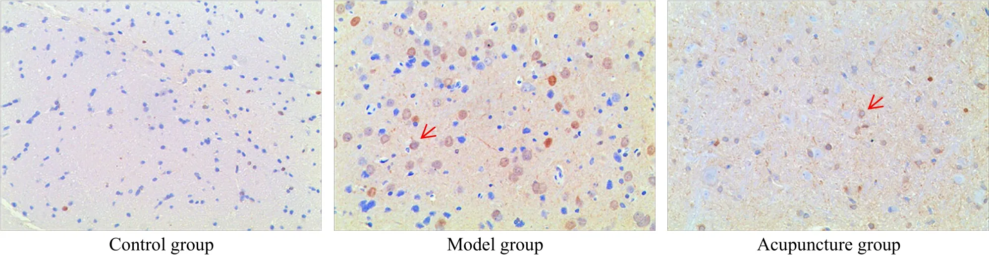
Figure 4. Expression of Ub protein in rat VTA (IHC, ×400)

Figure 5. Expression of E3 protein in rat VTA (IHC, ×400)
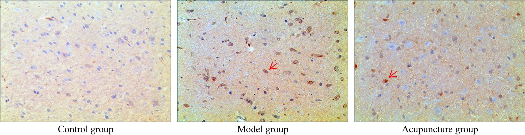
Figure 6. Expression of 26S protein in rat VTA (IHC, ×400)
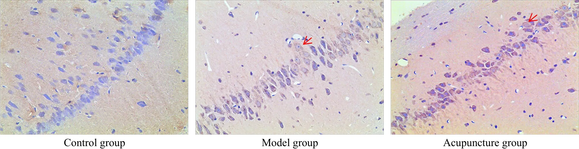
Figure 7. Expression of Ub protein in rat hippocampus (IHC, ×400)
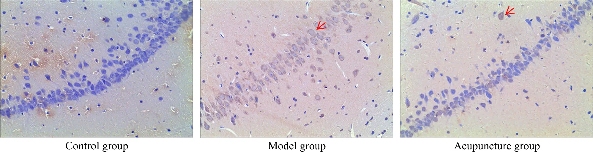
Figure 8. Expression of E3 protein in rat hippocampus (IHC, ×400)

Figure 9. Expression of 26S protein in rat hippocampus (IHC, ×400)
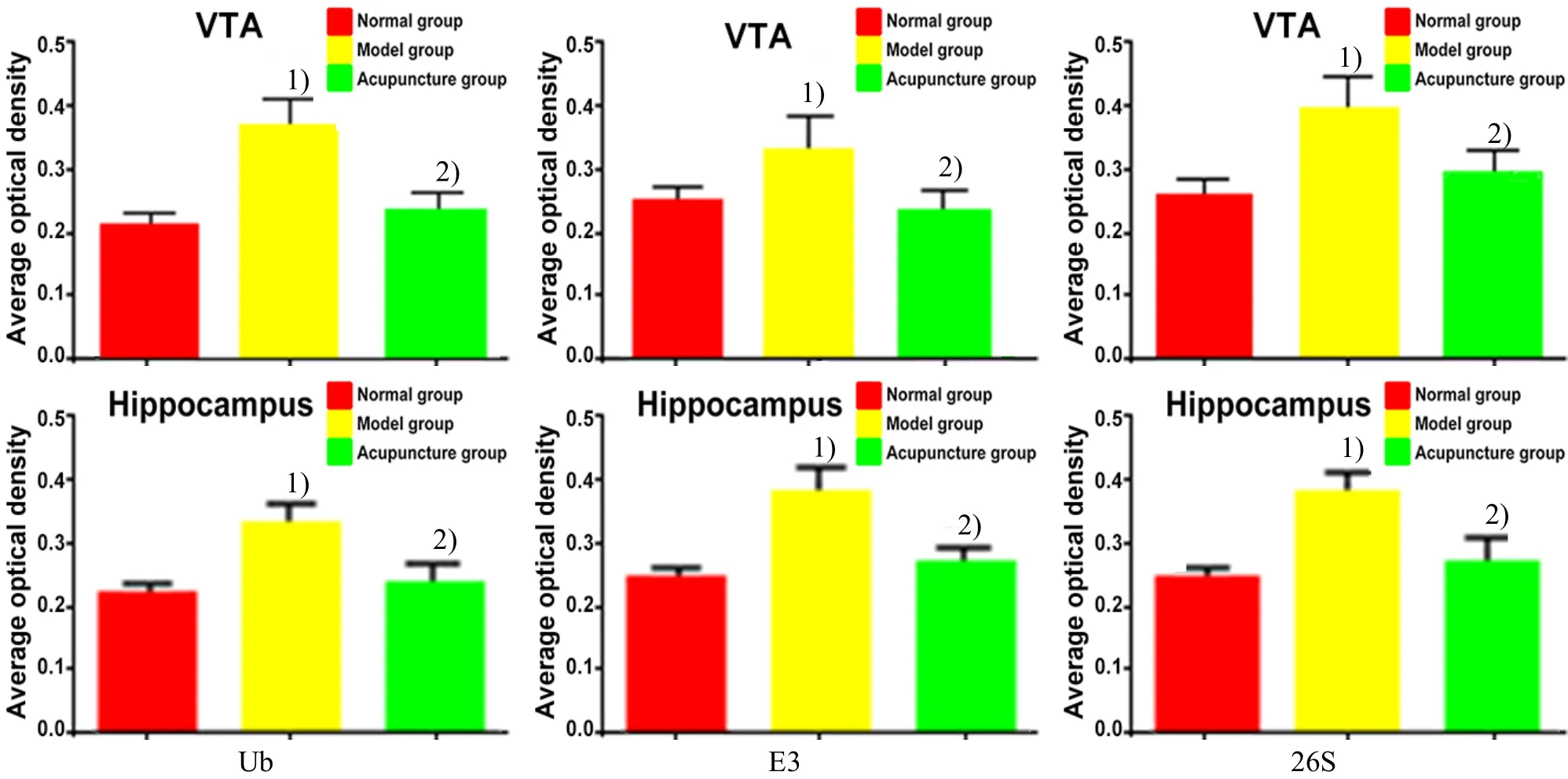
Figure 10. Comparison of the expressions of Ub, E3 and 26S proteins in rat VTA and hippocampus
3.3 Expressions of Ub, E3 and 26S mRNAs in rat VTA and hippocampus
The expressions of Ub, E3 and 26S mRNAs in rat VTA and hippocampus were significantly higher in the model group than in the control group (P<0.01,P<0.05); the expressions of Ub, E3 and 26S mRNAs in rat VTA and hippocampus were significantly lower in the acupuncture group compared with those in the model group (P<0.01,P<0.05), (Figure 11).
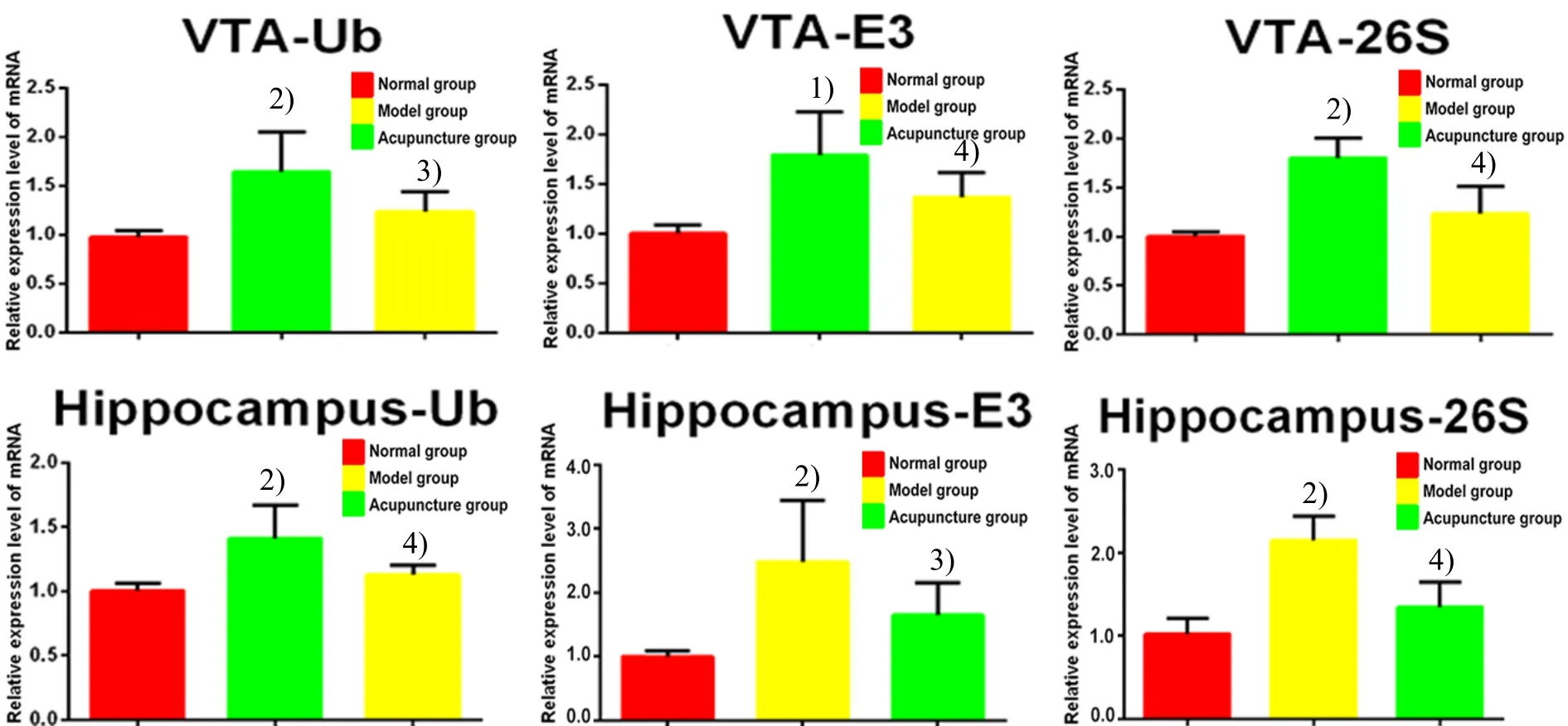
Figure 11. Comparison of the expressions of Ub, E3 and 26S mRNAs in rat VTA and hippocampus
4 Discussion
UPP is an important mechanism in modulating protein degradation[14], involved in the regulation of many aspects in the cell cycle, such as signal transduction, DNA repair, abnormal protein catabolism,and cell receptor function. UPP is significant in maintaining normal cell function and homeostasis.
Traditional Chinese medicine holds that the brain governs consciousness and mental activities. Heroin can cause damages to many systems of human body,especially the brain, usually manifested by disorders of consciousness, mental disorders, and attacks of convulsion[15]. The Governor Vessel travels through the spine and connects with the brain. The acupoints of this meridian often have functions to treat mental diseases.Baihui (GV 20) is located at the vertex with brains beneath it. Modern research has discovered that the superficial layer of Baihui (GV 20) is full of vessels and nerves, e.g. major occipital nerve and branch of the frontal nerve, and its deep layer resides motor cortex and paracentral lobule. Therefore, stimulating Baihui(GV 20) can directly improve the central nervous function and blood circulation. As the confluent point of the three yang meridians of both hand and foot and the Governor Vessel, Dazhui (GV 14) interiorly connects with the Governor Vessel and exteriorly connects with the three yang meridians, with function to clear pathogenic factors of yang nature and excess heat, open orifices and awaken brain, regulate the Governor Vessel and release convulsion. The two acupoints are significant in regulating brain function.
This study found that in the heroin relapse rats, the apoptotic central nervous cells increased, as well as the expressions of Ub, E3 and 26S mRNAs and proteins in rat VTA and hippocampus. It’s indicated that heroin re-addiction can cause certain damage to the central nervous system. Heroin-induced brain damage can trigger stress reactions[16], so that body will increase the UPP activity and expressions of Ub, E3 and 26S via autoregulation. However, the increase of UPP activity is not enough to inhibit cell apoptosis.
After acupuncture at Baihui (GV 20) and Dazhui(GV 14), the apoptotic rat central nervous cells decreased, and the expressions of Ub, E3 and 26S mRNAs and proteins in rat VTA and hippocampus dropped significantly. It’s suggested that acupuncture can inhibit the stress reactions via down-regulating the activity of UPP and cell apoptosis, so as to protect the brain. It’s widely known that the increase of CCAAT-enhancer-binding protein homologous protein (CHOP)is the signal of endoplasmic reticulum stress[17-19]. Our previous study showed that acupuncture can inhibit the expression of CHOP and promote the expression of glucose regulated protein 78 kD (GRP78), so as to modulate endoplasmic reticulum stress and inhibit nerve damage[20]. Meanwhile, acupuncture can also up-regulate the expressions of heat shock protein 70(HSP70), heat shock protein 105 (HSP105) and valosin-containing protein (Vcp), which can enhance protein-folding in endoplasmic reticulum, reduce cell apoptosis in the brain of heroin relapse rats, and prevent brain damage.
Another report told that the expression of E2 dropped after electroacupuncture in acute spinal cord injury rats[21]. It’s believed that the abnormal proteins produced during the injury triggered the increase of E2,which should be a protective reaction of the body;electroacupuncture may down-regulate the expression of E2 by reducing the expression of the abnormal proteins. It was suspected that electroacupuncture promoted the repair of the injured spinal cord through inhibiting nervous cell apoptosis, improving cell metabolism and regulating abnormal proteins. The results are in accordance with the outcomes of this study.
The action of UPP is rather complicated. Some reports suggest that it can promote cell apoptosis, while some other reports hold an opposite opinion[22-23].There still requires more valuable profound studies to support the effect of acupuncture in regulating UPP and intervening nerve injury. Therefore, researchers need to keep an eye on the novel research findings and progress.
Conflict of Interest
The authors declared that there was no potential conflict of interest in this article.
This work was supported by National Natural Science Foundation of China (国家自然科学基金项目, No.81173325, No. 81503658); Undergraduate Ⅰnnovation Training Program of Anhui Province (安徽省大学生创新训练计划项目, No. 2015043); Scientific Platform Construction Program of Anhui Colleges and Universities(安徽高校科研平台建设项目, No. 2015TD033).
Statement of Human and Animal Rights
The treatment of animals conformed to the ethical criteria in this experiment.
[1] Lai BQ, Pu HW, Cao QH, Jing HL, Liu XS. Activation of caspase-3 and c-Jun NH2-terminal kinase signaling pathways involving heroin-induced neuronal apoptosis.Neurosci Lett, 2011, 502(3): 209-213.
[2] Luo LM, Liu JF, Gong Q, Qin QY, Zhu H. The effect of heroin on the synaptic muber and structure of amygdaloid body in rats. Zhongguo Fayixue Zazhi, 2016, 31(2):134-136.
[3] Li YX, Liang WM. Expression of β-EP in brain ventral tegmental area of heroin dependent rats. Guiyang Yixueyuan Xuebao, 2013, 38(5): 458-460.
[4] Pi MS, Wu YX, Ru Q, Li CY. Research progress of the damage of common narcotics to neurons in the brain.Shenjing Sunshang Yu Gongneng Chongjian, 2014, 9(1):63-67.
[5] Hou XR, Zhang RJ, Lü H, Cai XH, Xie GC, Song XG.Acupuncture at Baihui (GV 20) and Dazhui (GV 14)reduces brain cell apoptosis in heroin readdicts. Neural Regen Res, 2014, 9(2): 164-170.
[6] Hou WG, Liang Y, Li Y, Cheng B, Xu J, Xu B, Zong L.Puncturing Jiaji (EX-B 2) points for protracted abstinence in heroin addicts. Shanghai Zhenjiu Zazhi, 2011, 30(6):357-359.
[7] Wang N, Zhang RJ, Hou XR, Song XG, Dong ZY, Xu T.Effect of acupuncture on brain nerve cell apoptosis in heroin relapse rats. Anhui Zhongyi Xueyuan Xuebao, 2013,32(5): 44-48.
[8] Chang TL, Lin SW, Wu SL, Hong CM. Regulation of ubiquitin and 26S proteasome mediated by phenolic compounds during oxidative stress. J Nutr Biochem, 2013,24(11): 1970-1981.
[9] Wang GK, Tian FX, Gong JF, Wang W. Programmed cell death mediated by ubiquitin-26S proteasome system.Shengming Kexue, 2011, 23(1): 26-31.
[10] He GZ. Establishment of the model of heroin addicted rats.Sichuan Jiepouxue Zazhi, 2005, 13(2): 4-6.
[11] Wei XL, Ye J, Zheng Y. Establishment of the model of heroin addicted rats. Guangxi Yixue, 2004, 26(6): 783-785.
[12] Hua XB. Atlas of Acupoints for Rat. Beijing: People’s Medical Publishing House, 1991: 121.
[13] Bao XM, Shu SY. The Stereotaxic Atlas of the Rat Brain.Beijing: People’s Medical Publishing House, 1991: 95-104.
[14] Wang X, Wei XF, Zhang HQ. Role of protein ubiquitimation and its functional improtance. Zhongguo Kexue: Shengming Kexue, 2015, 45(11): 1074-1082.
[15] Yang L, Zhang GS, Zhao X. The injury mechanism and reversibility on inhibitory control function of heroin addicts.Xinli Kexue Jinzhan, 2014, 22(3): 439-447.
[16] Gao YL, Zhang Y, Cao JP, Wu SB, Cai XH, Zhang YC,Zhang RJ, Song XG, Zhang LD. Regulation of the endoplasmic reticulum stress response and neuroprotective effects of acupuncture on brain injury caused by heroin addiction. Acupunct Med, 2017, 35(5): 366-373.
[17] Ferri KF, Kroemer G. Organelle-specific initiation of cell death pathways. Nat Cell Biol, 2001, 3(11): E255-E263.
[18] Gu SY, Chen CZ, Jiang XJ, Zhang ZZ. ROS-mediated endoplasmic reticulum stress and mitochondrial dysfunction underlie apoptosis induced by resveratrol and arsenic trioxide in A549 cell. Chem Biol Ⅰnteract, 2016,245: 100-109.
[19] Xie WY, Zhou XD, Li Q, Chen LX, Ran DH. Acid-induced autophagy protects human lung cancer cells from apoptosis by activating ER stress. Exp Cell Res, 2015, 339(2):270-279.
[20] Zhang Y, Cai XH, Zhang RJ, Hou XR, Song XG, Wu SB,Yu S, Cao JP. Acupuncture regulates the unfolded protein response and inhibits apoptosis in a rat model of heroin relapse. Acupunct Med, 2016, 34(6): 441-448.
[21] Yuan XC, Song JL, Tian GH, Shi SH, Li ZG. Proteome analysis on the mechanism of electroacupunture in relieving acute spinal cord injury at different time course in rats. Zhen Ci Yan Jiu, 2009, 34(2): 75-82.
[22] Wang YC, He F, Ma J, Zhou D, Liang YG, Yuan X. Ⅰmpacts of electroacupuncture on ubiquitin-proteasome system in rats with Parkinson’s disease. Zhongguo Zhen Jiu, 2013,33(8): 725-729.
[23] Tu Q, Liang Y, Ma J, Wang SJ, Shen F, Wang YC. Effect of electroacupuncture on 26S proteasome and nuclear factor kappa B in substantia nigra of rats with rotenone-induced Parkinson’s disease. Zhen Ci Yan Jiu, 2015, 40(4):259-264.
猜你喜欢
Chinese Physics B(2022年7期)2022-08-01
Chinese Physics B(2022年4期)2022-04-12
Chinese Physics B(2022年1期)2022-01-23
国际放射医学核医学杂志(2021年6期)2021-11-30
国际放射医学核医学杂志(2021年1期)2021-11-30
走向世界(2021年50期)2021-02-07
中国临床医学(2019年5期)2019-10-29
西安航空学院学报(2018年5期)2018-10-15
自动化学报(2018年2期)2018-04-12
中国气象科学研究院年报(2017年0期)2017-07-19
 Journal of Acupuncture and Tuina Science2018年2期
- Journal of Acupuncture and Tuina Science的其它文章
- Effect of acupuncture on hippocampal mitochondrial proteome expression in SAMP8 mouse model with Alzheimer disease
- Effect of An-pressing manipulation on the serum levels of T-AOC and CK-MM in volunteers with delayed onset muscle soreness in biceps brachii
- Effect of acupuncture plus Tai Ji Quan on the recovery of neurological function and depression state in post-stroke depression patients
- Yi Jin Jing (Sinew-transforming Qigong Exercises) for primary osteoporosis in the elderly: a clinical trial
- Observation on clinical effect of tuina plus Western medication for functional dyspepsia due to liver qi stagnation and spleen deficiency
- Clinical observation on cervical chiropractic for cervical spondylosis of vertebral artery type