通过棒状金属羧酸/羟基次级结构单元形成的锌金属有机骨架的低温余晖性质
2018-12-10 06:49汪鹏飞汪丽君
无机化学学报 2018年12期
汪鹏飞 汪丽君
(1池州学院化学与材料工程学院,池州 247000)
(2南京大学配位化学国家重点实验室,南京 210023)
As a new class of multifunctional crystalline materials,metal-organic frameworks (MOFs)or porous coordination polymers (PCPs)have attracted considerable attentions,not only because of their intriguing structures,but also for their potential applications in various fields such as gas storage and adsorption,catalysis,luminescence,magnetism,recognition of small molecules[1-8].MOFs can be conventionally designed and constructed from many metal ions and/or metal-cluster units,and organic ligands containing functional groups through many methods.On the other hand,MOFs can also be considered as the combination of organic and inorganic hybrid materials.Moreover,luminescent MOFs have also been extensively studied due to their facile synthesis methods,tunable structures and applications in light-emitting diodes,flat panel displays,and similar devices,chemical sensors[9-11].In particular,much interest has been extended to the luminescence performance of MOFs materials due to their inorganic-organic hybrid nature that offers unique advantages over other materials.Afterglow,also named long-lasting phosphorescence,which can last for an appreciable time after the removal of the excitation source,have attracted great attention since 2014[12-14]. However,the persistent luminescent materials have still relatively focused on rare-earth-containing inorganic materials,such as SrAl2O4∶Eu2+co-doped with Dy3+ions[15-16].An effective way of promoting phosphorescent properties on MOFs built of intrinsically fluorescent organic ligands is the inclusion of d10closed shell metal ions,as the resulting MOFs materials contain excited states of ligandcentered and/or ligand-to-metal charge transfer nature,which may produce a long-lived emission[17-18].Though many examples of fluorescent d10electronic configuration complexes have been reported,phosphorescent materials remain more challenging and only a few studies have been published so far[19-20].In this work we present the synthesis and characterization of a novel three-dimensional (3D)Zn-based MOF{[Zn4(OBTC)2(H2O)5]·2DMF·0.5H2O}n(1)based on a multifunctional ligand containing carboxylate and hydroxyl groups(HO-H3BTC=2-hydroxyl-1,3,5-benzenetricarboxylic acid),which features the afterglow property at low temperature (10 K).In addition,the thermal stability,solid-state UV-Vis absorption spectra of 1 also have been studied.
1 Experimental
1.1 Materials and methods
Theligand (HO-H3BTC)wassynthesized according to the literature[21],and analytically pure Zn(OAc)2·2H2O,N,N-dimethylformamide (DMF)were purchased from Alfa Aesar Co.Ltd.and used without further purification.The IR spectrum was recorded on a Nicolet IS10 spectrometer with a KBr disk in the range of 4 000~400 cm-1.Elemental analysis (C,H and N)was performed on a Thermo Fisher Flash 2000 elemental analyzer.Powder X-ray diffraction (PXRD)pattern was collected on a Rigaku Ultima-Ⅳdiffractometer with Cu Kα (λ=0.154 06 nm).The measurement was performed over the 2θrange of 5°~50°at room temperature.The operating power was set at 30 mA,40 kV.Thermogravimetric analysis (TGA)was carried out on a Shimadzu DTG-60 thermal analyzer under a N2atmosphere from room temperature to 800℃at a heating rate of 10℃·min-1.The solid-state UV-Vis absorption spectra were measured at room temperature using a Perkin-Elmer Lambda 900 UV-Vis spectrophotometer.Photoluminescence (PL)spectra,and time-resolved PL spectra experiments were conducted using an Edinburgh FLS980 fluorescence spectrometer.The PL quantum yield value at room temperature was estimated using a Teflon-lined integrating sphere in an Edinburgh FLS980 fluorescence spectrometer.
1.2 Synthesis of{[Zn4(O-BTC)2(H 2O)5]·2DMF·0.5H 2O}n(1)
Zn(OAc)2·2H2O (0.440 g,2.0 mmol),HO-H3BTC(0.228 g,1.0 mmol),N,N-dimethylformamide (DMF)(3.0 mL)and deionized water(27.0 mL)were added to a 50 mL Teflon reactor with stirring at room temperature (RT)for 60 min.Then it was sealed and kept under autogenous pressure at 140℃for 48 h.The mixture was cooled slowly to RT,the precipitate was filtered off and mother liquors were left to slowly evaporate at RT.Well shaped block crystals were grown 3 months later.Then,they were collected by filtration and washed several times with water and dried in air at RT.Yield:0.135 g,15%based on Zn(OAc)2·2H2O. Elemental analysis Calcd.for C24H29N2O21.5Zn4(%):C,30.31;H,3.07;N,2.95.Found(%):C,30.65;H,3.14;N,3.01.IR (KBr pellets,cm-1):3 420 (s),2 801 (m),1 667 (s),1 613 (vs),1 553 (s),1 470 (m),1 420 (s),1 370 (vs),1 280 (s),1 195 (w),1 102 (m),1 020 (w),940 (w),833 (m),795 (m),710(m),662 (w),564 (w),518 (w).
1.4 X-ray crystallography
The single-crystal diffraction data of 1 were collected on a Bruker APEXⅡarea detector with graphite monochromated Mo Kα (λ=0.071 073 nm)at 296 K.After data collection,in the case an empirical absorption correction (SADABS)was applied[22].The structure was then solved by the direct method using SHELXS-97[23]and refined on F2by full-matrix leastsquares using SHELXL-97 program[24].The coordinated water molecule (O1W)wasdisordered at two positions.The guest solvent molecules (DMF and H2O)are also disordered over two positions.All non-hydrogen atoms were refined with anisotropic thermal parameters.The hydrogen atoms attached to the phenyl group carbon atoms were put in calculated positions,and the coordinated water hydrogen atoms were located from the Fourier maps.The crystallographic data for 1 are listed in Table 1.Selected bond lengths and angles are given in Table 2.
CCDC:1856571.

Table 1 Crystal data and structure refinement for 1

Table 2 Selected bond lengths(nm)and angles(°)for 1
2 Results and discussion
2.1 Description of crystal structure of 1
The single-crystal X-ray analysis of 1 reveals that it crystallizes in the monoclinic P21/c space group,and there exist two crystallographically independent Znギions,one O-BTC4-anion,and two-half of coordinated water molecules (Fig.1).Interestingly,there are two types of five-coordinated Znギenvironment.The Zn1 atom is five-coordinated in a slightly distorted square-pyramidal coordination environment,which is coordinated by three carboxylate oxygen atoms(O2,O5E,O7A)from two O-BTC4-ligands and two hydroxyl oxygen atoms (O1,O1A).The Zn2 atom adopts a slightly distorted trigonal bipyramidal coordination environment.The five binding sites around Zn2 are provided by two carboxylate oxygen atoms (O3,O6F)from two O-BTC4-ligands and three coordinated water molecules (O1W,O2W,O3W).The Zn-Obond lengths fall in the 0.197 9(4)~0.217 3(4)nm range,and the O-Zn-O bond angles are 55.23(12)°~178.04(16)°.The O-BTC4-ligand with the deprotonated hydroxyl group exhibits an uncommon coordination mode using its(κ1-μ2)hydroxyl oxygen atom to bridge two Znギ ions,one (κ1)carboxylate group to link one Znギ ion,and two (κ1-κ1)-(κ1-κ1)-μ4carboxylate groups to bridge four Znギ ions (Scheme 1).As a result,the O-BTC4-ligand in 1 exhibits a unique (κ1-μ2)-(κ1)-(κ1-κ1)-(κ1-κ1)-μ5pentadentate coordination mode which is completely different from it in previous work[25-26].Zn1 and Zn1A ions are bridged by the hydroxyl oxygen atom (O1)to form a dimeric [Zn2O2]unit,which are further linked to Zn2 ions by the carboxylate groups to form an infinite Zn-carboxylate/hydroxyl secondary building unit (SBU)(Fig.S3).It can be described as an infinite rod-shaped SBU[27-28].In the rod-shaped SBU,the Zn1…Zn1A through the hydroxyl oxygen atom,Zn1…Zn2 through the carboxylate group,and Zn1A…Zn2C through the carboxylate group distances are 0.321 6,0.428 8 and 0.413 6 nm,respectively.Each rod-shaped SBU is linked to four adjacent rod-shaped SBUs through the O-BTC4-ligands resulted in a threedimensional framework with pcu type rod packing(Fig.2).As a result,the present 3D framework has 1D triangular channels that extend parallel to the crystallographic a-axis,in which the coordination molecules protrude and solvent molecules reside in and interact with host framework through weak interactions.The accessible volume of the solvent molecules is 52.5%calculated by PLATON[29].
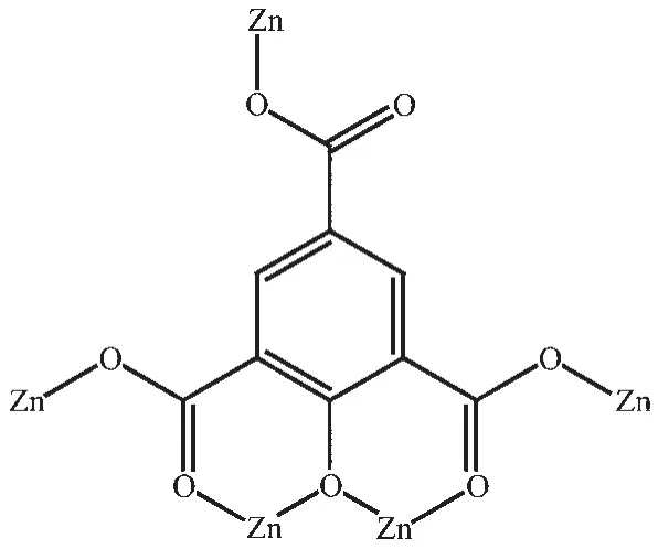
Scheme 1 Coordination mode of O-BTC4-anion in 1
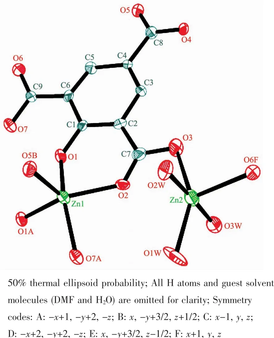
Fig.1 Molecular structure of 1 in ORTEPview

Fig.2 Space-filling view of the 3D framework structure of 1 along the a-axis
2.2 PXRD patterns,IR and thermal stability analysis of 1
The as-synthesized powder X-ray diffraction(PXRD)patterns obtained for the bulk sample of 1 are in good agreement with that of the simulated singlecrystal data,indicating the phase purity of the bulk products.Meanwhile,the sharp diffraction peaks confirm the high crystallinity of 1.The relatively high diffraction peaks of 2θfrom 30°to 40°can be related to the preferred orientation of 1 (Fig.S1).The broad peak of the IR spectrum centered between 3 400 and 3 460 cm-1is mainly attributed to the ν(OH)of the coordinated and lattice water molecules stretching vibrations (Fig.S2).The IR spectrum of 1 shows the characteristic bands of carboxylate groups of the OBTC4-ligand in 1 354~1 470 cm-1for the symmetric stretching vibration and in 1 552~1 674 cm-1for the asymmetric stretching vibration,all are in the general regions[30].In addition,the vanished bands in 1 690~1 720 cm-1for full deprotonation of the carboxylate groups of 1 implicate the full deprotonation of the HO-H3BTC ligand on the reaction with metal ions.All these results match well the structure analysis of 1.Thermogravimetric analysis (TGA)under N2atmosphere from 30 to 800℃was employed to under-stand the thermal stability of 1.As shown in Fig.3,sustained weight loss from room temperature to 600℃may be attributed to the loss of lattice DMF and H2O solvent as well as the coordinated water molecules,and the decomposition of the framework is completed.The ultimate residue may be zinc oxide(Obsd.28.12%,Calcd.32.10%).
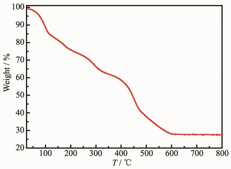
Fig.3 TG curve of 1 in nitrogen atmosphere
2.3 UV-Vis absorption spectra and luminescent properties of 1
The UV-Vis absorption spectroscopic properties for 1 and the organic ligand (HO-H3BTC)are investigated since the HO-H3BTC ligand is a chromophoric linker and present optical property.As given in Fig.4,the solid-state UV-Vis absorption spectra for the free HO-H3BTC ligand and 1 are different at room temperature.The free ligand molecule (Fig.4)shows a relatively high intensity and spin-allowed broad band at ca.262 nm.The main absorption can be ascribed to the π→π*transition within the S0→S1state of HOH3BTC molecule.The main absorption band also appears for 1 but has been red-shifted to ca.323 nm with broad band.The vanished shoulder peak and the red-shift phenomenon can be recognized as the results from the coordination interaction between the carboxylate groups of the HO-H3BTC ligand and Znバion[31].The fluorescence excitation and emission spectra of 1 were examined in the solid state at 298 K.The emission spectra (Fig.5)of 1 displays single broad emission band centered at 437 nm under 365 nm excitation (Fig.S4).Compared to the emission of the free HO-H3BTC ligand (λmax=440 nm),the emission of 1 may be attributed to the intraligand (π→π*)fluorescent emission as the similar emission to that of the free HO-H3BTC ligand[25],as reported for other Zn-MOFs constructed fromorganic ligands with conjugated systems[32].The absolute quantum yield was determined by means of an integrating sphere and the value obtained under an excitation of 365 nm is 8.5%.When the temperature is decreased from 298 K to 10 K,the maximum emission of 1 is red-shifted to 457 nm (Δλ=20 nm).The coordination of Znギions to the HOH3BTC ligand at low temperature (10 K)must allow more efficient intersystem crossing to the triplet state[33].Interestingly,at low temperature (10 K)1 shows an unexpected persistent emission after the removal of UV excitation.Time-resolved emission spectra (TRES)of 1 were recorded using different delays at 10 K(Fig.6).The normalized spectra confirm that the emission of 1 undergoes a progressive slight red-shift while it gradually decreases,whereas the similar phenomenon could not be observed at 298 K.

Fig.4 UV-Vis absorption spectra of 1 and HO-H3BTC ligand in the solid state at room temperature

Fig.5 Solid-state luminescence spectra of 1 at 298 K and 10 K withλex=365 nm
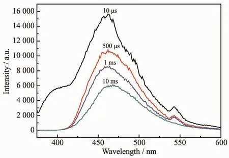
Fig.6 Time-resolved emission spectra at 10 K of 1 at different delays withλex=365 nm
3 Conclusions
In summary,herein we report the design,synthesis and characterization of the luminescent properties of a Zn-based MOF consisting Znギmetal and 2-hydroxyl-1,3,5-benzenetricarboxylic acid(HOH3BTC)ligand,{[Zn4(O-BTC)2(H2O)5]·2DMF·0.5H2O}n(1).Complex 1 presents a 3D framework structure based on a rod-shaped Zn-carboxylate//hydroxyl secondary building unit (SBU).The result demonstrates that the multifunctional HO-H3BTC ligand appears to be an appropriate candidate to generate diverse extended MOFs.In addition,1 shows the afterglow property at low temperature.
Supporting information isavailable at http://www.wjhxxb.cn
猜你喜欢
铁道标准设计(2021年9期)2021-09-26
海南医学(2021年5期)2021-03-25
武术研究(2019年11期)2019-04-20
制造技术与机床(2019年2期)2019-03-06
东坡赤壁诗词(2018年1期)2018-03-31
兵器装备工程学报(2015年5期)2015-12-23
科技视界(2015年27期)2015-10-08
现代电子技术(2014年13期)2014-07-09
中国石油大学学报(自然科学版)(2014年2期)2014-02-28
太原城市职业技术学院学报(2014年11期)2014-02-27