
Heterotopic pancreas adenocarcinoma in the stomach:A case report and literature review
2020-09-14 10:51YaoXiongYueXieDanDanJinXinYingWang
World Journal of Clinical Cases 2020年10期
Yao Xiong,Yue Xie,Dan-Dan Jin,Xin-Ying Wang
Yao Xiong,Yue Xie,Dan-Dan Jin,Xin-Ying Wang, Department of Gastroenterology,Zhujiang Hospital of Southern Medical University,Guangzhou 510280,Guangdong Province,China
Abstract
Key words:Stomach;Pyloric obstruction;Heterotopic pancreas;Adenocarcinoma;Case report
INTRODUCTION
Heterotopic pancreas,also known as ectopicor aberrant pancreas,is defined as pancreatic tissue lying outside its normal location and lacking anatomic or vascular connections with the pancreas[1].The majority of heterotopic pancreatic masses are small(<1 cm)and the patients appear to be asymptomatic[2].The frequency of this disease has been reported to be 0.5% in upper abdominal operations and 0.55%-13.7%at autopsy[3,4].It has been observed in areas such as the gastrointestinal tract[5-7],biliary duct[8],liver[9],spleen[10],and mesentery[11].The most common location of heterotopic pancreas is the stomach and the lesion is usually discovered by endoscopy,appearing as a submucosal mass with an endoluminal growth pattern[12].
Although heterotopic pancreatic tissue is relatively common in the stomach,malignancy arising within it is extremely rare,with only 16 well-documented cases being reported in the English language literature[4,13-27].Herein,we present a case of this rare disease and review the series of other case reports describing malignant transformation of aberrant pancreas in the stomach indexed in the PubMed database(up to December 2019).To our knowledge,our case is the first of its kind reported in a Chinese patient with malignant aberrant pancreas in the stomach;our literature review provides a better understanding of the clinicopathological features,treatment and prognosis of this rare clinical entity.
CASE PRESENTATION
Chief complaints
A 44-year-old female presented to our department with symptoms of intermittent abdominal distension for 6 months and repeated vomiting over the last 3 days.
History of present illness
Because of the abdominal distension,the patient presented to a local hospital for assessment and underwent a gastroscopy 2 months later.The gastroscopy revealed a protruding lesion in the gastric antrum,with its distal margin invading the pyloric ring.Since no malignant tissue was found in the biopsy specimen obtained from the lesion,the patient was treated with acid-suppressive and mucosa-protecting drugs.At 2 months later,gastroscopic re-examination was carried out at the same local hospital and showed gastric retention and pyloric stenosis.An ultrasound gastroscopy was then performed and detected significant thickening of the gastric antrum wall,with a heterogenous hypoechoic lesion where the layered structure had disappeared.
As the abdominal distension symptom persisted and progressed to repeated vomiting,the patient came to our hospital for further evaluation.
History of past illness
The patient had no other significant medical history.She denied history of hypertension,diabetes,coronary heart disease,and any other chronic disease.
Personal and family history
The patient had no significant personal and family history.
Physical examination upon admission
Physical examination showed no abnormality.
Laboratory examinations
The results of complete blood count and routine biochemical investigations were unremarkable.Blood tumor marker tests showed elevated level of carbohydrate antigen 72-4(CA724)(50.1 kU/L;normal range:0-6.9 kU/L)but normal levels of carcinoembryonic antigen(CEA)and carbohydrate antigen 19-9(CA199).
Imaging examinations
Gastroscopy showed that the antrum mucosa was congestive and edematous,and its part near the pylorus appeared rough,with a nodular protruding lesion causing obstruction of the gastric outlet(Figure 1A).Endoscopic passage through the site was difficult,and only an ultrathin electronic gastroscope could pass to the duodenum.The duodenum bulb and descending duodenum appeared unremarkable.Biopsies were not representative for a neoplastic lesion.Abdominal computed tomography showed marked wall thickening of the antrum,which was enhanced by contrast agent(Figure 1B),while the remainder of the abdomen,including the pancreas,was unremarkable.Barium meal examination showed filling defect at the antrum and stenosis of the lumen(Figure 1C).
Pathological findings
A distal gastrectomywas performed.Gross examination of the surgically resected specimen revealed that the gastric pylorus was thickened and rigid.Histopathological examination confirmed the presence of a well-differentiated adenocarcinoma and the coexisting presence of pancreatic heterotopia composed of ducts(Heinrich type III)(Figure 2A and B),thus supporting the diagnosis of a malignancy arising from pancreatic heterotopia in the stomach.The lesions developed predominantly in the submucosal layer and infiltrated the muscular layer.The lesions also invaded and broke through the serosal membrane and invaded bundles of nerves(Figure 2C).The resected margins were free of tumor tissue.
None of the 20 identified lymph nodes was positive.Immunohistochemistry analysis of the cancer cells showed positivity for cytokeratin 7(Figure 3A),cytokeratin,CEA(Figure 3B),CDX-2(Figure 3C),cytokeratin20(partially)(Figure 3D),P53(about 10%)and Ki-67(about 7%),and negativity for Her-2.
FINAL DIAGNOSIS
The final diagnosis was malignant transformation of aberrant pancreas in the stomach.
TREATMENT
At 1 month after surgery,the patient received chemotherapy using the combination FOLFOX6 regimen(folinic acid,fluorouracil,and oxaliplatin),which is a protocol for treatment of resected stomach cancer.However,after three cycles,adverse effects(e.g.,hepatic and mucocutaneous damage)and an increased serum level of CA724(107 kU/L)were observed.The chemotherapy was switched to gemcitabine,which is recommended in pancreatic cancer,and the CA724 serum level decreased gradually(19 kU/L).
OUTCOME AND FOLLOW-UP
The follow-up course was uneventful at 12 months postoperatively.
DISCUSSION
Heterotopic pancreas is often found incidentally during surgery or endoscopic examination and is clinically silent and benign.Malignant transformation of the heterotopic pancreas tissue is extremely rare,thus it is usually neither suspected nor included in the diagnostic work-up of different subepithelial tumors.Most cases are diagnosed by pathological findings after surgery.Guillouet al[28]has proposed the following three criteria for the diagnosis of carcinoma arising from a heterotopic pancreas:(1)The tumor must be within or near the heterotopic pancreatic tissue;(2)A direct transition between the pancreatic structures and carcinoma must be present;and(3)The non-neoplastic heterotopic pancreatic tissue must comprise,at least,fully developed acini and/or ductal structures.The lesion in the present case was compatible with all the three criteria.

Figure 1 Radiologic and endoscopic features of the tumor.A:Gastroscopy showed a nodular protruding lesion at the gastric antrum and stenosis of the pyloric ring;B:Abdominal computed tomography showed that the wall of the antrum was thickened and enhanced(arrow);C:Barium meal examination showed filling defect at the antrum and stenosis of the lumen.
A search of the English and Chinese language literature revealed 16 reports of welldocumented cases of neoplasms arising in the heterotopic pancreas in the stomach,which met the three diagnostic criteria mentioned above(Table 1)[4,13-27].Including the case reported herein,the patients' ages ranged from 31 to 85 years(56.7 years,on average).There was no sex predilection.Except for two patients who were asymptomatic,the others had common symptoms,including epigastralgia,dyspepsia,and vomiting.With respect to tumor markers,excluding eight cases that were not described in detail,33% of the remaining cases had increased CA199 or CEA levels,and increased CA724 level was reported only in our case.Most of the tumors are located in the antropyloric region(70%),which is likely to produce gastric outlet obstruction.A subepithelial tumor-like appearance is most frequently observed,followed by a stenotic or ulcerated appearance.
The diagnosis of malignant transformation in the heterotopic pancreas in the stomach may be difficult by endoscopic biopsy because of the intramural location of the mass and the delayed involvement of the overlying mucosa[29].However,distinction between benign and malignant lesion is critical for patient management.It has been suggested that asymptomatic heterotopic pancreas of less than 2 cm can be monitored without specific therapy.In our review of well-documented cases,the sizes of malignant heterotopic pancreas in the stomach ranged from 1.7 cm to 12.5 cm,with a mean of 4.2 cm.However,in one patient,the tumor was only 0.5 cm×2 cm×2 cm in size but with extensive metastasis to the bone[16].Actually,37.5% of these cases had a tumor of no more than 2 cm.In our case,the tumor appeared as a protruding lesion,causing stenosis and with irregular shape or unclear boundary,making it difficult to accurately measure its size.Therefore,we suggest early treatment for heterotopic pancreas to prevent progression to carcinogenesis,especially when the lesion size is more than 1 cm or if there are signs of obstruction,ulceration or weight loss.Endoscopic ultrasound(EUS)was performed in our patient and other 4 patients in the literature[13,17,25,26].In the case reported by Uraet al[17],EUS showed a swollen perigastric lymph node and changes in mass shape and size during dynamic monitoring,which strongly indicated a malignant lesion.EUS in other cases including ours showed nonspecific features such as heterogenous hypoechoic lesion or thickness of the gastric wall.So using single EUS to detect the lesion itself can hardly predict the histologic diagnosis,but dynamic monitoring and detecting for perigastric lymph nodes by EUS could help distinguishing malignant and benign lesions.As for the interval of endoscopic examination for surveillance of malignant change of heterotopic pancreas,there is still no consensus.Dynamic monitoring by endoscopy was only reported in one case[17].In this case,a mass of 1.8 cm×1.3 cm was detected by EUS and it showed no change 1 year later,but 2 years after the second examination the mass was found to have increased to 3.3 cm×3.0 cm and ultimately diagnosed as an invasive adenocarcinoma extending to the peritoneal surface with lymph node metastases.This case suggests that the interval of endoscopic surveillance of malignant change of heterotopic pancreas should be less than 2 years.Positron emission tomography(PET)has an important role in clinical imaging diagnosis of malignancy.PET findings were reported in only one case[25].In this case,PET scan showed hot spots in gastric wall which indicated a malignant lesion.As the lesion size was large(7.5 cm×4.4 cm),whether PET scan is helpful to diagnose small malignant lesion still need to be further studied.
Histologically,malignant heterotopic pancreas in the stomach has been classified predominantly as adenocarcinomas,with only one report of neuroendocrine carcinoma[20].The Heinrich classification divides pancreatic heterotopia into three types,with type I encompassing typical pancreatic tissue with acini,ducts and islet cells,type II encompassing only exocrine components with numerous acini,few ducts and no islet cells,and type III encompassing numerous ducts,few to no acini,and no islet cells[13].The modification by Gasparet al[30]added a fourth type(type IV),comprising pure endocrine heterotopia containing only islet cells.In the majority of pancreatic heterotopia cases with documented classification,the malignancy arose within a type I heterotopia(46.7%).The next most frequent type was II(33.3%).Our case arose within a type III heterotopic pancreas,which is the least common background for the origin of adenocarcinoma in heterotopic pancreas.
There is no clear evidence of the efficacy of chemotherapy for malignant heterotopic pancreas in the stomach,and the type of adjuvant treatment is not clear because of the small number of reported cases.In our literature review,chemotherapy regimen was reported for only two cases,with one having used gemcitabine[25]and the other having used S-1 and cisplatin[26].We selected the FOLFOX6 regimen based on its use in treatment of resected stomach cancer;however,the patient's serum level of CA724 increased after receiving three cycles of the chemotherapy.We then changed the treatment to gemcitabine monotherapy based on its use in primary pancreatic cancer,and the serum level of CA724 decreased gradually.While gemcitabine seems to be effective in our case,more cases need to be investigated to provide strong evidence to support its use in these rare cases.
The prognosis of malignancies arising from heterotopic pancreas in the stomach is also unknown.In our literature review,28.6% of the patients with a description of the clinical course were alive without evidence of recurrence at least 1 year following resection of the malignant heterotopic pancreas.In addition,21.4% of the patients died from cancer-related complications,and 42.9% of the patients had metastasis to other organs at the time of diagnosis or during the follow-up period.These observations suggest that the prognosis may be poor.
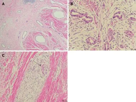
Figure 2 Hematoxylin-eosin histopathological examination of the surgically resected specimen.A:A well-differentiated adenocarcinoma was revealed,coexisting with pancreatic heterotopia composed of ducts(×100);B:High-power view of duct-like structures of adenocarcinoma(×400);C:Invasion of bundles of nerves was seen(arrow)(×200).
CONCLUSION
We have described a rare case of adenocarcinoma arising from heterotopic pancreas in the stomach.It should be noted that heterotopic pancreas is a source of potentially malignant lesion and endoscopic features of its malignant change in the stomach are nonspecific,so we suggest early treatment or close monitoring of the heterotopic pancreas.The rarity of this condition makes the treatment a herculean challenge,and more detailed and in-depth studies on this topic are required.

Figure 3 Immunohistochemical staining.A:Cytokeratin 7(×200);B:Carcinoembryonic antigen(×200);C:CDX-2(×400);D:Cytokeratin 20(×200).

Table 1 Published cases of carcinoma arising from heterotopic pancreas in the stomach
ACKNOWLEDGEMENTS
The authors thank Mr.Yong-Jian Deng,Department of Pathology,Nanfang Hospital of Southern Medical University,for his technical assistance for this study.
 World Journal of Clinical Cases2020年10期
- World Journal of Clinical Cases的其它文章
- French Spine Surgery Society guidelines for management of spinal surgeries during COVID-19 pandemic
- Prophylactic and therapeutic roles of oleanolic acid and its derivatives in several diseases
- Macrophage regulation of graft-vs-host disease
- Antiphospholipid syndrome and its role in pediatric cerebrovascular diseases:A literature review
- Remotely monitored telerehabilitation for cardiac patients:A review of the current situation
- Keystone design perforator island flap in facial defect reconstruction