
Unexplained huge liver infarction presenting as a tumor with bleeding:A case report
2020-09-14 10:51FuHaiWangNingNingYangFengLiuHuTian
World Journal of Clinical Cases 2020年10期
Fu-Hai Wang,Ning-Ning Yang,Feng Liu,Hu Tian
Fu-Hai Wang,Feng Liu,Hu Tian, Department of General Surgery,The First Affiliated Hospital of Shandong First Medical University,Jinan 250014,Shandong Province,China
Ning-Ning Yang, Department of Respiratory and Critical Care Medicine,Shandong Provincial Chest Hospital Affiliated to Shandong University,Jinan 250013,Shandong Province,China
Abstract
Key words:Liver infraction;Liver lesions;Surgery;Imaging;Diagnosis;Case report
INTRODUCTION
Primary liver cancer is one of the most common malignant tumors,and ranks third with respect to tumor mortality worldwide.The incidence of primary liver cancer is much higher in China due to hepatitis B infection[1].The precise diagnosis of liver lesions is helpful for the application of targeted therapeutic options and a good prognosis,while misdiagnosis is sometimes inevitable due to the similar imaging appearances of liver cancer and other benign lesions.We describe a patient with a huge liver infarction and bleeding that was initially considered to be liver cancer with bleeding.A review of the literature on liver infarction was also performed.
CASE PRESENTATION
Chief complaints
A 31-year-old man was admitted with persistent right upper abdominal pain,fever,and anorexia for 6 d.
History of present and past illness
He was healthy without a history of perso nal or family tumors.Abdominal ultrasound revealed fatty liver and mixed echoes in the right lobe of the liver,and a non-enhanced computed tomography(CT)scan of the abdomen also suggested a huge lesion in the right lobe of the liver with mixed density.Antibiotics had been administered in a local hospital to help relieve the patient's symptoms.
Physical examination upon admission
Upon admission,his intermittent fever peaked at 38.5 °C without symptoms of respiratory infection.Percussion pain in the liver area was detected with no other abnormalities upon physical examination.
Laboratory examinations
Laboratory studies excluded hepatitis B and C infection,but showed leukocytosis,neutrophilia,elevated aspartate aminotransferase at 76 U/L,and alanine transaminase at 477.4 U/L.The triglyceride,total cholesterol,carcinoembryonic antigen,alpha-fetoprotein,and carbohydrate antigen-199 levels were in the normal range.
Imaging examinations
An enhanced CT scan indicated fatty liver,and a huge irregular lesion with a maximum diameter of 12.7 cm was observed in the right lobe of the liver.On nonenhanced CT,a slightly high density was also found in the lesion(Figure 1A),which was consistent with previous CT images.The entire tumor was not enhanced,and the peripheral tissues were delayed enhanced(Figure 1B and D).The typical CT image of hepatocellular carcinoma was enhanced at the early stage and with peripheral vascular contrast enhancement.However,for some unusual hepatic tumors,the image feature was delayed enhanced,such as liver metastasis from gastric cancer.The imaging findings suggested a malignant liver tumor with bleeding.Threedimensional reconstruction was performed to identify the relationship between the lesion and adjacent hepatic vessels.No significant interruption of the main liver artery and portal vein was observed(Figure 2A-C).
FINAL DIAGNOSIS
The surgical specimen showed a huge necrotic lesion,and multiple adipose lesions were also observed(Figure 3A).Pathological examination indicated a rare liver infarction(Figure 3B-D).
TREATMENT
The antibiotics ceftriaxone sodium and levofloxacin,as well as hepatoprotective drugs,were administered to control the fever and improve liver function.
OUTCOME AND FOLLOW-UP
On the 9thday after admission,the patient underwent laparoscopic right hepatectomy.He recovered well following treatment with antibiotics,nutritional support,and human albumin,and was discharged on postoperative day 21.No fever or abnormal liver function were subsequently reported.
DISCUSSION
Liver infarction is a type of hepatic necrosis caused by the obstruction of vessels,which is rare due to the hepatic dual blood supply,the tolerance of hepatocytes to low oxygen,and the immediate opening of collateral vessels within the liver[2,3].Simultaneous occlusion of the hepatic artery and portal vein is sometimes considered to be essential in a huge liver infarction[4];however,the abrupt truncation of a single hepatic artery can also lead to severe liver infarction and even death[5].Liver infarction is usually reported at autopsy.Seeleyet al[6]investigated 19 autopsies of patients with hepatic infarcts,and found that ten autopsies showed arterial occlusion,four showed only portal vein thrombosis,and no vascular occlusion was found in the other five patients.In our case,we found no obvious hepatic vessel lesions on the CT scan and three-dimensional reconstruction of the liver.In a subsequent study,Saegusaet al[7]reported 20 cases of hepatic infarction and 17 had circulatory failure.Furthermore,15 had portal vein thrombosis,which indicated the importance of portal vein disturbance in the development of liver infarction.Based on these reports,simultaneous or single occlusion of the hepatic artery and portal vein appears to be the anatomical basis of hepatic infarction.
The early diagnosis of liver infarction is difficult due to its rarity and absence of typical symptoms.It can initially present as chest or right upper abdominal pain,fever,or with associated nausea and vomiting[8-10].In addition,related laboratory studies for hematological and biochemical markers are not specific,which makes the diagnosis challenging[11,12].In our report,the patient had right upper abdominal pain and fever,and related serum tests showed leukocytosis,neutrophilia,and elevated aspartate aminotransferase and alanine transaminase,but it is difficult to differentiate between liver abscess or a tumor with bleeding or infection.Thus,a CT scan was performed,which indicated the areas of liver infarction with low attenuation that were circumscribed and wedge-shaped,and extended to the periphery of the liver[11].The CT images in our patient showed an irregular lesion with an inside of slightly high density,which was considered to be bleeding.It was reported that gadophrin-2 displayed a persistent necrosis-specific contrast enhancement on magnetic resonance imaging,and was considered useful for diagnosis in a rat model of reperfused liver infarction[13].Hepatobiliary scintigraphy is also considered to be a sensitive method for detecting early hepatic infarction even before ultrasonographic changes occur,which is helpful in improving prognosis[14].
The causes of liver infarction are diverse,although primary lesions in hepatic vessels are rare.It has been suggested that liver infarction may be secondary to circulatory shock,sepsis,anesthesia,or biliary disease[8,10].We reviewed the literature on liver infarction(Table 1)and concluded that the common causes of this disorder are as follows:(1)Iatrogenic injury of hepatic vessels,mainly the hepatic artery.In normal liver,approximately 75% of the blood supply is from the portal vein,and 25%is from the hepatic artery.An anatomical abnormality of the hepatic artery is frequently observed,such as the right hepatic artery that can be derived from the proper or common hepatic artery or superior mesenteric artery.Wonget al[15]reported liver infarction due to injury to the right hepatic artery and portal vein during laparoscopic cholecystectomy.Surgeons should avoid hepatic vessel injuries during gallbladder,bile duct,liver,and pancreas surgery.(2)Systemic diseases:Churg-Strauss syndrome is characterized by granulomatous vasculitis of multiple organ systems,which can lead to irregular narrowing of hepatic arteries and even liver infarction[3].In pregnant or postpartum woman,antiphospholipid syndrome(APS)and systemic lupus erythematosus(SLE)are also considered causes of liver infarction due to multiple thrombosesin vivo[11,16,17].Related serum studies on anti-nuclear antibodies,anti-double-stranded DNA,anti-cardiolipin antibodies,and lupus anticoagulant are useful for the diagnosis of APS and SLE[18].In the present report,the above markers were investigated and found to be in the normal range.A hypercoagulable state,such as disseminated intravascular coagulation caused by severe postpartum hemorrhage,can induce thrombosis formation in hepatic vessels,hepatic infarction,and even acute hepatic failure[19,20].(3)Treatment of liver diseases includes transarterial chemoembolization(TACE)and transjugular intrahepatic portosystemic shunt(TIPS).TIPS is performed for the treatment of variceal bleeding and refractory ascites in patients with portal hypertension[21],while TACE is used for the treatment of liver cancer,liver bleeding,or for diagnostic purposes[22].TIPS can induce hepatic hypoperfusion in the portal vein,while TACE interrupts liver artery blood flow.Therefore,the risk of liver infarction due to TACE is increased by TIPS[9,23].Before TACE or TIPS is undertaken,it is important to evaluate the condition of the hepatic artery and portal vein for a better prognosis.And(4)Other causes:Ashidaet al[2]reported a case of liver infarction due to liver abscess,without the occlusion of hepatic vessels.In our case,liver infarction was not due to the above three common causes;however,the patient had fatty liver and severe fatty degeneration of hepatocytes.Whether this was the origin of liver infarction requires further investigation.
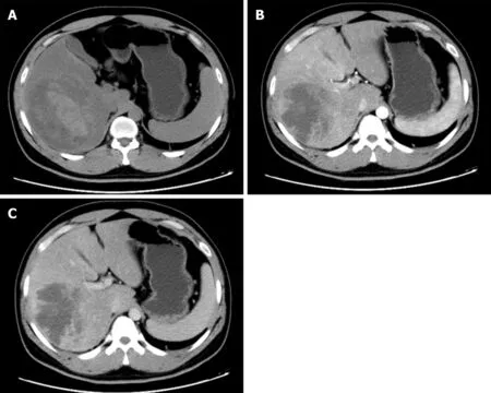
Figure 1 Computed tomography imaging of liver infarction.A:Plain scan phase of computed tomography(CT)detected an irregular lesion in the right lobe of the liver;B:Arterial phase of CT showed no obvious enhancement;C:Venous phase of CT showed no obvious enhancement.
CONCLUSION
The causes of liver infarction are complex with an outcome that is sometimes systemic,and can even be fatal.Simple imaging,such as ultrasound,CT scan,or magnetic resonance imaging,is sometimes insufficient,and at least two imaging methods are required.In addition,detailed history taking,physical examination,and related serum studies are also crucial.The treatment of primary diseases,such as APS and SLE,is essential,and treatment with antibiotics,hepatoprotective drugs,thrombolysis,hormones,and even surgery are also significant.We should also be aware of the clinical characteristics,imaging appearance,and serum results of other liver lesions,such as liver cancer and abscess,as the early precise diagnosis of liver infarction is difficult and important for controlling further progression and improving prognosis.

Figure 2 Three-dimensional reconstruction of liver and lesion.A:Arterial reconstruction of the liver revealed the relationship between lesion and hepatic artery;B:Venous reconstruction of the liver revealed the relationship between lesion and portal vein;C:Mixture of arterial and venous reconstruction.
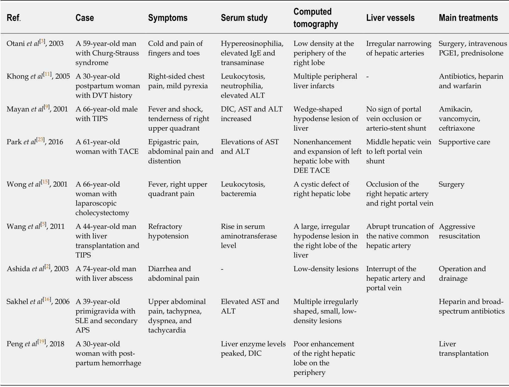
Table 1 Literature review for liver infraction
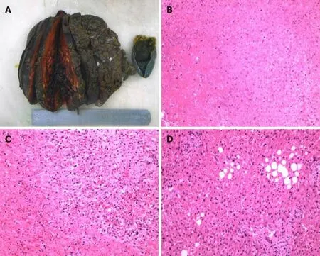
Figure 3 Pathological examination of lesion.A:Necrotic lesion was 16 cm×12 cm×8 cm in size without an envelope;B:Pathological section analysis of liver infarction;C:Pathological section analysis of tissue adjacent to lesion;D:Pathological section analysis of normal liver tissue,revealing fatty degeneration of hepatocytes.
 World Journal of Clinical Cases2020年10期
- World Journal of Clinical Cases的其它文章
- French Spine Surgery Society guidelines for management of spinal surgeries during COVID-19 pandemic
- Prophylactic and therapeutic roles of oleanolic acid and its derivatives in several diseases
- Macrophage regulation of graft-vs-host disease
- Antiphospholipid syndrome and its role in pediatric cerebrovascular diseases:A literature review
- Remotely monitored telerehabilitation for cardiac patients:A review of the current situation
- Keystone design perforator island flap in facial defect reconstruction