
Cryptococcal pneumonia in a human immunodeficiency virusnegative patient:A case report
2020-09-14 10:51XueQinJiangYanBeiZhang
World Journal of Clinical Cases 2020年10期
Xue-Qin Jiang,Yan-Bei Zhang
Xue-Qin Jiang,Yan-Bei Zhang,Department of Geriatric Respiratory and Critical Care Medicine,The First Affiliated Hospital of Anhui Medical University,Hefei 230022,Anhui Province,China
Abstract
Key words:Pneumonia;Cryptococcosis;Voriconazole;Case report
INTRODUCTION
Cryptococcosis is a form of opportunistic invasive mycosis that is driven by infection withCryptococcus neoformans(C.neoformans),which is a relatively common environmental microorganism.Cryptococcal infections are most common in immunocompromised patients,but they can also infect immunocompetent individuals.PathogeniccryptococcihaveC.neoformans var neoformansandC.neogormans var gattii,which are the two most common causes of this infection in humans[1,2].The mechanistic basis for such cryptococcal infections remains to be fully elucidated[3],withC.neoformansinfections being most common in HIV-positive patients and Cryptococcus gattiiinfections being more commonly observed in immunocompetent persons.Inhalational exposure is thought to be the most common mode of infection,although bird droppings are also believed to be a potential source of infection in some cases[4].While exposure toC.neoformansis relatively common,only patients with dysfunctional cell-mediated immune responses typically suffer from invasive forms of cryptococcal disease[5].Infections withcryptococcican result in skin lesions,or in more serious conditions including meningitis and pneumonia[6].Pulmonary cryptococcosis often presents with a series of variable and nonspecific physical symptoms and imaging findings,leading it to often be incorrectly diagnosed as a more typical form of pneumonia,or as another condition such as diffuse lung disease or lung cancer.Herein,we describe the case of an immunocompetent patient who suffered from pulmonary cryptococcosis complicated by fluconazole resistance and voriconazole sensitivity.
CASE PRESENTATION
Chief complaints
A 42-year-old man was admitted to our hospital suffering from a non-resolving case of pneumonia.The patient had suffered from slight cough for 1 mo,without any associated headache,pleuritic,fever,or sputum production.Two weeks prior to admission,the patient had undergone a routine physical examination,during which a chest computed tomography(CT)scan detected the presence of infiltrative pneumonia in the upper-left lung(Figure 1).The patient had no history of allergies or pulmonary tuberculosis,and he was not a smoker.
History of illness
The patient had a free previous medical history.
Physical examination
At the time of initial admission,the patient had a heart rate of 84 bpm,respiratory rate of 20 breaths per minute,body temperature of 36.5 °C,and blood pressure of 180/120 mmHg.
Laboratory examinations
Upon physical examination,the patient exhibited no sighs of wheezing or crackling in the lungs,and no neck lymph nodes were palpable.A complete blood count examination revealed leukocyte numbers to be in the normal range(6.85×10⁹/L).Normal liver and renal function and normal electrolyte levels were also detected during routine laboratory testing.The patient was also found to be seronegative for an anti-human immunodeficiency virus antibody.Sputum was first analyzed for acidfast bacteria,with this examination failing to detect any microorganisms.CT-guided lung puncture was next conducted,and pathological examination of the collected tissue revealed the presence of granulomatous lesions containing both fungal spores and multinucleated giant cells.Hematoxylin and eosin and periodic-acid-Schiff staining of these tissue samples confirmed the presence of yeast-like fungi both in intercellular spaces and within the observed giant multinucleated cells(Figure 2).

Figure 1 Multiple modes and areas of patchy increased density were evident in the upper left lung.
FINAL DIAGNOSIS
The final diagnosis of the present case is cryptococcal pneumonia.
TREATMENT
The antibiotic regimen on which the patient had been placed was subsequently replaced with a once-daily injection of fluconazole 400 mg(doubling the first dose)for 1 wk,after which the patient was discharged and prescribed oral fluconazole 400 mg once a day.However,no improvements in respiratory symptoms or radiographic findings were detected after a 6-wk treatment period(Figure 3).The patient was found to have a serum cryptococcal antigen titer > 1:80 after this 6-wk period.The patient was thereafter administered with 200 mg oral voriconazole twice per day for 10 wk.
OUTCOME AND FOLLOW-UP
The patient's overall condition improved,with chest X-rays demonstrating a steady decrease in the size of the left lung mass(Figure 4).Following a 9-mo voriconazole course,> 90% lesion absorption was observed(Figure 5).
DISCUSSION
Cryptococcosis is a form of invasive infectious disease caused by pathogeniccryptococci.Soil contaminated with bird droppings is one of the most common sources ofC.neoformans[7-9],with human infections often occurring due to microbial inhalation.However,there are some documented cases of person-to-person or animal-to-person cryptococcal transmission.The patient described in the present case report lived on an apartment block wherein 40 chickens were kept.Innate and T cell-mediated immunities are the primary mediators of host immune responses toC.neoformans[10],resulting in the elevated incidence of cryptococcal infections observed among the immunocompromised.
When symptomatic,pulmonary cryptococcosis can present as a form of pneumonia associated with fever,weight loss,chest pain,coughing,and hemoptysis.However,in up to 30% of patients,these infections are asymptomatic and are only detected incidentally[11].Typically,C.neoformansinfections can be safely and reliably treatedviaoral administration of fluconazole.Any patients suffering from serious diseaseassociated symptoms,multiple lung nodules,extensive lung infiltration,and/or positive serum cryptococcal antigen levels represent good candidates for therapeutic intervention.Patients are typically administered with 400 mg of fluconazole per day for 6-12 mo,with an initial 4-wk induction dose of 600-800 mg[12].
CONCLUSION
In the present study,we observed no significant improvements in patient condition after 6 wk of treatment with fluconazole 400 mg once a day(doubling the first dose).The patient was therefore administered with voriconazole(200 mg,twice daily).After 10 wk,the patient exhibited general improvements and a reduction in mass size,with 90% lesion absorption having been achieved following a 9-mo treatment period.Maybe,the failure of initial treatment with fluconazole may be caused by insufficient initial dosing of fluconazole.Meanwhile,the present case highlights the potential for the successful treatment of pulmonary cryptococcosis using voriconazole.Further patient follow-up is ongoing.

Figure 2 Hematoxylin and eosin and periodic-acid-Schiff-stained lung tissue sections highlighted the presence of granulomatous inflammation containing yeast-like microbes that were surrounded by clear halos within multinucleated giant cells and in intercellular spaces.

Figure 3 Multiple modes and areas of patchy increased density were evident in the upper left lung,with no significant changes relative to Figure 1.
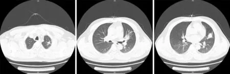
Figure 4 Multiple modes and areas of patchy increased density were evident in the upper left lung,with significant reductions relative to Figure 3.
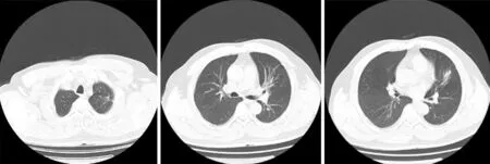
Figure 5 A small cable-like area of increased density was evident in the upper left lung,consistent with a > 90% reduction in the mass relative to the previous examination.
 World Journal of Clinical Cases2020年10期
- World Journal of Clinical Cases的其它文章
- French Spine Surgery Society guidelines for management of spinal surgeries during COVID-19 pandemic
- Prophylactic and therapeutic roles of oleanolic acid and its derivatives in several diseases
- Macrophage regulation of graft-vs-host disease
- Antiphospholipid syndrome and its role in pediatric cerebrovascular diseases:A literature review
- Remotely monitored telerehabilitation for cardiac patients:A review of the current situation
- Keystone design perforator island flap in facial defect reconstruction