
CD24+CD44+胰腺癌细胞的获取及其干细胞特性的初步鉴定
2011-05-28 08:56虞先濬刘辰徐近龙江倪泉兴
中国癌症杂志 2011年2期
虞先濬 刘辰 徐近 龙江 倪泉兴
复旦大学附属肿瘤医院胰腺肝胆外科,复旦大学上海医学院肿瘤学系,上海 200032
CD24+CD44+胰腺癌细胞的获取及其干细胞特性的初步鉴定
虞先濬 刘辰 徐近 龙江 倪泉兴
复旦大学附属肿瘤医院胰腺肝胆外科,复旦大学上海医学院肿瘤学系,上海 200032
背景与目的:近年研究发现,肿瘤内存在极小一部分细胞,决定肿瘤的恶性表型及生物学特性,被称为“肿瘤干细胞”。本研究拟从人原发胰腺癌组织中找到具有干细胞特性的胰腺癌细胞,为今后靶向干预胰腺癌干细胞,解决胰腺癌治疗的临床难题提供研究基础。方法:将人原发胰腺癌组织种植于NOD/SCID小鼠成瘤,对移植瘤细胞进行流式细胞分选;将流式细胞仪分选获得的细胞用无血清、含有生长因子的DMEM/F12培养基培养,观察细胞的生长、传代及体外干细胞球形成能力;将细胞接种于非肥胖型糖尿病/重症联合免疫缺陷型 (Nonobese diabetic/severe combined immunodeficiency,NOD/SCID)小鼠,观察、比较成瘤率,免疫组化法检测干细胞移植瘤肿瘤分化相关抗原Ki67、CK7及甲基化调控因子MBD1的表达;免疫荧光检测细胞Hedgehog、BMI-1的表达。结果:人原发胰腺癌组织种植于NOD/SCID小鼠可部分成瘤,对移植瘤细胞进行流式细胞分选,获得CD24+CD44+细胞(获得率0.6%~1.8%);将CD24+CD44+细胞用无血清、含有生长因子的DMEM/F12培养基培养,细胞呈悬浮球状生长,可连续传代并能再次形成干细胞球;将102个CD24+CD44+细胞接种于NOD/SCID小鼠,成瘤率12.5%(1/8小鼠成瘤),将103个CD24+CD44+细胞接种于NOD/SCID小鼠,4周后100%成瘤,而同样数量的CD24-CD44-胰腺癌细胞则无法成瘤(P<0.05),免疫组化法检测干细胞移植瘤中部分细胞表达Ki67、CK7及MBD1阳性;免疫荧光检测证实CD24+CD44+细胞阳性表达Hedgehog、BMI-1,表达量分别高于CD24-CD44-细胞9.95倍和2.74倍(P<0.05)。结论:所获得的CD24+CD44+细胞具有胰腺癌干细胞特性,可作为今后胰腺癌干细胞研究的实验基础。
胰腺癌; 干细胞; Hedgehog; BMI-1
近年来,胰腺癌的发病率明显上升。胰腺癌恶性程度高,生物学行为特异,容易发生早期转移,且对放、化疗不敏感,手术切除率低,发病率接近死亡率,预后很差[1]。因此,深入研究胰腺癌的特性,探求胰腺癌发生、发展的根源,具有重要的意义。研究表明,多数恶性肿瘤中存在极少部分的细胞群体,能够无限生长、永久生存,并具备自我更新、多向分化的能力,是恶性肿瘤失控生长、转移、耐药和预后差的根源[2]。本研究拟从人原发胰腺癌组织中找到具有干细胞特性的胰腺癌细胞,为今后靶向干预胰腺癌干细胞,解决胰腺癌治疗的临床难题提供研究基础。
1 资料和方法
1.1 人原发胰腺癌组织的异种种植
取手术切除的新鲜人原发胰腺癌组织10例,其中6例男性,4例女性,平均年龄(54.7±10.3)岁(41~70岁),TNM分期(UICC,2010)均为Ⅲ/Ⅵ期,病理证实为导管腺癌,术前均未行化疗。将肿瘤组织在无菌RPMI 1640培养基内制成2 mm×2 mm大小的肿瘤组织条,用无血清的PBS溶液冲洗,种植于8周龄的非肥胖型糖尿病/重症联合免疫缺陷型 (Nonobese diabetic/severe combined immunodeficiency,NOD/SCID) 雌性小鼠(8~10周龄,SPF级环境饲养)背部皮下,成瘤后取出移植瘤,分别用胶原酶消化后制成单细胞悬液。
1.2 流式细胞分选
取对数生长期的细胞(1×10⁶/100 μL),用胰酶消化2~3 min,加入HBSS/2%FBS溶液冲洗,重悬细胞,DAPI(浓度为1 μg/mL)染色。加入PE标记的抗人CD44,FITC标记的抗人CD24(美国PharMingen公司,浓度均为1∶40),冰上温育20 min。用流式细胞仪(美国BD公司)进行分选。细胞分选重复2次,分选后各亚群细胞纯度>95%。
1.3 CD24+CD44+细胞的体外培养
收集对数生长期的细胞,用无血清、含生长因子EGF、FGF10的DMEM-F12培养基重悬、培养,收集细胞制成单细胞悬液,接种于无血清DMEM-F12培养基传代。
1.4 CD24+CD44+细胞体内成瘤实验
取8周龄NOD/SCID雌性小鼠24只,饲养于SPF级环境。分为3组,每组8只,分别接种102、103、104数量级的细胞。将CD24+CD44+细胞接种于小鼠右侧背部, CD24-CD44-细胞接种于小鼠左侧背部,每周观察成瘤情况及肿瘤大小,12周后处死小鼠。移植瘤取出后切片,进行HE及免疫组化染色。
1.5 免疫组化
采用二步法免疫组化染色(EnVision System),Ki67(购自美国DAKO公司)、CK7、MBD1(均购自美国Santa Cruz公司)一抗的工作浓度均为1∶100。常规脱蜡、水化、抗原修复,一抗和二抗分别温育、DAB显色、苏木精复染,中性树脂封片观察。用PBS代替一抗作为阴性对照。
1.6 免疫荧光
将细胞分为CD24+CD44+和CD24-CD44-2组。取对数生长细胞,制备细胞涂片,固定、通透、封闭后加入一抗(抗人Hedgehog、抗人BMI-1,均购自美国Santa Cruz公司,浓度为1∶100)温育1 h,PBS冲洗,加入荧光标记的二抗(浓度为1∶50),冲洗、封片,荧光显微镜观察。MoticFluo1.0图像分析系统进行图像分析,根据荧光显示的亮度,测定其灰度值,代表蛋白的相对表达量。
1.7 统计学处理
采用SPSS 19.0统计软件进行分析,计数资料用χ2检验或Fisher’s确切概率检验,计量资料的比较用t检验。P<0.05为有统计学意义。
2 结 果
2.1 CD24+CD44+胰腺癌细胞的获取
10例人原发胰腺癌组织异种种植2周后开始成瘤,4周后共有3例成瘤,瘤体大小(995.8±46.8) mm3,未见明显转移灶,移植瘤经HE染色证实为导管腺癌。将3例移植瘤分别进行流式细胞分选,CD24+细胞的比例为2.5%~26.8%,CD44+细胞的比例为2.8%~9.5%,CD24+CD44+细胞的比例为0.6%~1.8%(图1)。
2.2 CD24+CD44+细胞的体外成球能力
将CD24+CD44+细胞用无血清的含生长因子的DMEM/F12培养基培养,4~5 d后细胞呈悬浮球状生长(图2),12 d左右将细胞传代,传代细胞仍能悬浮球状生长。


2.3 CD24+CD44+细胞的体内成瘤能力
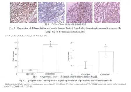
将CD24+CD44+细胞接种于NOD/SCID小鼠右侧背部,CD24-CD44-细胞接种于小鼠左侧背部,观察小鼠成瘤率。102组:CD24-CD44-细胞均未成瘤(0/8),CD24+CD44+细胞有1例成瘤(1/8);103组:CD24-CD44-细胞均未成瘤(0/8),CD24+CD44+细胞均成瘤(8/8)(P<0.05);104组:CD24-CD44-细胞有2例成瘤(2/8),CD24+CD44+细胞均成瘤(8/8)。移植瘤取出后切片,进行HE染色及Ki67、CK7、MBD1免疫组化染色(图3)。
2.4 肿瘤干细胞“自我更新”相关信号通路因子的表达
免疫荧光检测CD24+CD44+和CD24-CD44-2组细胞Hedgehog、BMI-1的表达,发现CD24+CD44+细胞Hedgehog、BMI-1的表达较CD24-CD44-细胞分别上调了9.95倍和2.74倍(P<0.05,图4)。
3 讨 论
胰腺癌作为一个日趋多见的消化道恶性肿瘤,有其特有的生物学特性:容易发生早期转移;对化、放疗耐药;进展迅速、预后极差。因此,深入研究胰腺癌的特性,探求胰腺癌发生、发展的根源,具有重要的意义。
2007年,Simeone等首次报道发现了胰腺癌干细胞,这些细胞仅占癌细胞总数的0.2%~0.8%,具有自我更新及多向分化潜能,并能不断适应各种恶劣环境的变化而永久生存,被认为是胰腺癌无限增殖、早期转移和化、放疗耐药的根源。100个具有干细胞特性的肿瘤细胞就可使半数以上的小鼠成瘤,其致癌性是普通肿瘤细胞的100倍[3]。有理由相信,如果能够靶向干预胰腺癌的干细胞特性,就有可能在这恶性顽疾的治疗上获得重大的突破。
对于胰腺癌干细胞的表面标志物,目前尚无一致的定论。2007年Li等[4]用流式细胞仪分离出CD44、CD24、ESA三阳性的胰腺癌干细胞。同年,Hermann等[5]报道CD133+的胰腺癌细胞具有强大的致瘤、耐药和转移等干细胞特性。
甲基化结合域蛋白1 (methyl-CpG binding domain protein 1,MBD1)是一个重要的转录调控因子,它通过特异地结合DNA上甲基化的CpG位点而发挥活跃的转录抑制作用。我们前期的研究发现,MBD1在胰腺癌中存在高表达,并与胰腺癌的生长增殖、淋巴转移以及上皮-间质转化过程密切相关[6-9]。本研究在胰腺癌干细胞移植瘤中检测发现MBD1表达阳性,提示胰腺癌干细胞特性可能与甲基化调控相关,为今后深入研究甲基化调控与胰腺癌干细胞特性之间的联系及相关机制提供启示。
胰腺癌干细胞的核心本质为自我更新及无限生长能力,多种信号传导通路的异常激活参与了胰腺癌干细胞的自我更新特性,如Hedgehog、Notch、BMI-1、PTEN等[10-13]。研究发现,Hedgehog与BMI-1的活化可使肿瘤干细胞球数量增多、体积增大,增强干细胞的多向分化及传代后再次形成干细胞球的能力,在体内,下调Hedgehog与BMI-1表达可抑制NOD/SCID小鼠成瘤[14]。Li等[4]发现,胰腺癌干细胞中Hedgehog的表达高于普通胰腺癌细胞近10倍。异常表达的Hedgehog信号可促使正常胰腺导管发生癌前病变,并向胰腺癌逐渐发展,活化的Hedgehog还可维持胰腺癌的恶性表型,而阻断Hedgehog表达则会减缓胰腺癌增殖、促进细胞凋亡,进而抑制肿瘤转移、改善肿瘤耐药[15-16]。BMI-1同样与胰腺癌的增殖、转移、预后有关[17],本研究中,具有肿瘤干细胞特性的CD24+CD44+细胞表达Hedgehog、BMI-1均明显高于普通胰腺癌细胞,证实Hedgehog和BMI-1的表达与胰腺癌干细胞特性相关。
本研究将人原发胰腺癌组织种植于NOD/SCID小鼠成瘤,对移植瘤细胞进行流式细胞分选,获得CD24+CD44+细胞(获得率0.6%~1.8%); 将CD24+CD44+细胞用无血清、含有生长因子的DMEM/F12培养基培养,细胞呈悬浮球状生长,可连续传代并能再次形成干细胞球;将103个CD24+CD44+细胞接种NOD/SCID小鼠,4周后100%成瘤,而同样数量CD24-CD44-胰腺癌细胞则无法成瘤;免疫荧光检测证实CD24+CD44+细胞阳性表达Hedgehog、BMI-1,表达量显著高于CD24-CD44-细胞。证明我们所获得的CD24+CD44+细胞具有胰腺癌干细胞特性,为今后靶向干预胰腺癌干细胞,解决胰腺癌治疗的临床难题提供研究基础。
[1]Li DH, Xie KP, Wolff R, et al. Pancreatic cancer [J].Lancet, 2004, 363(9414):1049-1057.
[2]Reya T, Morrison SJ, Clarke MF, et al. Stem cells, cancer, and cancer stem cells[J]. Nature, 2001, 414(6859): 105-111.
[3]Lee CJ, Dosch J, Simeone DM. Pancreatic cancer stem cells[J]. J Clin Oncol, 2008, 26(17): 2806-2812.
[4]Li C, Heidt DG, Dalerba P, et al. Identification of pancreatic cancer stem cells [J]. Cancer Res, 2007, 67(3): 1030-1037.
[5]Hermann PC, Huber SL, Herrler T, et al. Distinct populations of cancer stem cells determine tumor growth and metastatic activity in human pancreatic cancer [J]. Cell Stem Cell,2007, 1(3): 313-323.
[6]Yu XJ, Long J, Fu DL, et al. Analysis of gene expression profiles in pancreatic carcinoma by using cDNA microarray[J]. Hepatobiliary Pancreat Dis Int, 2003, 2(3): 467-470.
[7]Liu C, Chen Y, Yu X, et al. Proteomic analysis of differential proteins in pancreatic carcinomas:Effects of MBD1 knockdown by stable RNA interference [J]. BMC Cancer, 2008,8: 121.
[8]Xu J, Liu C, Yu XJ, et al. Activation of multiple tumor suppressor genes by MBD1 siRNA in pancreatic cancer cell line BxPC-3[J]. Zhonghua Yi Xue Za Zhi, 2008, 88(28):1948-1951.
[9]Luo G, Jin C, Long J, et al. RNA interference of MBD1 in BxPC-3 human pancreatic cancer cells delivered by PLGA-poloxamer nanoparticles[J]. Cancer Biol Ther, 2009, 8(7):594-598.
[10]Simeone DM. Pancreatic cancer stem cells: implications for the treatment of pancreatic cancer [J]. Clin Cancer Res,2008, 14(18): 5646-5648.
[11]Park IK, Morrison SJ, Clarke MF. Bmi1, stem cells, and senescence regulation [J]. J Clin Invest, 2004, 113(2):175-179.
[12]Androutsellis-Theotokis A, Leker RR, Soldner F, et al. Notch signaling regulates stem cell numbers in vitro and in vivo[J]. Nature, 2006, 442 (7104): 823-826.
[13]Lindgren AG, Natsuhara K, Tian E, et al. Loss of PTEN causes tumor initiation following differentiation of murine pluripotent stem cells due to failed repression of nanog[J]. PLoS One,2011, 6(1): e16478.
[14]Liu S, Dontu G, Mantle ID, et al. Hedgehog signaling and Bmi-1 regulate self-renewal of normal and malignant human mammary stem cells [J]. Cancer Res, 2006, 66(12): 6063-6071.
[15]Thayer SP, di Magliano MP, Heiser PW, et al. Hedgehog is an early and late mediator of pancreatic cancer tumorigenesis[J]. Nature, 2003, 425(6960): 851-856.
[16]Olive KP, Jacobetz MA, Davidson CJ, et al. Inhibition of Hedgehog signaling enhances delivery of chemotherapy in a mouse model of pancreatic cancer[J]. Science, 2009,324(5933): 1457-1461.
[17]Song W, Tao K, Li H, et al. Bmi-1 is related to proliferation,survival and poor prognosis in pancreatic cancer[J]. Cancer Sci, 2010, 101(7): 1754-1760.
The isolation and functional verification of the cells with CD24CD44 double positive expression from primary human pancreatic adenocarcinomas
YU Xian-jun,LIU Chen,XU Jin,LONG Jiang,NI Quan-xing(Department of Pancreatic & Hepatobiliary Surgery, Fudan University Shanghai Cancer Center; and Department of Oncology, Shanghai Medical College, Fudan University,Shanghai 200032, China)
NI Quan-xing E-mail:blurlc@hotmail.com
Background and purpose:Emerging evidence has shown that cancer stem cells, which is a few subset of cells in tumor, could determine the malignant phenotype and biological behavior of the tumor, This study aimed to isolate a highly tumorigenic subpopulation of cells from primary human pancreatic adenocarcinomas and verify the biological function of the cells.Methods:Primary human pancreatic adenocarcinomas was planted in immunocompromised (NOD-SCID) mice, and then fl ow cytometry was used to sort the cells into several subpopulation.The sorted cells were cultured in a serum-free DMEM/F12 medium with EGF and FGF to observe the proliferation,passage and spheroid forming capacity of the cells, the cells with those unique features were injected s.c. into NOD/SCID mice to determine the tumorigenicity. The expression of differentiation markers like Ki67, CK7 and DNA methylation regulating factor MBD1 in xenograft tumors was examined by immunohistochemistry, and the expression of Hedgehog and BMI-1 was observed by immunof l uorescence method.Results:Through the processes, a highly tumorigenic subpopulation of pancreatic cancer cells were isolated and had been shown positive expression of CD44 and CD24 of cell surface markers. CD24+CD44+cells could form tumor spheroids in nonadherent culture conditions,and the spheroids also had CD24+CD44+features and could be passaged multiple times without loss of tumor spheroids forming capability. In vivo study, the tumorigenesis was dependent on the number of the cells that implanted into mice.Some of CD24+CD44+tumor cells had been observed to also express the differentiation markers Ki67, CK7 and DNA methylation regulating factor MBD1, and the expression of Hedgehog and BMI-1 were significantly up-regulated about 9.95 fold and 2.74 fold, respectively, in CD24+CD44+cells compared with CD24-CD44-cells.Conclusion:The CD24+CD44+cells are highly tumorigenic subpopulation of pancreatic cancer cells and might be the stem cells of pancreatic adenocarcinomas.
Pancreatic carcinoma; Stem cell; Hedgehog; BMI-1
10.3969/j.issn.1007-3969.2011.02.002
R735.9;R73-35
A
1007-3639(2011)02-0086-05
国家自然科学基金资助项目(No:81001058)。
倪泉兴 E-mail:blurlc@hotmail.com
2010-08-25
2010-10-23)
猜你喜欢
保健医苑(2022年6期)2022-07-08
植物保护(2021年5期)2021-10-12
浙江医学(2020年9期)2020-07-01
浙江中西医结合杂志(2019年4期)2019-05-05
浙江医学(2019年2期)2019-01-23
石河子大学学报(自然科学版)(2018年6期)2018-04-12
癌变·畸变·突变(2018年2期)2018-04-09
天津医药(2016年9期)2016-10-20
郑州大学学报(医学版)(2016年4期)2016-08-11
中国继续医学教育(2015年1期)2016-01-06
